System and method for differentiating benign from malignant contrast-enhanced lesions

a contrast enhancement and magnetic resonance imaging technology, applied in the field of dynamic contrast enhancement magnetic resonance imaging, can solve the problems of small positive predictive value (ppv) of biopsies, and the inability to use the presence of enhancement alone to differentiate benign from malignant lesions

Inactive Publication Date: 2011-08-04
BOARD OF TRUSTEES OPERATING MICHIGAN STATE UNIV
View PDF1 Cites 26 Cited by
  • Summary
  • Abstract
  • Description
  • Claims
  • Application Information

AI Technical Summary

Problems solved by technology

Although DCE-MRI demonstrates high sensitivity to invasive breast cancers, one major limitation is the low specificity caused by the overlap in enhancement between benign and malignant le...

Method used


Image

  • System and method for differentiating benign from malignant contrast-enhanced lesions
  • System and method for differentiating benign from malignant contrast-enhanced lesions
  • System and method for differentiating benign from malignant contrast-enhanced lesions

Examples


Embodiment Construction

Materials and Methods

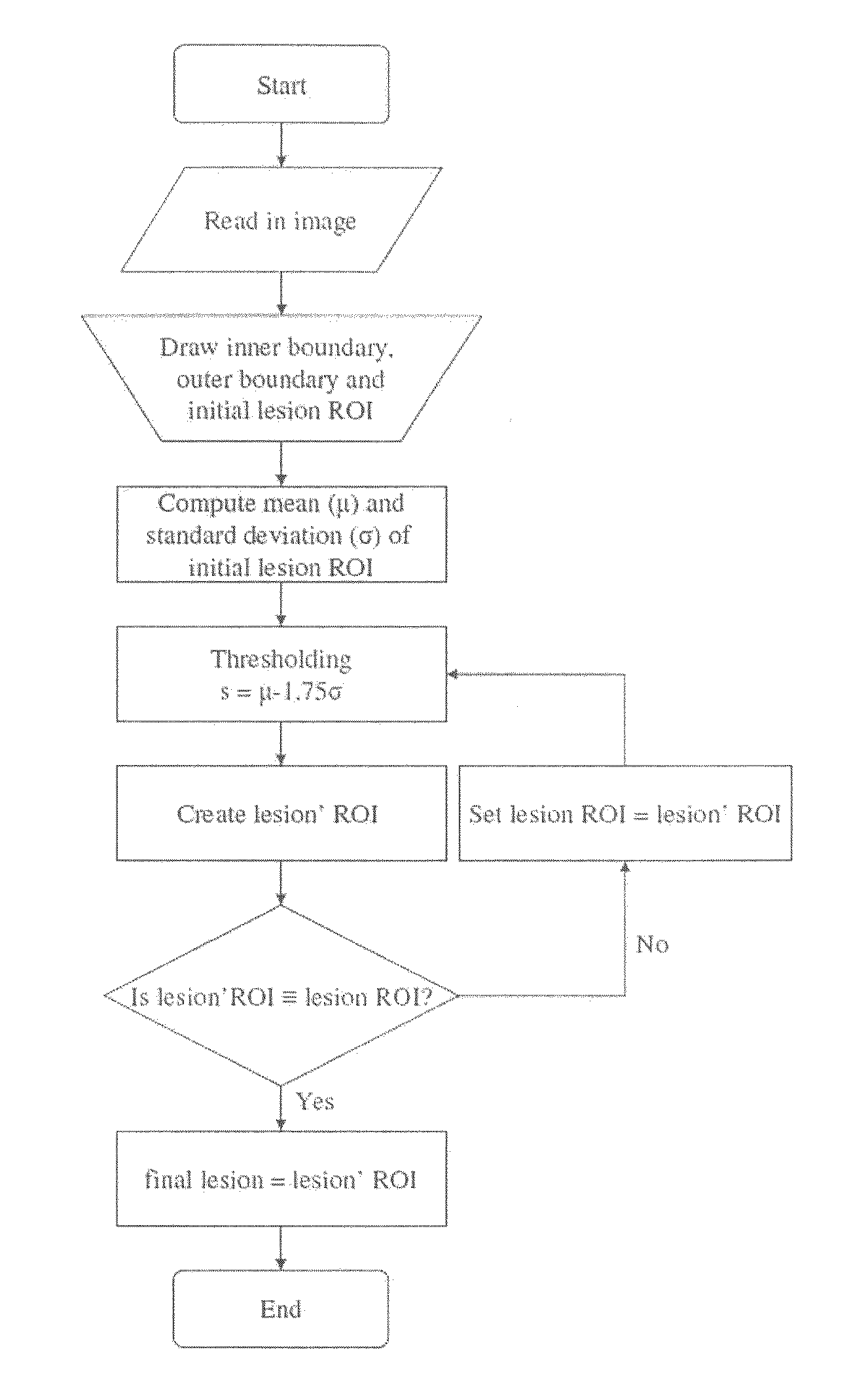
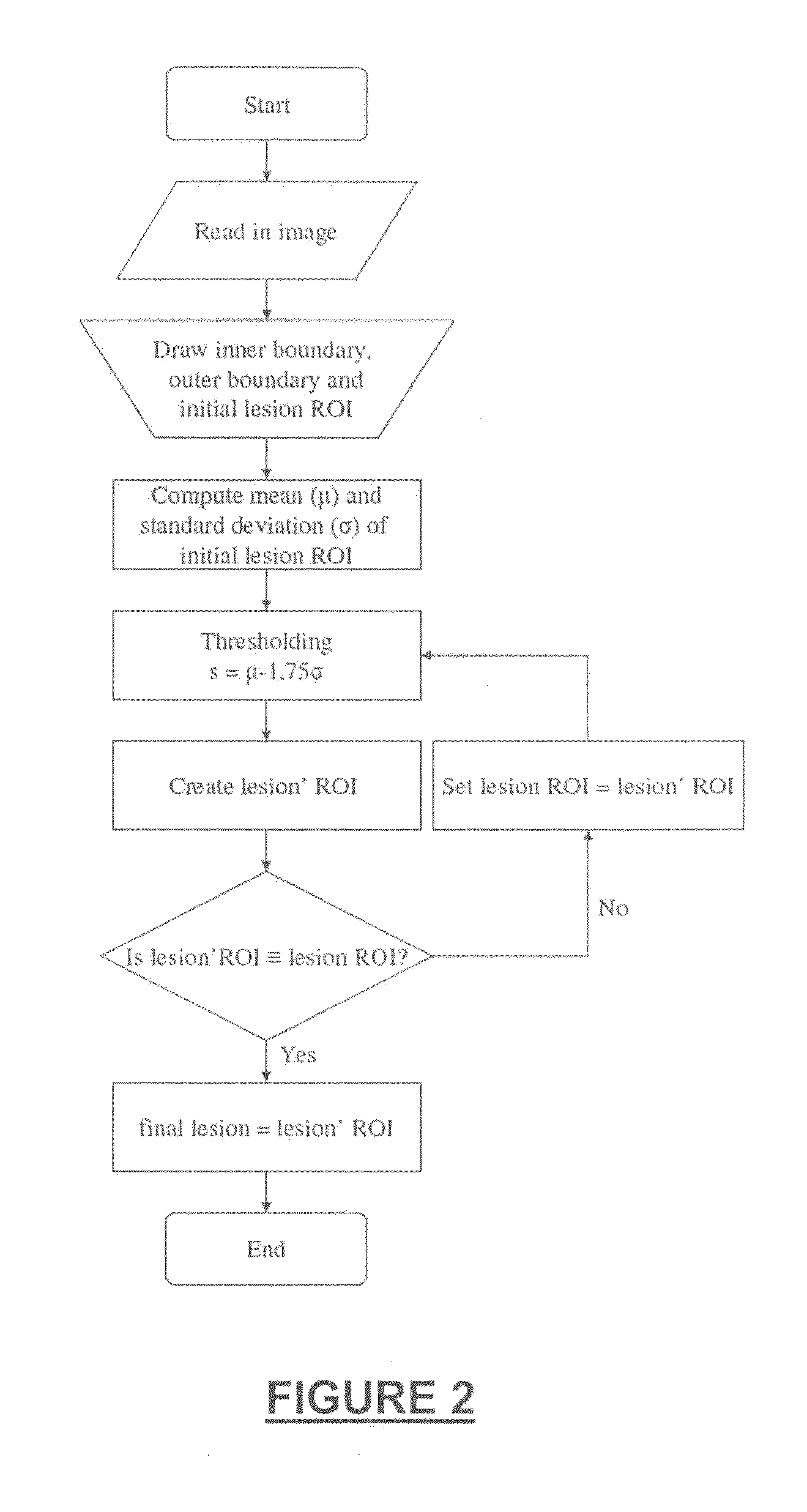
[0055]Patient Selection

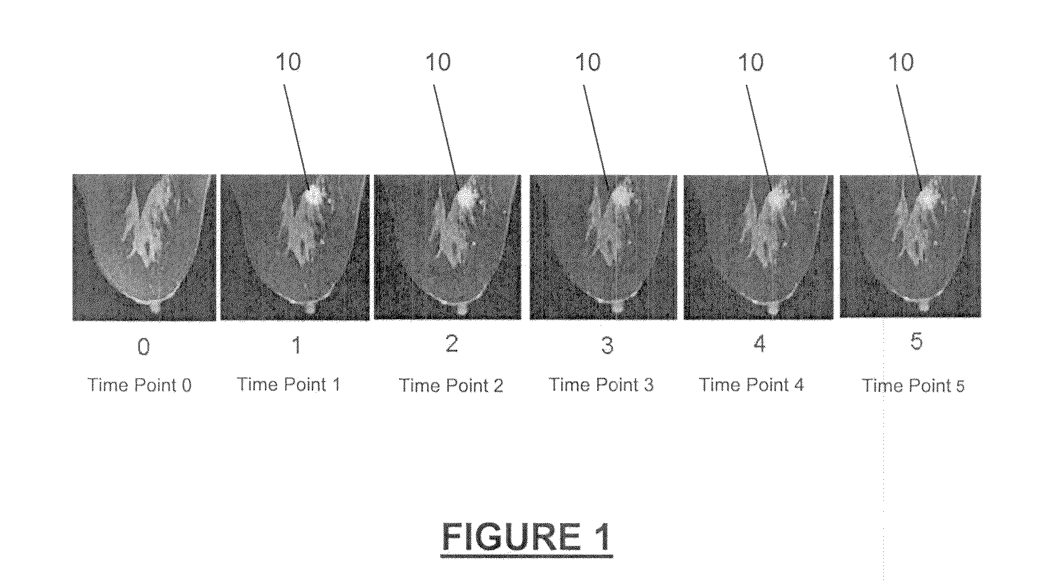
[0056]Patients who underwent standard clinical breast MRI examination at Michigan State University (MSU) Radiology were screened for abnormal contrast-enhancing breast lesions. A lesion was included in this study if it met the following criteria: (1) It was radiologically reported as suspicious for malignancy; (2) it was larger than 7 mm in size and (3) its pathology report was available for comparison. The study included two primary classifications of lesions: (1) malignant tumors histologically diagnosed as infiltrating invasive ductal carcinoma and (2) benign lesions diagnosed as either fibrocystic disease or fibroadenoma. A total of eleven malignant tumors and six benign lesions involving fifteen patients were included in this study. One patient had two lesions: one malignant tumor on one breast and one benign lesion on the other breast. The study was approved by the MSU Institutional Review Board for Research Involving Human Subjects....


PUM


Abstract

Methods for assessing contrast-enhanced lesions using a dynamic contrast-enhancing magnetic resonance imaging system are provided. A boundary of a contrast-enhanced lesion is objectively and automatically determined. The kinetic behavior of voxels is quantitatively examined. The wash-out volume fraction relative to the lesion volume is used as a biomarker to characterize the lesion as malignant or benign.

Description

CROSS-REFERENCE TO RELATED APPLICATIONS[0001]This application is a continuation-in-part of International Application No. PCT / US2009 / 052108, filed on Jul. 29, 2009, which claims the benefit of U.S. Provisional Application No. 61 / 084,384, filed on Jul. 29, 2008. The entire disclosures of the above applications are incorporated herein by reference.INTRODUCTION[0002]The present disclosure relates to dynamic contrast-enhancement magnetic resonance imaging for differentiating benign lesions from malignant lesions.[0003]Magnetic resonance imaging (MRI) is a clinical diagnostic tool that allows for non-invasive imaging of internal structures of a subject. Dynamic contrast-enhancement magnetic resonance imaging (DCE-MRI) combines magnetic resonance imaging principles with the effects of paramagnetic contrast agents on a magnetic resonance signal to track the entrance of the diffusible contrast agents into tissue over time.[0004]DCE-MRI has been shown to be very sensitive, particularly for sm...

Claims


Application Information

Patent Timeline
IPC IPC(8): G06K9/00
CPCG01R33/5608G01R33/56
Inventor HUANG, JIE
Owner BOARD OF TRUSTEES OPERATING MICHIGAN STATE UNIV
Why Eureka
Try Eureka