Endoscope system and imaging device thereof

a technology of endoscope and imaging device, which is applied in the field of endoscope system and imaging device, can solve the problems of reducing the image quality of fluorescence image, increasing the burden on patients, and compromising convenience, so as to prevent the deterioration of convenience, increase the burden of patients, and prevent the increase of the diameter of the distal end

Inactive Publication Date: 2011-09-01
FUJIFILM CORP
View PDF8 Cites 8 Cited by
  • Summary
  • Abstract
  • Description
  • Claims
  • Application Information

AI Technical Summary

Benefits of technology

[0016]According to the present invention, since the distance between the first and second plates is regulated so that the light of the same wavelength as that of the excitation light passes through the spectral characteristics variable optical element, the spectral characteristics variable optical element can remove a component of the excitation light from the image light incident from the section to be examined. Accordingly, in fluorescence endoscopy using a plur

Problems solved by technology

The excitation light reflected back becomes noise in production of the fluorescence image, and causes degradation in image quality of the fluorescence image.
This compromises convenience, and increases a burden on a patient.
As for the automatic exchange of the filter by the rotation mechanism, on

Method used


Image

  • Endoscope system and imaging device thereof
  • Endoscope system and imaging device thereof
  • Endoscope system and imaging device thereof

Examples


Embodiment Construction

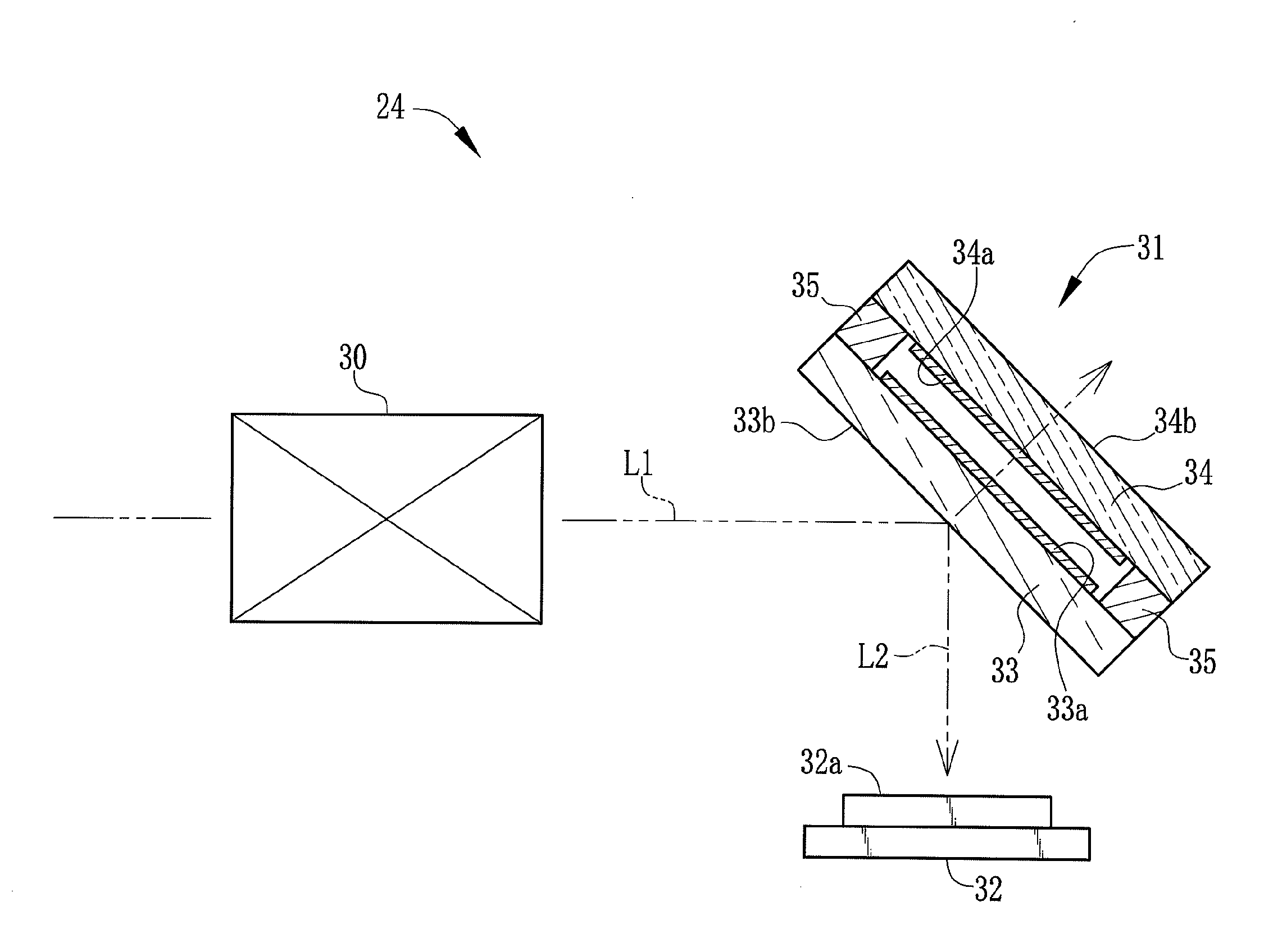
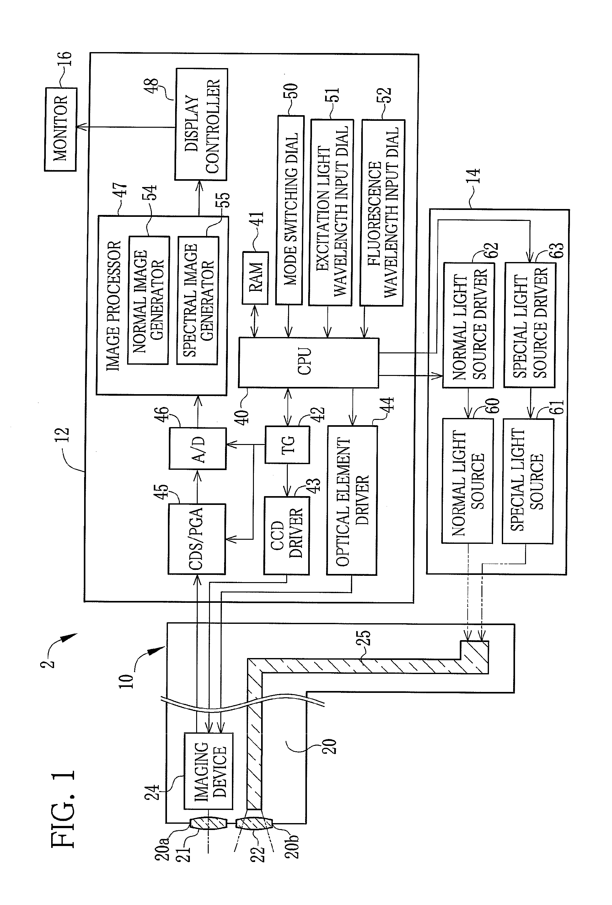
[0021]As shown in FIG. 1, a fluorescence endoscope system 2 is constituted of an electronic endoscope 10 for imaging the inside of a patient's body, a processing device 12 for producing an endoscope image, a light source device 14, and a monitor 16 for displaying the endoscope image. The light source device 14 selectively supplies to the electronic endoscope 10 one of white light (normal light) for lighting the inside of the body and excitation light for exciting a fluorescent labeling agent administered on or injected into an internal body part.

[0022]This endoscope system 2 has a normal imaging mode and a fluorescence imaging mode. In the normal imaging mode, the normal light is supplied to the electronic endoscope 10 to image the inside of the body part with visible light. In the fluorescence imaging mode, the excitation light is supplied to the electronic endoscope 10 so as to image with fluorescence a tumor or the like to which the administered or injected fluorescent labeling a...


PUM


Abstract

An imaging device for an endoscope includes an image pickup lens, a spectral characteristics variable optical element, and a CCD. The spectral characteristics variable optical element passes only light of a specific wavelength in accordance with the distance between first and second plates, and reflects light of the other wavelengths. The CCD converts the light reflected from the variable optical element into an image signal. In fluorescence endoscopy, a fluorescent labeling agent is administered on an internal body part to be examined. Application of excitation light to the body part causes the fluorescent labeling agent to emit fluorescence. The variable optical element removes the excitation light from the fluorescence by regulation of the distance between the first and second plates.

Description

BACKGROUND OF THE INVENTION[0001]1. Field of the Invention[0002]The present invention relates to an endoscope system and an imaging device used therein.[0003]2. Description Related to the Prior Art[0004]There is known an endoscope system that takes a fluorescence image of an internal body part to be examined where a fluorescent labeling agent is administered or injected (refer to Japanese Patent Laid-Open Publication No. 2009-291554, for example). The fluorescent labeling agent is selectively bonded to specific living tissue containing a lesion or the like. Only upon application of excitation light having a specific wavelength, the fluorescent labeling agent makes a transition to an excited state by the excitation light, and emits fluorescence having a wavelength different from that of the excitation light. Fluorescence endoscopy using such a fluorescent labeling agent makes it possible to find out the minute lesion including a cancer or a tumor that is difficult to find out in norm...

Claims


Application Information

Patent Timeline
IPC IPC(8): A61B1/045
CPCA61B1/05A61B1/043A61B1/00186A61B1/0638A61B1/0655
Inventor KURODA, OSAMU
Owner FUJIFILM CORP
Why Eureka
Try Eureka