Biopsy device

a biopsy device and sampler technology, applied in medical science, ovulation diagnostics, vaccination/ovulation, etc., can solve the problems of inability to provide consistent cartilage harvesting material, extremely user-dependent biopsy quantities obtained with this instrument, and lack of standardization

Inactive Publication Date: 2011-12-08
DOKTER YVES FORTEMS
View PDF25 Cites 76 Cited by
  • Summary
  • Abstract
  • Description
  • Claims
  • Application Information

AI Technical Summary

Problems solved by technology

Currently available biopsy devices are not designed to give consistent cartilage harvest material without contaminations by other tissues or the risk of loosing the biopsy during the procedure, or are restricted to only being used at specific sites (e.g. the notch).
Biopsy quantities obtained with this instrument are extremely user dependent and lack standardization.
Even with the same surgeon (user), a lack of consistent reproducibility has been observed despite long-term experience.
Furthermore, the devic...

Method used


Image

  • Biopsy device
  • Biopsy device
  • Biopsy device

Examples


Embodiment Construction

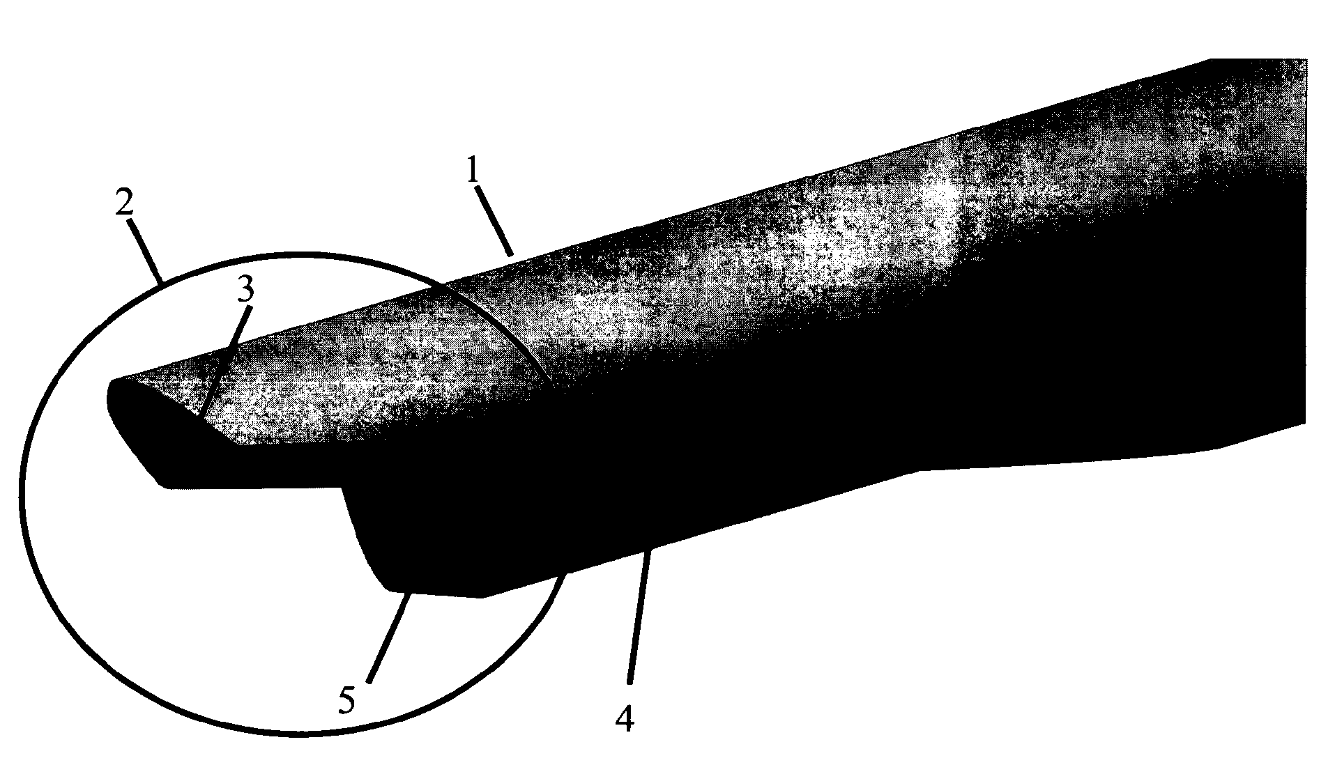
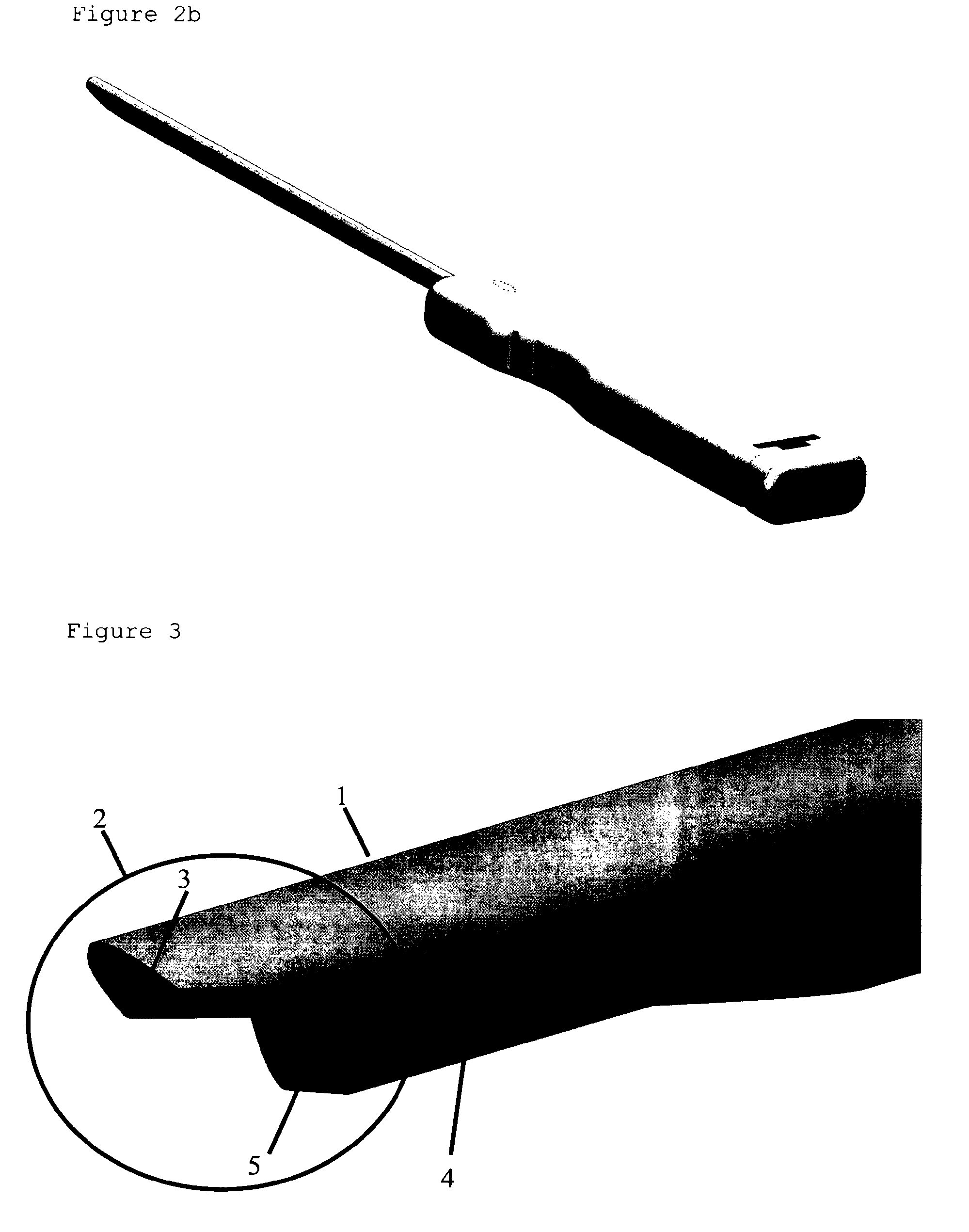
[0025]The biopsy device of the present invention solves the problems associated with the aforementioned prior art devices in that it provides:[0026]1) the ability to control and select the biopsy length (shape and size) in relation to the defect size;[0027]2) the ability to standardized biopsy harvesting at all locations in the knee joint, with in particular the lateral and medial intercondylar notch;[0028]3) the ability to avoid osteochondral defects for reasons of patient safety and product contamination with non-chondrogenic cells;[0029]4) a cartilage insertion into the biopsy needle with minimal tissue damage;[0030]5) a capturing chamber for the biopsy sample to minimize risk of loss;[0031]6) a measurable and visible positioning of the device;[0032]7) a user-friendly and safe use;[0033]8) a single-use to reduce the risk of contamination and / or infection and to maintain its sharpness.

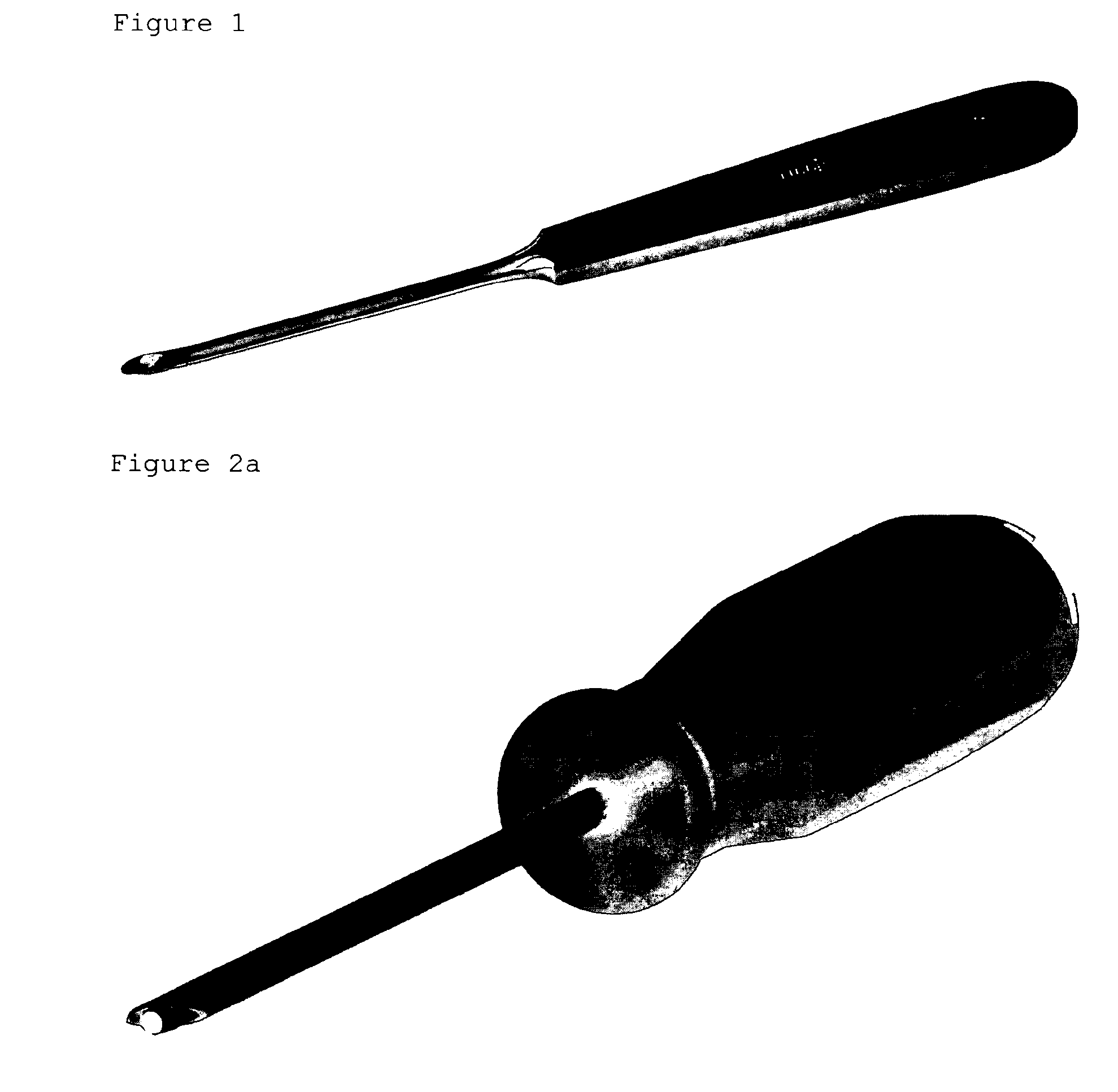
[0034]The invention relates to a biopsy device as shown in FIGS. 2a, 2b, 7, 8a, 8b, 9b, 12 and 13...


PUM


Abstract

This invention relates to a biopsy device consisting of an inner cannula (4) and an outer hollow tube (1), a handle (7) which may be removably attached to the outer hollow tube, a locking system to secure the inner cannula and/or the attenuator in the outer hollow tube, and characterized in that the tip of the outer hollow tube is ellipse shaped and extends beyond the inner cannula, the latter ending in a blunt edge. The blunted tip of the outer hollow tube together with the sharpened ending of the inner cannula determines the cutting edge of the device. In combination the distal ends of inner cannula and outer hollow tube determine the biopsy depth size and shape of the biopsy sample in a reproducible way. In one embodiment of the present invention, the length of the inner cannula can be controlled, allowing varying the aforementioned sample parameters as desired.

Description

FIELD OF THE INVENTION[0001]The present invention relates to biopsy sampling and in particular to a device for such sampling in hard tissue on e.g. humans or animals. The biopsy device as provided herein is more in particular for sampling cartilage tissue, such as at the non-weight bearing areas of the superomedial or superolateral edge of the femoral condyle or the lateral and medial intercondylar notch.BACKGROUND TO THE INVENTION[0002]Autologous chondrocyte implantation (ACI) is a clinically relevant treatment to repair articular cartilage in patients with knee cartilage defects. This repair method is based on the introduction of adult chondrogenic cells into the defect area. To accomplish this, chondrocytes are first isolated from a limited amount of articular cartilage harvested arthroscopically from a minor weight-bearing area of the injured knee of the same patient. The cells are released from the cartilage tissue by enzymatic digestions and expanded in culture medium until a ...

Claims


Application Information

Patent Timeline
IPC IPC(8): A61B10/02
CPCA61B10/025A61B10/0233A61B2019/462A61B2090/062
Inventor MAENHOUT, SVENMOTMANS, KRISFORTEMS, YVESJAHRAUS, ANDREAOPREA, WANDA
Owner DOKTER YVES FORTEMS
Why Eureka
Try Eureka