
Methods for multiplex analyte detection and quantification

a multiplex analyte and quantification technology, applied in combinational chemistry, chemical libraries, instruments, etc., can solve the problems of increasing the delay in treatment time, increasing the cost and possibility of analytical errors, and major limitation of detecting diseases with more, so as to achieve fast and cost-effective methods

Inactive Publication Date: 2011-12-15
SQI DIAGNOSTICS SYST
View PDF1 Cites 19 Cited by
  • Summary
  • Abstract
  • Description
  • Claims
  • Application Information

AI Technical Summary

Benefits of technology

[0016]The present invention provides a fast and cost effective method for detecting and quantifying multiple target analytes in test sample using a single reaction vessel. The method disclosed herein allows for the simultaneous detection of multiple target analytes without the need for separate assays or reaction steps for each target analyte.

Problems solved by technology

Current immunoassay methods are limited as they only detect one target per detection test cycle within a single reaction well.
This required multitude of tests and samples increases delay in time to treatment, costs and possibility of analytical error.
This is a major limitation for detecting diseases with more than one marker or transgenic organisms which express more than one transgenic product.
The result is a need for two separate washing steps which defeats the purpose of the direct assay.
These bead based systems' capability is limited to distinguishing between simultaneous detection of capture antigens.
While methods for detecting and quantifying multiple analytes are known, these methods require the use of separate assaying steps for each of the analytes of interest and as such, can be time consuming and costly, especially in the context of a clinical setting.

Method used


Image

  • Methods for multiplex analyte detection and quantification
  • Methods for multiplex analyte detection and quantification
  • Methods for multiplex analyte detection and quantification

Examples


Embodiment Construction

Detection and Quantification of Three Different Target Antibodies in a Serum Sample

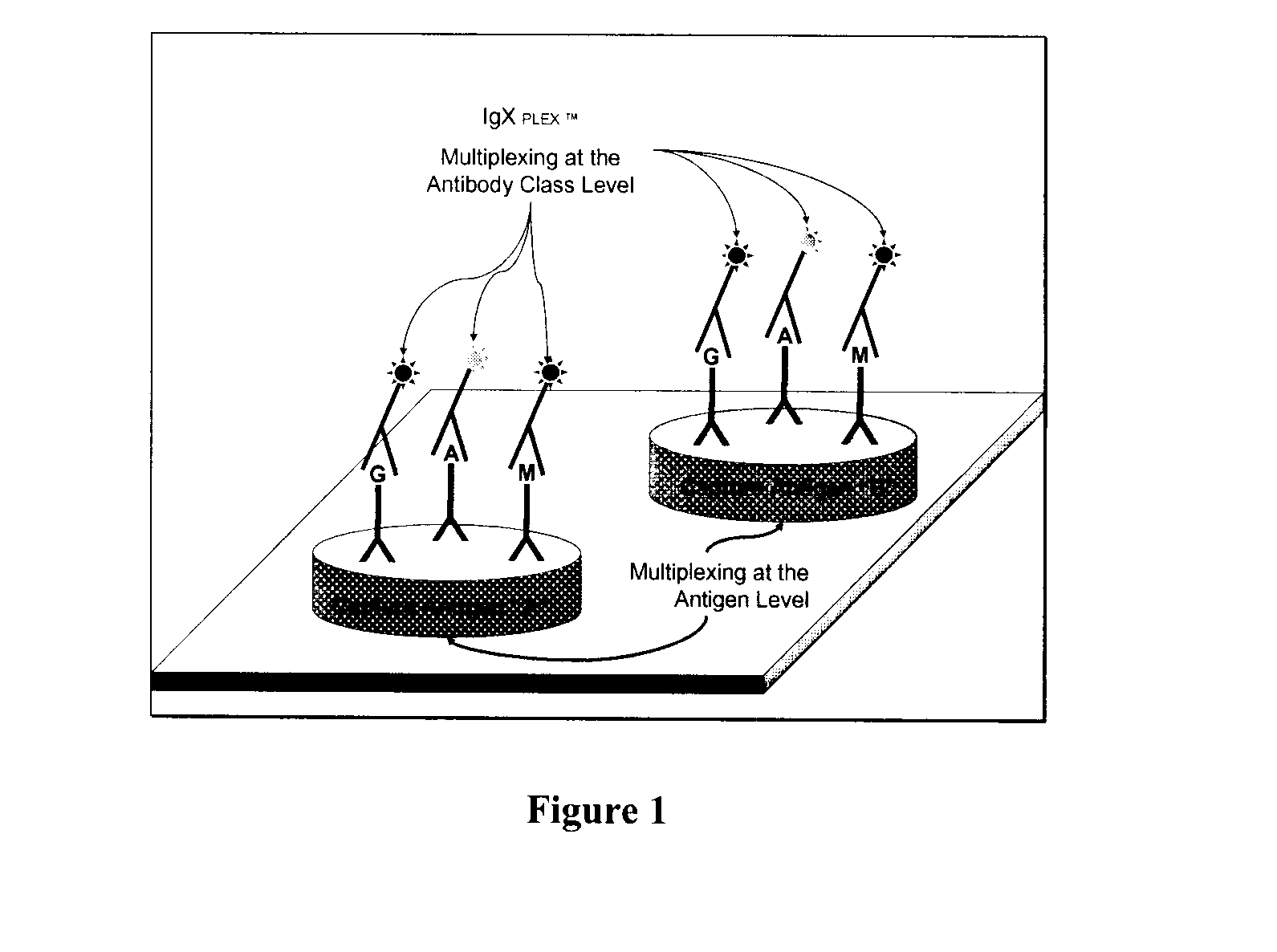
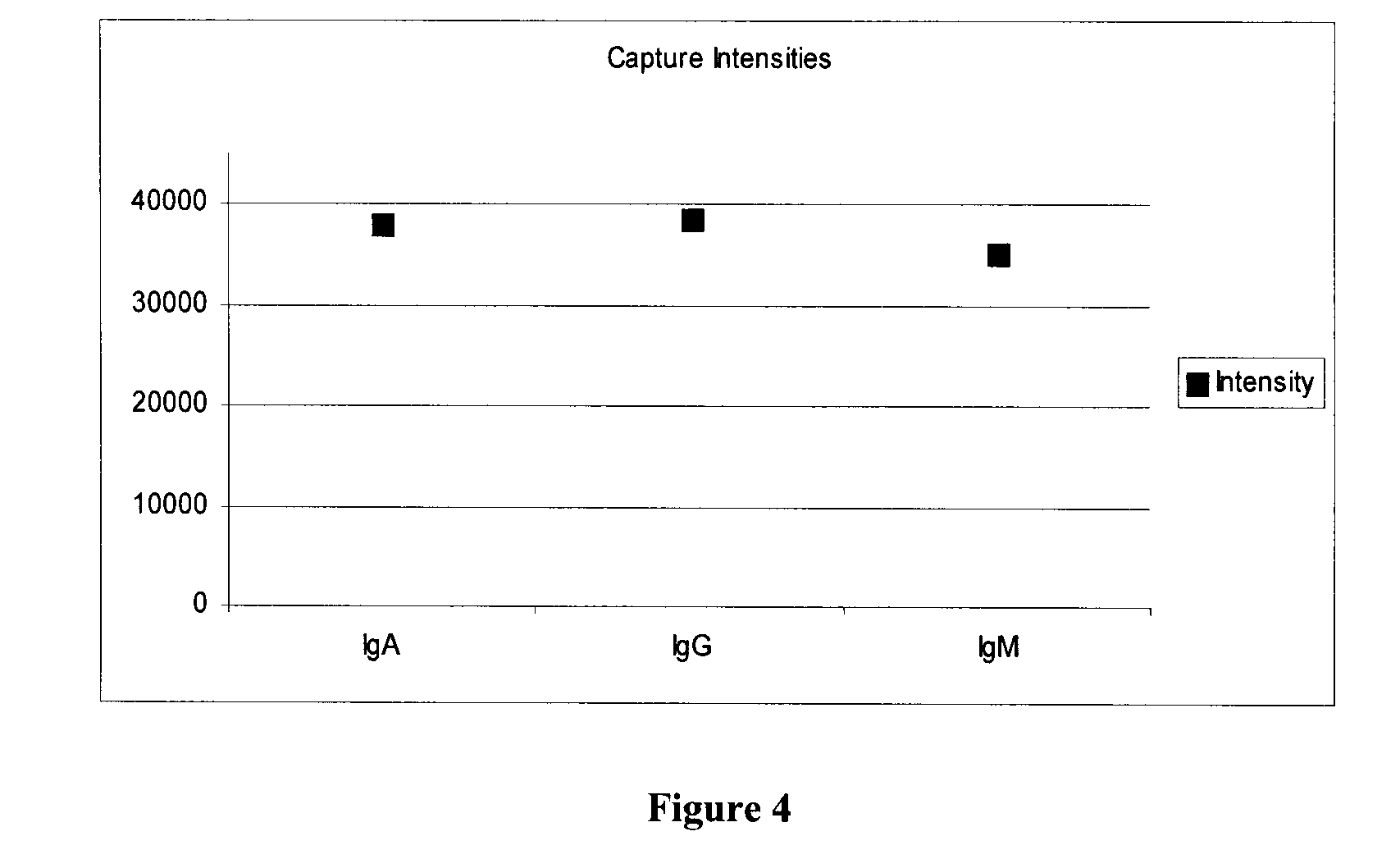
[0068]Four concentrations each of human IgM, IgG, IgA are printed in the same sample well on a 16-well slide, pretreated to create an epoxysilane substrate surface. The protein printed slides were incubated overnight with fish gelatin to block unreacted epoxysilane binding sites in the well.

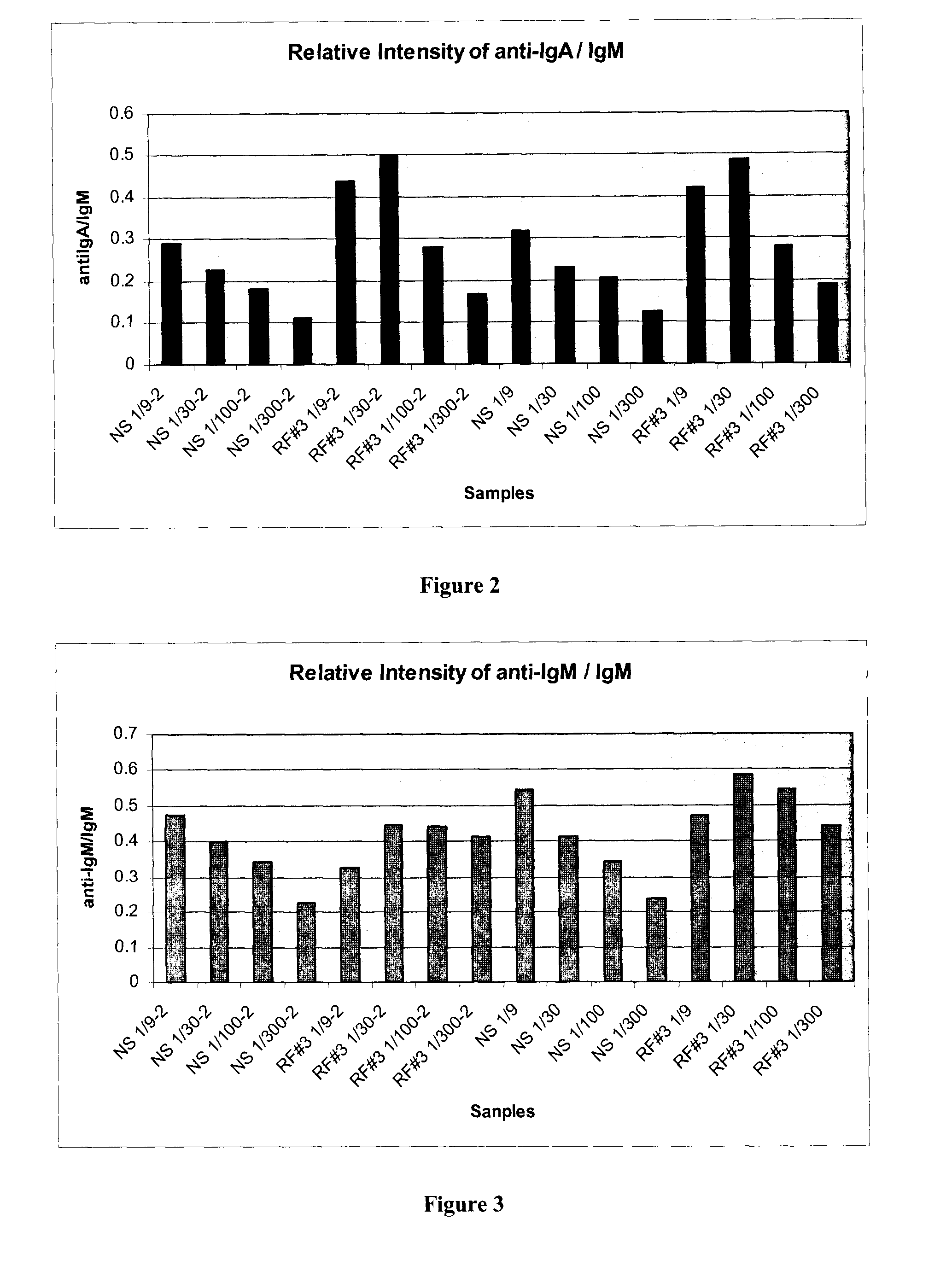
[0069]To perform the assay, serum samples were diluted 1 in 9 to 1 in 200 in buffers containing fish gelatin. Each sample was diluted to four dilutions, 1:9, 1:30, 1:100, 1:300 in duplicate. The two diluted samples (named NS and RF #3, see FIGS. 2 and 3) were incubated for 45 min. The slide was washed five times, in Tris buffered saline. A cocktail of goat antihuman antibody conjugated to FITC, two mouse antihuman IgA antibodies conjugated to DY652 (Dyomics, Germany), and a mouse antihuman IgG antibody conjugated to Cy3 dye, each in about 1 μg / ml concentration, was added to all wells of the slide.

[0070]The reagent ...


PUM


Abstract

The application refers to a method for detecting and quantifying multiple target analytes in a test sample using a single reaction vessel. The method uses a reaction vessel (a multi-well plate), which comprises a microarray of: (a) calibration spots, each having a predetermined quantity of the target analyte; and (b) capture spots, each having an agent (antibody) that selectively binds the target analyte. The captured analytes and the calibration spots are detected with fluorescently labelled antibodies specific for each different target analyte. The calibration spots are used to generate calibration curves that allow the measurement of the concentration of the different target analytes. The application also refers to a method for detecting and quantifying biomarkers that are useful for diagnosing rheumatoid arthritis. More specifically, the application discloses the use of rheumatoid factor (RF) and cyclic citrullinated peptide (CCP), as capture spots. Finally, based on the above method, it is proposed a method for diagnosing or monitoring rheumatoid arthritis.

Description

FIELD OF INVENTION[0001]The present invention relates to methods for the quantification of analytes, in particular, the invention relates to improved microarray methods for the detection and quantification of multiple analytes in a single sample.BACKGROUND[0002]Current immunoassay methods are limited as they only detect one target per detection test cycle within a single reaction well. It is common for several antigenic substances or bio-markers to be associated with detection and diagnosis of any pathological or physiological disorder. To confirm the presence of multiple markers, each marker within a test sample requires a separate and different immunoassay to confirm the presence of each target molecule to be detected. This required multitude of tests and samples increases delay in time to treatment, costs and possibility of analytical error. The current state of the art for quantitative multiplexing of proteins / antibodies, especially biomarkers expressed in auto-immune diseases, ...

Claims


Application Information

Patent Timeline
IPC IPC(8): C40B30/04
CPCB01L3/5085G01N33/564G01N33/543
Inventor LEA, PETER
Owner SQI DIAGNOSTICS SYST
Learn More