
Method and system for enhancing microscopy image

a microscopy and image technology, applied in the field of methods and systems for enhancing microscopy images, can solve the problems of light penetration problem, acute degradation by light attenuation effects, and other light microscopy techniques, such as single-plane illumination microscopy and wide-field microscopy, and achieve the same problems

Inactive Publication Date: 2011-12-29
AGENCY FOR SCI TECH & RES
View PDF12 Cites 9 Cited by
  • Summary
  • Abstract
  • Description
  • Claims
  • Application Information

AI Technical Summary

Benefits of technology

[0007]Provided the values of the scattering parameter are found accurately, the enhanced image should be

Problems solved by technology

However, degradation by light attenuation effects is acute in confocal microscopy.
The fundamental problem in confocal microscopy is the light penetration problem.
Other light microscopy techniques, such as the single plane illumination microscopy and wide-field microscopy suffer the same problem.
The classical space invariant deconvolution approaches [2], [3], [4] cannot cope with this problem of microscopy imaging.
Both techniques are inadequate and have drawbacks: increasing laser power accelerates photo-bleaching effects whereas increasing the sensitivity of the photo-multiplier tube adds noise.
A fundamental issue with these restoration techniques is the amplification of noise.

Method used


Image

  • Method and system for enhancing microscopy image
  • Method and system for enhancing microscopy image
  • Method and system for enhancing microscopy image

Examples


Embodiment Construction

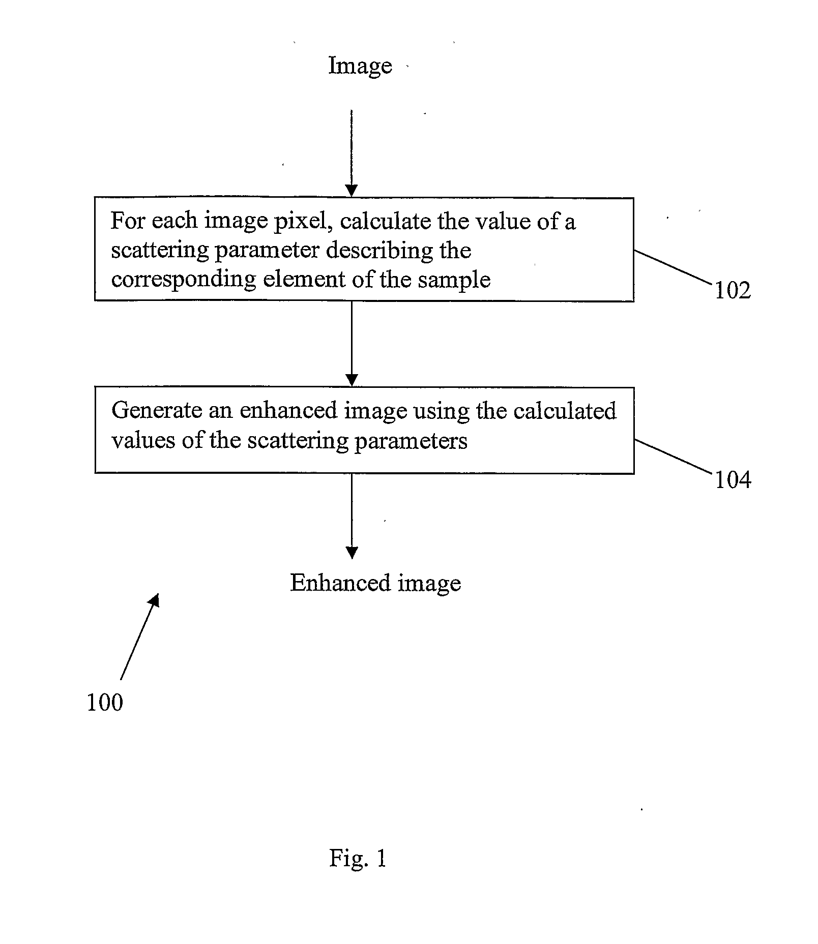
[0023]Referring to FIG. 1, the steps are illustrated of a method 100 which is an embodiment of the present invention, and which is a method for enhancing a microscopy image.

[0024]The input to method 100 is an image of a sample acquired using a microscope which illuminates the sample and collects light absorbed and then scattered by the elements of the sample. Pixels of the image correspond to elements of the sample. The intensity at each point in the sample is the sum of a component of incident light (gradually attenuated as it passes through the sample) and a scattering component due to the scattering. The scattering of incident light by a given element of the sample is described by the value of a scattering parameter, which is typically an emission coefficient ρem or else equal to an absorption coefficient ραb. In step 102, for each image pixel, the value of the scattering parameter of the corresponding element is calculated using a mathematical expression linking the values of th...


PUM


Abstract

A microscopy image, formed by illuminating a sample by shining light onto it in an illumination direction and capturing scattered light, is used to produce an enhanced image. This is done using an expression which links the intensity of the portions of the image to respective values of a scattering parameter at multiple respective elements of the sample. The scattering parameter may be an emission coefficient ρem or else equal to an absorption coefficient ραb. This expression is solved to find the values of the scattering parameter. The scattering parameter is used to construct an enhanced image, for example an image which maps the variation of the scattering parameter itself. Provided the scattering parameter is found accurately, the enhanced image should be less subject than the original image to degradation due to non-uniform light attenuation and scattering.

Description

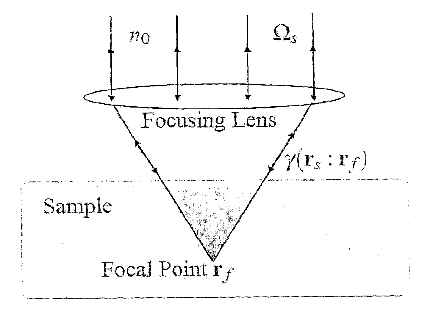
FIELD OF THE INVENTION[0001]The present invention relates to a method and system for enhancing a microscopy image, that is an image acquired using a microscope. In particular, the enhanced image is one which suffers less from degradation due to non-uniform light attenuation and scattering.BACKGROUND OF THE INVENTION[0002]Microscopy [1] is an important optical imaging technique for biology. While there are many microscopy techniques such as two-photon excitation microscopy and single plane illumination microscopy, confocal microscopy [1] has become one of the most important tools for bioimaging. In confocal microscopy, out-of-focus light is eliminated through the use of a pin-hole. Incident illuminating light passes through the pin-hole and gets focused onto a small region in the sample, where it is scattered by the sample. Only scattered light travelling along the same path as the incident illuminating light passes back through the pin-hole, and such light gets focused again at a li...

Claims


Application Information

Patent Timeline
IPC IPC(8): H04N7/18
CPCG02B21/365
Inventor LEE, HWEE GUANUDDIN, MOHAMMAD SHORIF
Owner AGENCY FOR SCI TECH & RES
Learn More