Trans-perineal prostate MR elastography

a trans-perineal prostate and elastography technology, applied in the field of diagnostic imaging, can solve the problems of difficult induction of mechanical deformation non-invasively into the abdominal region for elastography imaging, insufficient tissue penetration, and inability to achieve large mechanical shear waves in the prostate,

Inactive Publication Date: 2012-03-01
SALCUDEAN SEPTIMIU +2
View PDF14 Cites 22 Cited by
  • Summary
  • Abstract
  • Description
  • Claims
  • Application Information

AI Technical Summary

Benefits of technology

[0011]To generate mechanical shear waves in the prostate with a large enough amplitude, this invention proposes that vibrations be applied at the perineum via a coupling surface that is attached to a mechanical exciter. In a preferred embodiment a coupling surface of the mechanical exciter undergoes a rec

Problems solved by technology

Inducing mechanical deformation non-invasively into the abdominal region for elastography imaging is challenging in particular in the prostatic tissue.
As a result, applying mechanical excitations to the pubic b

Method used


Image

  • Trans-perineal prostate MR elastography
  • Trans-perineal prostate MR elastography
  • Trans-perineal prostate MR elastography

Examples


Embodiment Construction

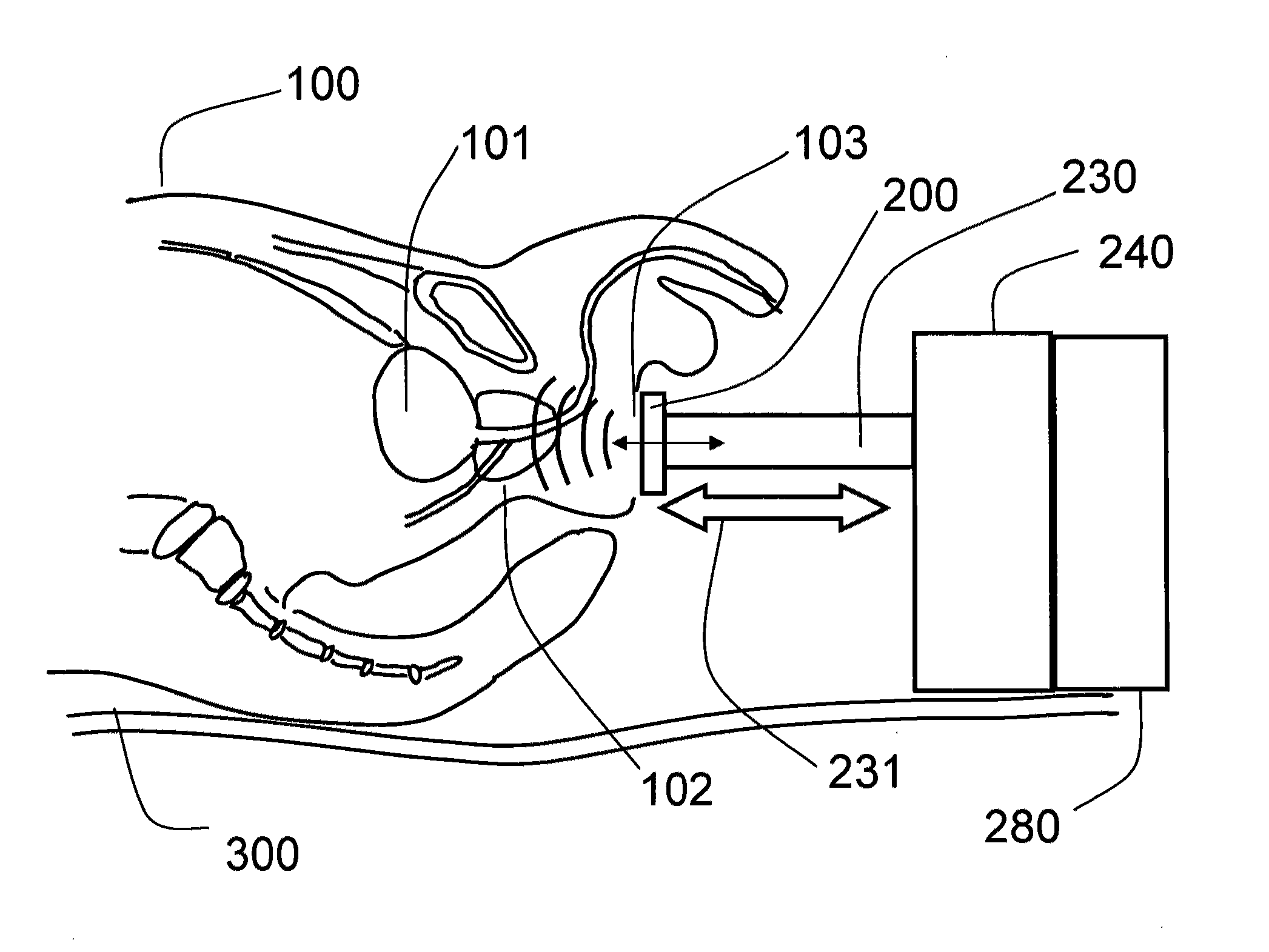
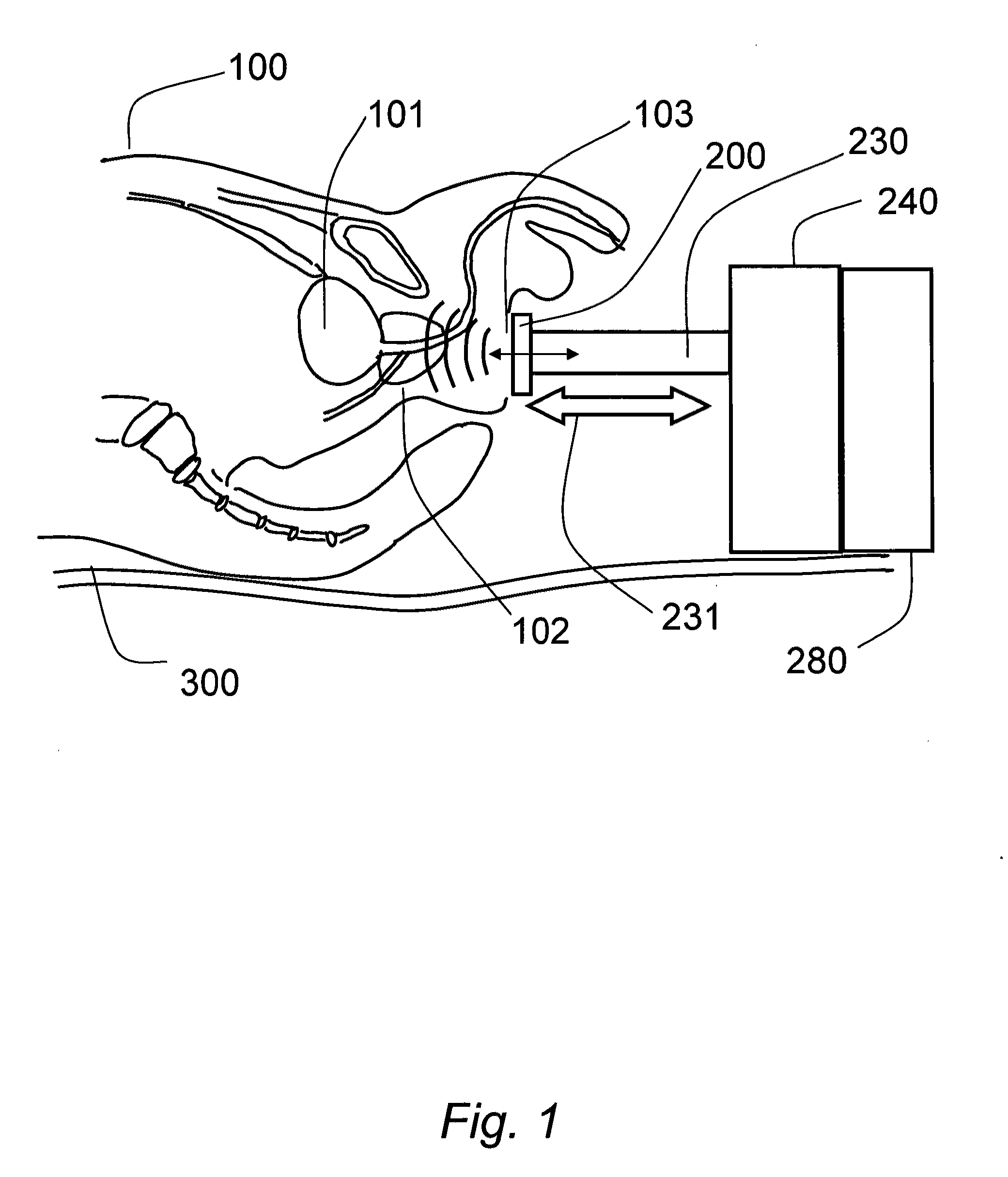
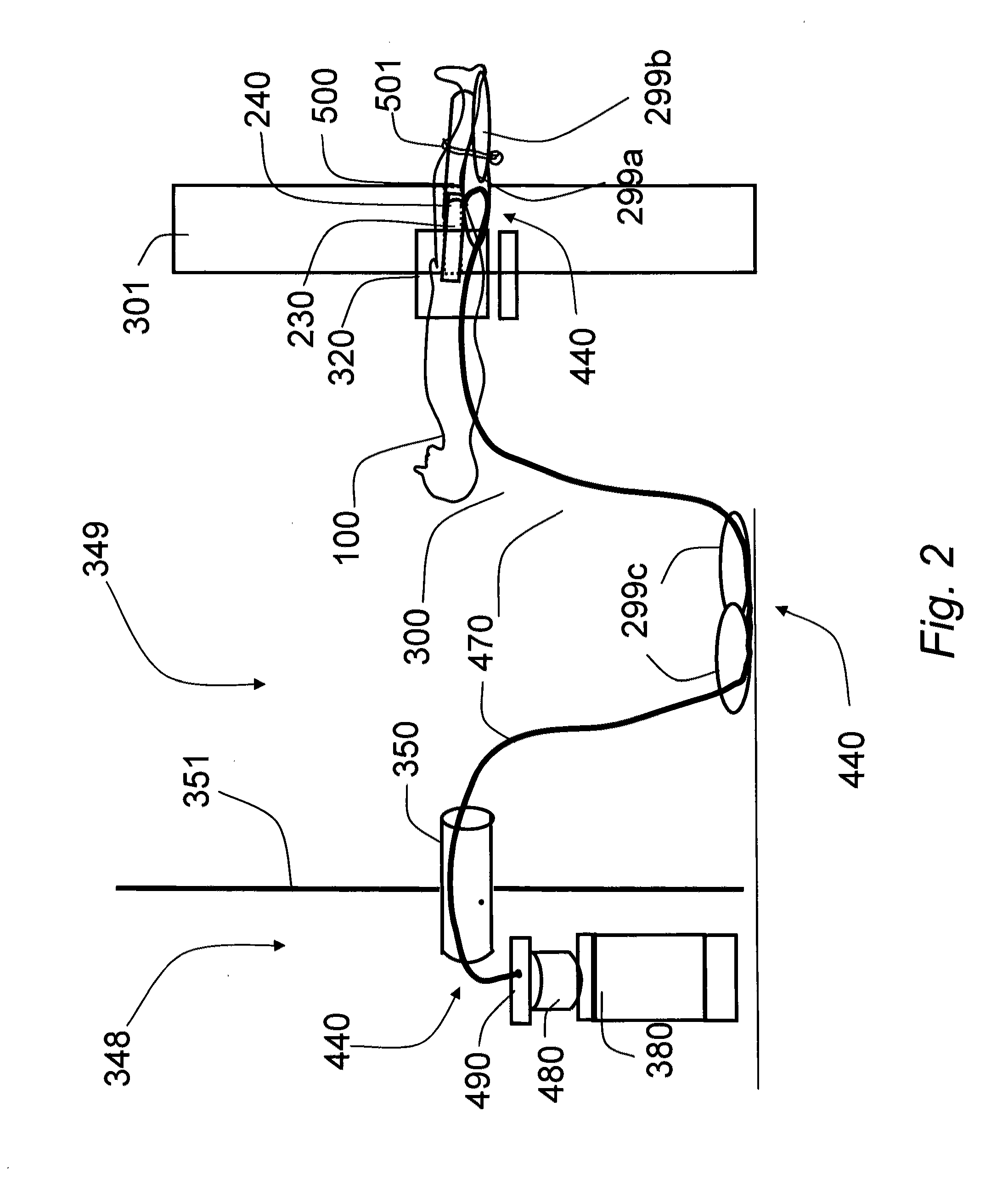
[0032]The embodiments described herein relate to a method and apparatus of a magnetic resonance elastography (MRE) image acquisition apparatus for the measurement of the mechanical properties of a tissue region in a patient using a mechanical transducer. Particularly, the embodiments relate to inducing or transferring mechanical shear and longitudinal vibrations into the lower abdomen, specifically in the prostate area for male patients. This is achieved by applying a mechanical exciter to the perineal region of the patient that generates mechanical longitudinal vibrations.

Anatomy, Positioning of Exciter at Patient End

[0033]FIG. 1 shows the abdominal section of a patient lying in a typical MR scanner being prepared for a MRE scan. The patient 100 rests in the supine position (facing up) on the table of the magnetic resonance imaging (MRI) scanner 300. The patient is positioned such that the feet enter the MR magnet (feet-first) while the head stays out. In other possible embodiments...


PUM


Abstract

The present invention relates to a method and apparatus for imaging the mechanical properties of the prostate of a patient non-invasively. The apparatus generally comprises a magnetic resonance scanner, a vibration assembly coupled to the perineal region of the patient, and a driver that drives the mechanical exciter. The method generally comprises positioning the vibration assembly against the perineal region of the patient, vibrating the mechanical exciter to cause deformational excitation of a tissue region contacted in the perineum, capturing a series of images in time (snapshots) of the tissue region using the MR scanner, and finally processing the displacement images to generate maps of mechanical properties of images tissue.

Description

[0001]This application claims the benefit of U.S. Provisional Patent Application No. 61 / 395,058, filed on May 10, 2010, which is incorporated herein by reference.FIELD OF THE INVENTION[0002]The present invention relates to diagnostic imaging generally, and measurement of the mechanical properties of the prostate using magnetic resonance elastography specifically.BACKGROUND OF THE INVENTION[0003]The basis of medical imaging is the measurement of a property of tissue that varies with tissue composition. Medical images are formed by displaying intensities as a function of these properties measured at various locations in the body. Mechanical properties of tissue are important indicators of disease potential. Indeed, palpation techniques are commonly used by medical doctors to determine the potential for disease, for example, stiffer tissue regions that can be felt as harder objects can indicate the presence of cancer. This is the basis for a number of clinical examinations such as the ...

Claims


Application Information

Patent Timeline
IPC IPC(8): A61B5/055
CPCA61B5/0051G01R33/56358A61B5/4381A61B5/055
Inventor SALCUDEAN, SEPTIMIUSAHEBJAVAHER, RAMIN S.SINKUS, RALPH
Owner SALCUDEAN SEPTIMIU
Why Eureka
Try Eureka