Method for removal of retained lens fragments using small diameter fragmatome

a fragmatome and small diameter technology, applied in the field of small diameter fragmatome, can solve the problems of large incisions, large trauma to patients, and large design of conventional fragmatomes, and achieve the effect of reducing trauma to patients as a result of the operation and improving recovery tim

Inactive Publication Date: 2012-04-05
FOSTER WILLIAM J
View PDF2 Cites 9 Cited by
  • Summary
  • Abstract
  • Description
  • Claims
  • Application Information

AI Technical Summary

Benefits of technology

[0019]A further technical advantage of the invention is that trauma to the patient as a result of the operation is reduced, and recovery time is improved.

Problems solved by technology

If lens fragments, and especially portions of the center of the lens, also known as the nucleus or the nuclear lens, shift into the back or posterior chamber of the eye, those lens fragments may cause severe trauma (including inflammation and blindness) if left unremoved.
Relatively large incisions are suffered by the patient, requiring suturing.
A drawback to conventional removal of lens fragments from the back of the eye, as described above, is that conventional fragmatomes are comparatively large in design.
However, when lens fragments are left in the posterior eye cavity, removal of those fragments through those same small incisions is made difficult by the unavailability of a small gauge (23-gauge or less) fragmatome.

Method used


Image

  • Method for removal of retained lens fragments using small diameter fragmatome
  • Method for removal of retained lens fragments using small diameter fragmatome
  • Method for removal of retained lens fragments using small diameter fragmatome

Examples


Embodiment Construction

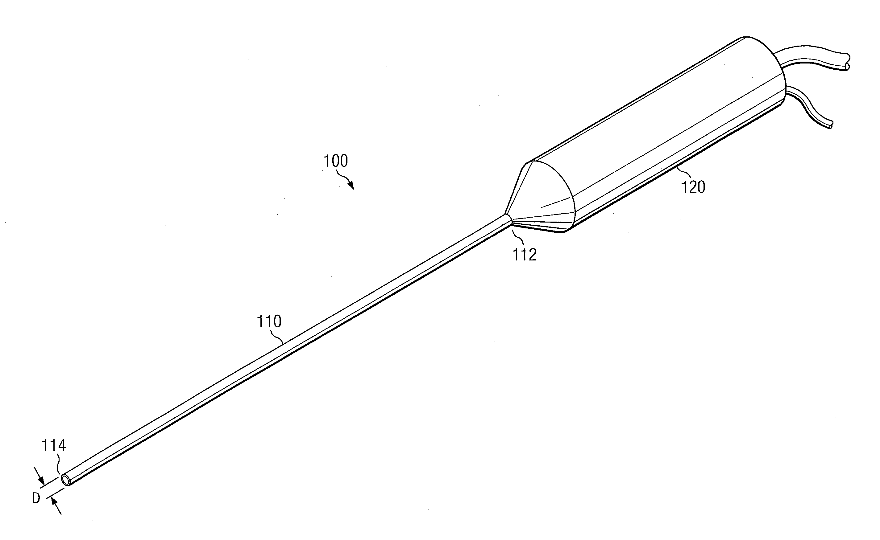
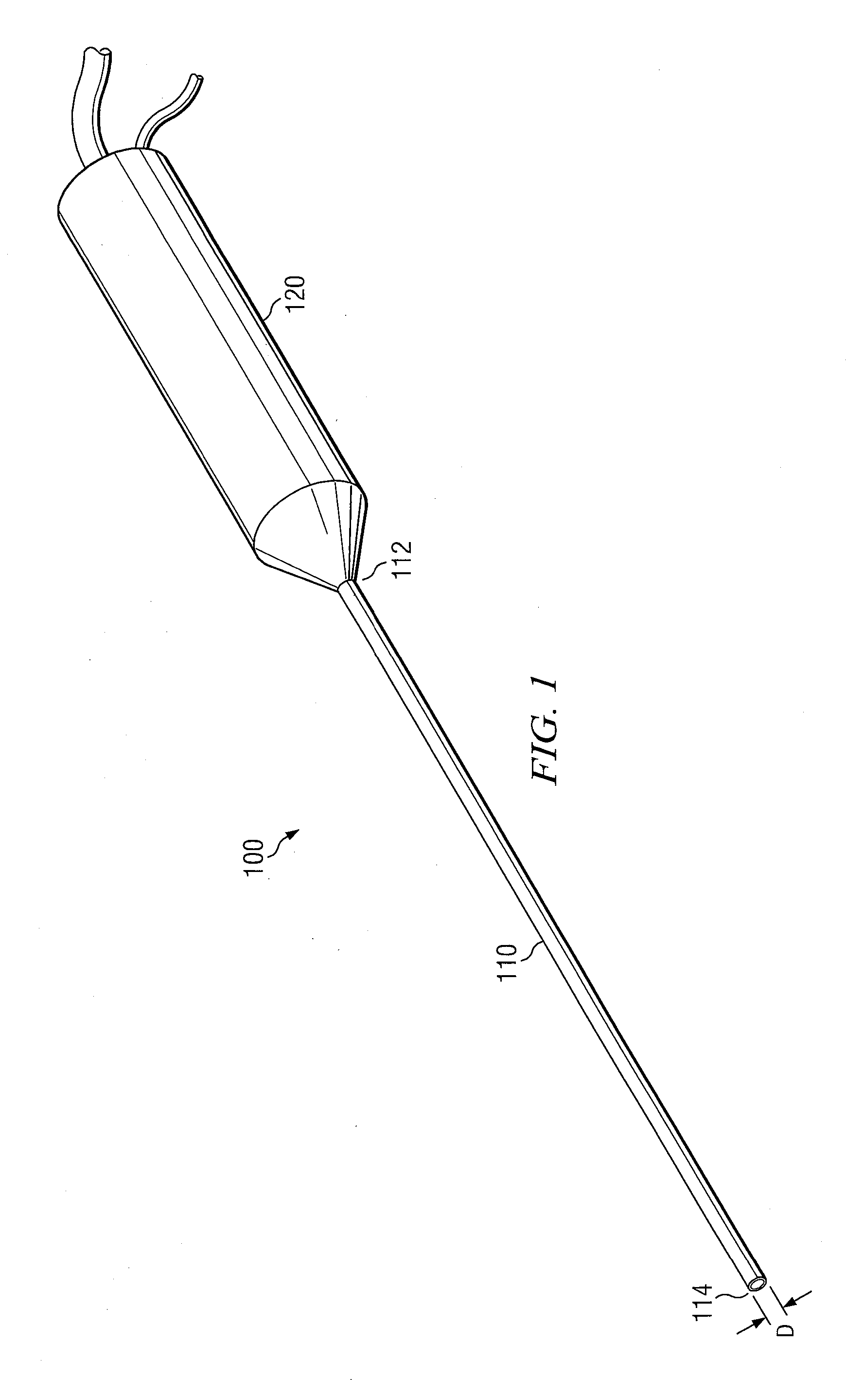
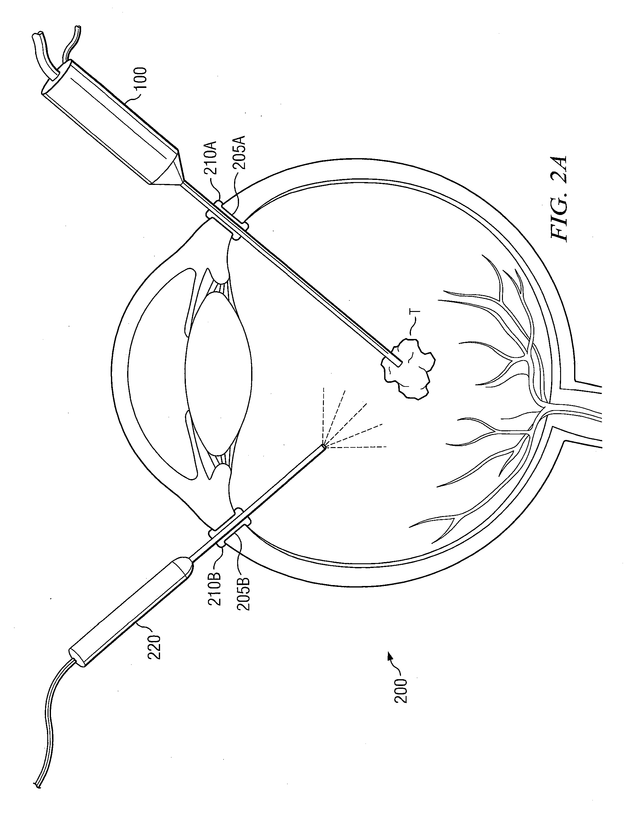
[0026]FIG. 1 is an outline illustration of a fragmatome 100 in accordance with a first aspect of the present invention. Fragmatome 100 comprises a tubular member 110 with a substantially constant outside diameter D. Tubular member 100 provides proximal end 112 and distal end 114. Fragmatome 100 may be constructed conventionally. However, in accordance with the invention, D is no more than 23-gauge (about 0.57 mm) in diameter. With further reference to FIG. 1, handle 120 is attached to a proximal end 112. Although not specifically illustrated, it will be understood that fragmatome 100 is disposed to deliver emulsification energy at distal end 114. Such emulsification energy may be, for example and without limitation, ultrasonic energy or laser energy. The source of emulsification energy may be conventional, and may be deployed in handle 120 or elsewhere in emulsification energy communication with distal end 114.

[0027]FIG. 1 shows tubular member 110 being of substantially constant dia...


PUM


Abstract

A medical instrument is disclosed, comprising a tubular member with proximal and distal ends. The distal end of the tubular member provides an elongated section having a substantially constant outside diameter of no more than 23-gauge (about 0.57 mm). The tubular member is in energy communication with an emulsification energy source disposed to deliver emulsification energy at the distal end of the tubular member. The tubular member may also be in suction communication with a suction source disposed to deliver suction at the distal end of the tubular member. The medical instrument may be used in a method to remove lens fragments from the eye, through incisions in the eye that are small enough not to require suturing after the operation.

Description

CROSS-REFERENCE TO RELATED APPLICATIONS[0001]This application claims the benefit of, and priority to, U.S. provisional patent application No. 61 / 389,311 filed Oct. 4, 2010, the disclosure of which provisional application is incorporated herein by reference.[0002]This application is also a divisional application of U.S. patent application Ser. No. 13 / 106,478 filed May 12, 2011.BACKGROUND OF THE INVENTION[0003]The present invention generally relates to eye surgery, and more particularly relates to a method and an apparatus, advantageously a small-diameter fragmatome, for removing retained lens fragments in a minimally traumatic fashion.[0004]During cataract surgery, the natural lens may be removed from inside of the patient's eye. Conventional procedure for removal of the natural lens typically involves making a large (2.9 mm, or larger) incision into the eye and a second, smaller incision. Typically, two incisions are required to accommodate conventional instrumentation into the eye....

Claims


Application Information

Patent Timeline
Patent Type & Authority Applications(United States)
IPC IPC(8): A61F9/00
CPCA61F9/00745A61F2009/00887A61F2009/0087A61F9/008
Inventor FOSTER, WILLIAM J.
Owner FOSTER WILLIAM J
Why Eureka
Try Eureka