Method and device to assist in dose reduction of x-ray radiation applied to a patient

a technology for reducing assisting in the patient's dose reduction, which is applied in the field of methods and devices to assist in the reduction of the dose of x-ray radiation applied to the patient, can solve the problems of not being able to automatically provide the existing protocols, image quality, and only rarely adapted by the users of the computed tomography apparatus to their own requirements and ideas, etc., so as to reduce the time cost of application training, simplify the work flow, and reduce operating errors

Active Publication Date: 2012-05-24
SIEMENS HEALTHCARE GMBH
View PDF1 Cites 5 Cited by
  • Summary
  • Abstract
  • Description
  • Claims
  • Application Information

AI Technical Summary

Benefits of technology

[0009]The invention thus displays to a user of an x-ray device that measure or those measures installed at the apparatus or its software that lead(s) to an additional reduction of the dose of x-ray radiation to be applied to the patient during the acquisition of the x-ray projections of the defined tissue, whenever a defined protocol that he has selected by the user for the examination of that defined tissue of the patient (such as for the acquisition of x-ray projections of the defined tissue) does not exploit all measures available (installed) at the x-ray device for the reduction of that dose of x-ray radiation to be applied to said patient during the acquisition of said x-ray projections of said defined tissue. In this way the user is informed of the additional available measures for dose reduction of the patient, particularly in the case of the user intending to operate the x-ray device after a software update with protocols adapted to the user's requirements before the software update, or at an entirely new apparatus type of the x-ray device. The user is thus given the opportunity to activate these additional measures. An advantage is thereby achieved not only for the patient (in that a smaller dose of x-ray radiation is applied to the patient by the activation of newly available measures) but also for the user. Not only is the workflow simplified for the user, but also operating errors are reduced and the time cost of the application training to be conducted for the user given a software update or the change to a new apparatus type can be decreased.
[0010]The method can be applied to all protocols of the x-ray device so that, before its intended use, every protocol is always checked as to whether it is current with regard to the activation of measures for dose reduction for the patient. This also enables the user to supplement each of his or her protocols that have been individually adapted to his or her requirements with newly available measures for dose reduction, such that after an apparatus change or software update the adaptation of the user-defined or user-adapted protocols can take place successively without running the risk of overlooking protocols that are to be adapted.

Problems solved by technology

The scan protocols of a computed tomography apparatus that are included at the factory or by the manufacture, however, are only rarely adapted by the users of the computer tomography apparatuses to their own requirements and ideas, in particular with regard to the image quality.
If a user of the computed tomography apparatus receives a new apparatus, or even just a software version including new protocols, even though all protocols installed by the manufacturer are provided with all new or improved measures for dose reduction, it is most often technically impossible to automatically provide the existing protocols, that have already (previously) been adapted by the user to his own requirements (which protocols the user would like to continue to use) with the new or improved measures for dose reduction.
Therefore the risk exists that the user at least occasionally makes no use of the new or improved measures for dose reduction, either due to a lack of knowledge as to how to manually embody them in the user's previously adapted protocols, or due to the lack of an automated adaptation of the protocols that user has previously adapted to his or her requirements before the software update.

Method used


Image

  • Method and device to assist in dose reduction of x-ray radiation applied to a patient
  • Method and device to assist in dose reduction of x-ray radiation applied to a patient
  • Method and device to assist in dose reduction of x-ray radiation applied to a patient

Examples


Embodiment Construction

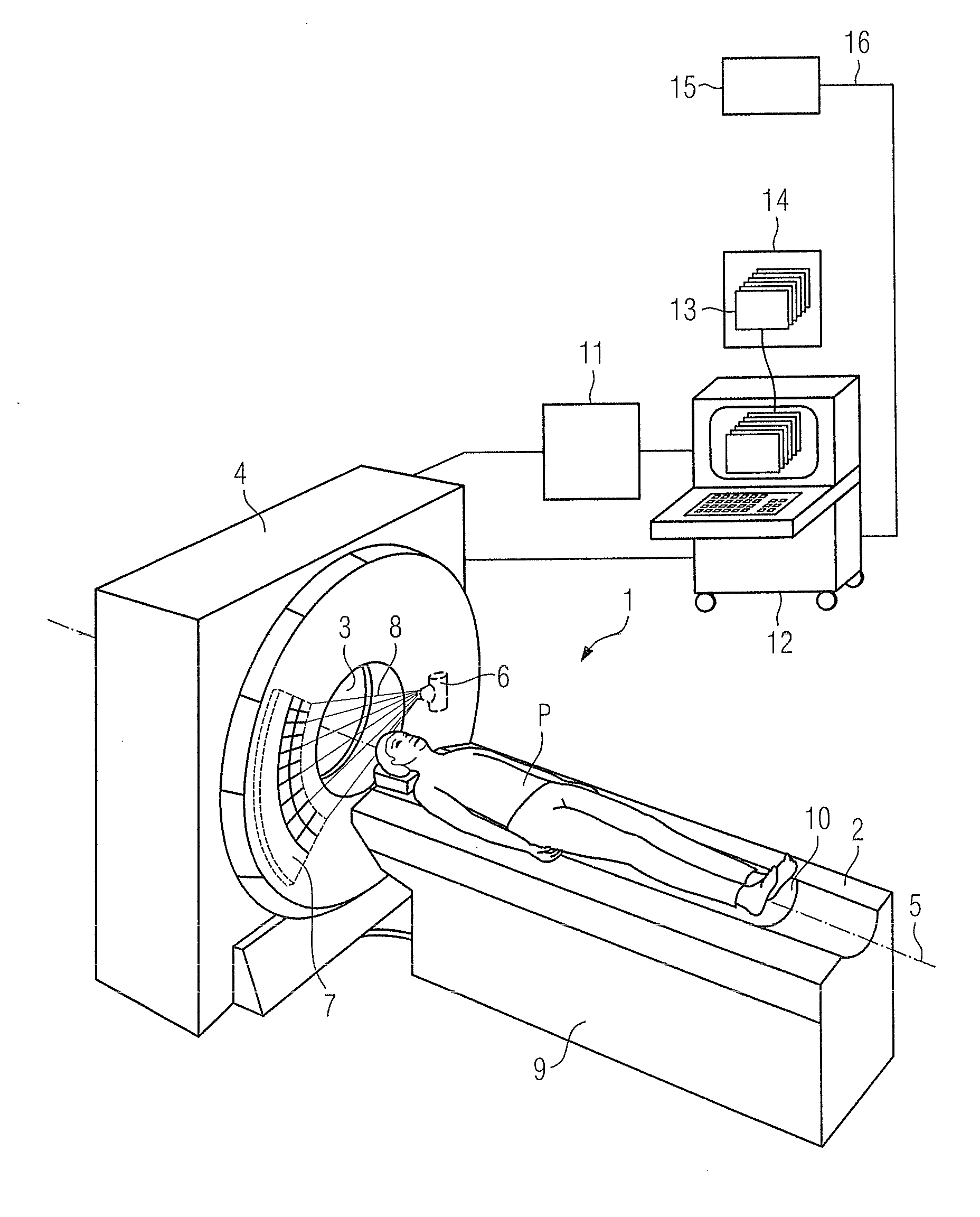
[0019]Shown in FIG. 1 is a computed tomography apparatus 1 that is suitable to execute the method according to the invention. The computed tomography apparatus 1 has a patient bed 2 to support a patient P to be examined. The computed tomography apparatus 1 also has a gantry 4 with a tube / detector system borne such that it can rotate around a system axis 5. The tube / detector system has an x-ray tube 6 and an x-ray detector unit 7 situated opposite one another. In operation, x-ray radiation 8 emanates from the x-ray tube 6 in the direction of the x-ray detector unit 7 and is detected by the detector unit 7.

[0020]The patient bed 2 has a bed base 9 on which is arranged a patient support plate 10 provided to actually support the patient P. The patient support plate 10 is adjustable relative to the bed base 9 such that the patient support plate 10 with the patient P thereon can be introduced into the opening 3 of the gantry 4 to acquire x-ray projections of the patient P, for example for ...


PUM


Abstract

In a method and x-ray device to assist in the reduction of the dose of x-ray radiation applied to the patient in the acquisition of at least one x-ray projection of the patient, a selected protocol is entered by a user into an x-ray device, the selected protocol being defined for the acquisition of the at least one x-ray projection and including a measure or measures for reduction of the dose of x-ray radiation to be applied to the patient in the acquisition of the at least one x-ray projection. The does reduction measure or measures in the selected protocol are automatically compared in a processor of the x-ray device, with the dose reduction measures that are installed at the x-ray device for reduction of the dose of x-ray radiation to be applied to a patient in the acquisition of the at least one x-ray projection. If the result of the comparison indicates a discrepancy between the dose reduction measure or measures in the selected, defined protocol and those installed in the x-ray device, at least one such measure installed at the x-ray device is visualized as a measure that can be set or activated by the user, that is not yet included in the selected protocol.

Description

BACKGROUND OF THE INVENTION[0001]1. Field of the Invention[0002]The invention concerns a method and an x-ray device to assist in the reduction of the dose of x-ray radiation applied to a patient in the acquisition of at least one x-ray projection of the patient. The invention also concerns a non-transitory computer-readable data storage medium encoded with programming instructions for implementing such a method.[0003]2. Description of the Prior Art[0004]In medical technology using imaging with x-ray radiation, for example in computed tomography, it is always sought to apply an optimally low dose of x-ray radiation to the patient to generate one or more images of tissue of the patient. However, a defined image quality must also be achieved in order to be able to answer clinical question at issue using the generated image or images, which requires a minimum dose.[0005]In order to assist a user (of a computed tomography apparatus, for example) in the examination of defined tissue of a ...

Claims


Application Information

Patent Timeline
Patent Type & Authority Applications(United States)
IPC IPC(8): H05G1/60H05G1/64
CPCA61B6/542A61B6/107
Inventor FEUERLEIN, UTEFREUND, JANGEHRSITZ, SEBASTIAN
Owner SIEMENS HEALTHCARE GMBH
Why Eureka
Try Eureka