Devices, systems, and methods for measuring and evaluating the motion and function of joints and associated muscles

a technology of motion and function, applied in the field of devices, systems and methods for measuring and evaluating the motion and function of joints and associated muscles, can solve the problems of inability to accurately assess pathologies, inability to detect many types of pathologies, and significant number of patients with joint problems to be diagnosed and treated

Active Publication Date: 2012-05-24
AECC ENTERPRISES
View PDF12 Cites 9 Cited by
  • Summary
  • Abstract
  • Description
  • Claims
  • Application Information

AI Technical Summary

Problems solved by technology

However, currently available diagnostic tools are limited in the level of detail and analysis that can be achieved.
However, currently available diagnostic techniques provide measurement data that is imprecise and often inconclusive which results in an inability to detect many types of pathologies or to accurately assess pathologies that might be considered borderline.
As a result, a significant number of patients having joint problems remain undiagnosed and untreated using current techniques, or worse are misdiagnosed and mistreated due to the poor clinical efficacy of these techniques.
There are currently no reliable techniques for identifying soft tissue injury.
Additionally, the level of entrenchment of muscle guarding behavior cannot currently be determined.
There has not however, been a corresponding improvement in the diagnostic methods used to identify proper candidates for these interventions.
As a result, the potential impact and utility of the improvements in orthopedic intervention has been limited.
However, these applications yield poor diagnostic performance with an unacceptably high proportion of testing events yielding either inconclusive or false positive / false negative diagnostic results (Lawrence, J. S. (1969) Annals of Rheumatic Diseases 28: 121-37; Waddell, G.
Using uncontrolled, weightbearing motion to derive quantitative measurements of joint motion confounds the diagnostic interpretation of such measurements so as to render them diagnostically useless.
Not controlling the motion that is being studied introduces variability into these comparative analyses due to differences that exist across testing subjects with respect to each subject's individual range of motion, symmetry of motion, and regularity of motion.
These references do not, however, provide for any form of measurement or identification of objectively defined motion abnormalities, and therefore is of very limited diagnostic value other than in the detection of grossly and visibly obvious abnormalities that would be detectable using static image analysis methods.
Without any quantitative or objective measurement parameters defined, it is impossible to utilize such approaches in comparative analyses across wide populations of subjects, which is required for the purpose of the producing definitive diagnostic interpretations of the results as being either “normal” or “unhealthy”.
Further, there have been no diagnostically useful validations of qualitative motion patterns that are generally absent in non-sufferers but present in subjects suffering from known and specific joint functional derangements or symptoms, or vice versa.
Prior attempts at controlling the motion to derive quantitative measurements have provided a more accurate measurement of joint motion, however, some aspects of the motion have continued to be uncontrolled.
For example, while some researchers have been able to control the gross motion of the subject, they have not been able to control for the rotation of the patient at their hips while undergoing the controlled motion sweep.
This uncontrolled rotation during the controlled sweep of trunk bending confounds the goal of controlling the motion, but also can confound the quantitative imaging results.
When the subject rotates out of plane during the trunk bending, the definition of the vertebra in the image becomes compromised, making the vertebra harder to track throughout the image sequence, and thus confounding the diagnostic measurement results.
However RSA requires the surgical implantation of these markers into subjects' internal joint structures, requires the use of two radiographic units simultaneously, and requires a highly complicated calibration process for every single test, and therefore is too invasive and too cumbersome a process for practicable clinical application.
A method for determining vertebral body positions using skin markers was developed (Bryant (1989) Spine 14(3): 258-65) but could only measure joint motion at skin positions and could not measure the motion of structures within the joint.
There have been many examples skin marker based spine motion measurement that have all been similarly flawed.
This method is, however, inherently impractical for clinical diagnostic use.
However RSA requires the surgical implantation of these markers into subjects' internal joint structures, requires the use of two radiographic units simultaneously, and requires a highly complicated calibration process for every single test, and therefore is too invasive and too cumbersome a process for practicable clinical application.
Taken together, all of these sources of variability serve to confound diagnostic conclusions based on comparative analyses by making the ranges of “normal” and those of “abnormal” difficult to distinguish from one another other in a statistically significant way.
Such an inability to distinguish between “normal” and “unhealthy” subjects based on a specific diagnostic measurement renders such a measurement diagnostically useless, as has been the case heretofore known which has focused on measurements of uncontrolled joint motion measured in subjects in weight-bearing postures and moving their joints through the power of their own muscles and in an uncontrolled fashion.
These electromyographical readings of “unhealthy” subjects were then compared to those of a “normal” population so as to be able to identify those subjects with abnormal readings, however does not provide for a method to report the results as a degree of departure from an ideal reading, instead can only say whether the reading is “abnormal”.
However this approach studied joint motion that was uncontrolled and required an invasive surgical procedure to place the metal markers, and thus were neither useful nor feasible for clinical diagnostic application.
This approach however does not address the issue of which discrete muscle group or groups might account for the difference between activation patterns in subjects with joint dysfunctions and those without.
Furthermore, this method does not take into consideration the internal structural joint motions and thus provides an incomplete set of information upon which to draw diagnostic conclusions.

Method used


Image

  • Devices, systems, and methods for measuring and evaluating the motion and function of joints and associated muscles
  • Devices, systems, and methods for measuring and evaluating the motion and function of joints and associated muscles
  • Devices, systems, and methods for measuring and evaluating the motion and function of joints and associated muscles

Examples


example 1

Hypothetical Example 1

A Weight Bearing Joint

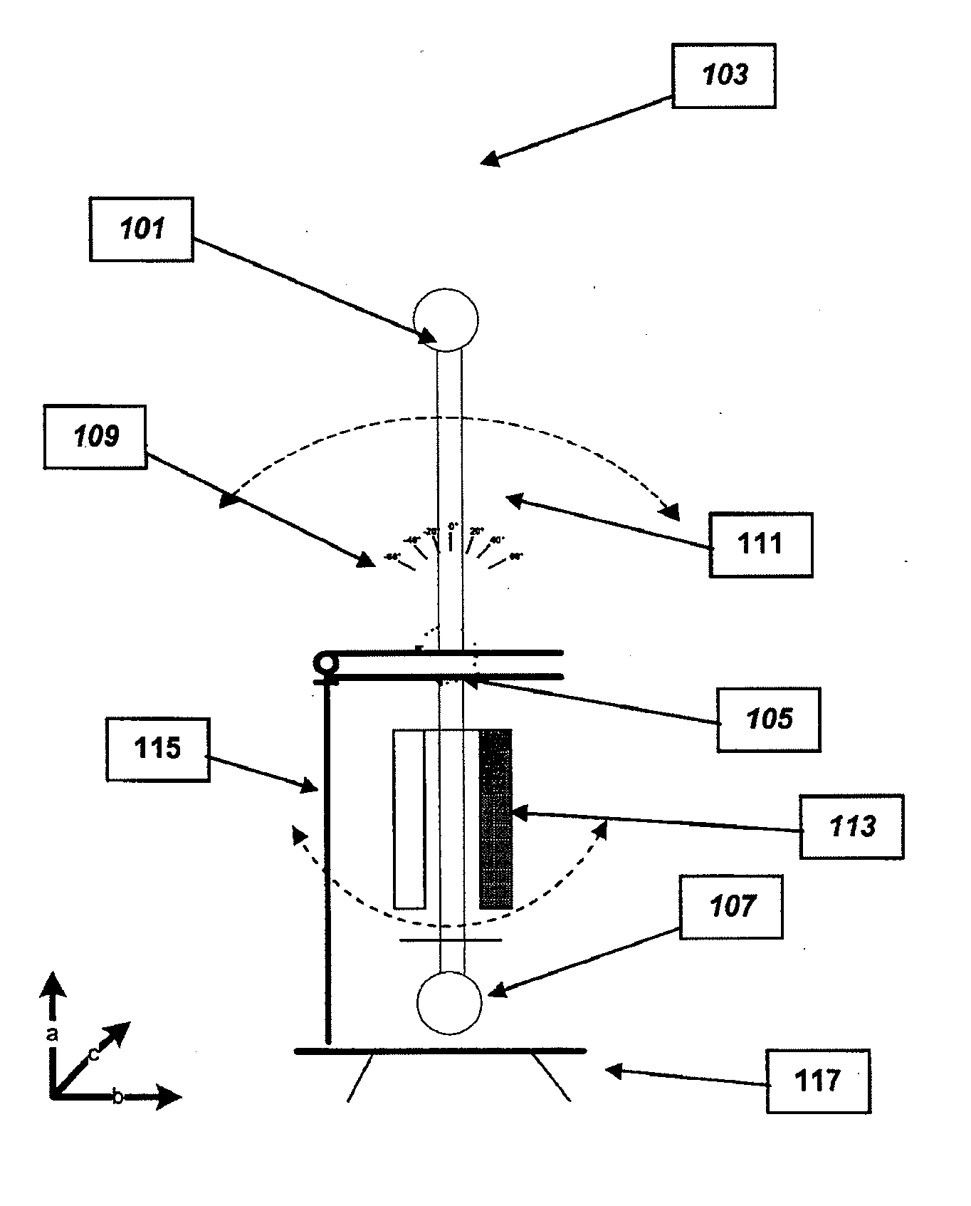
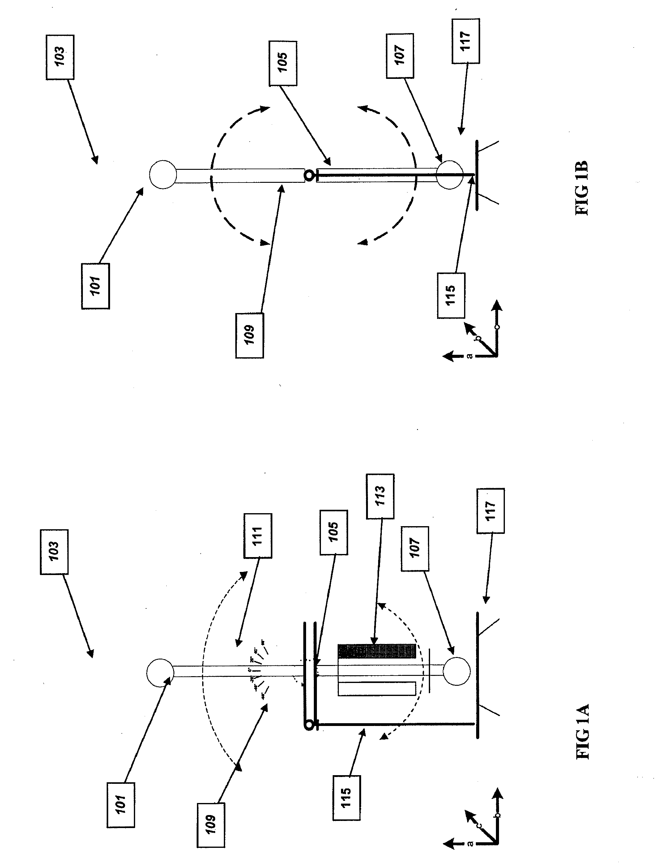
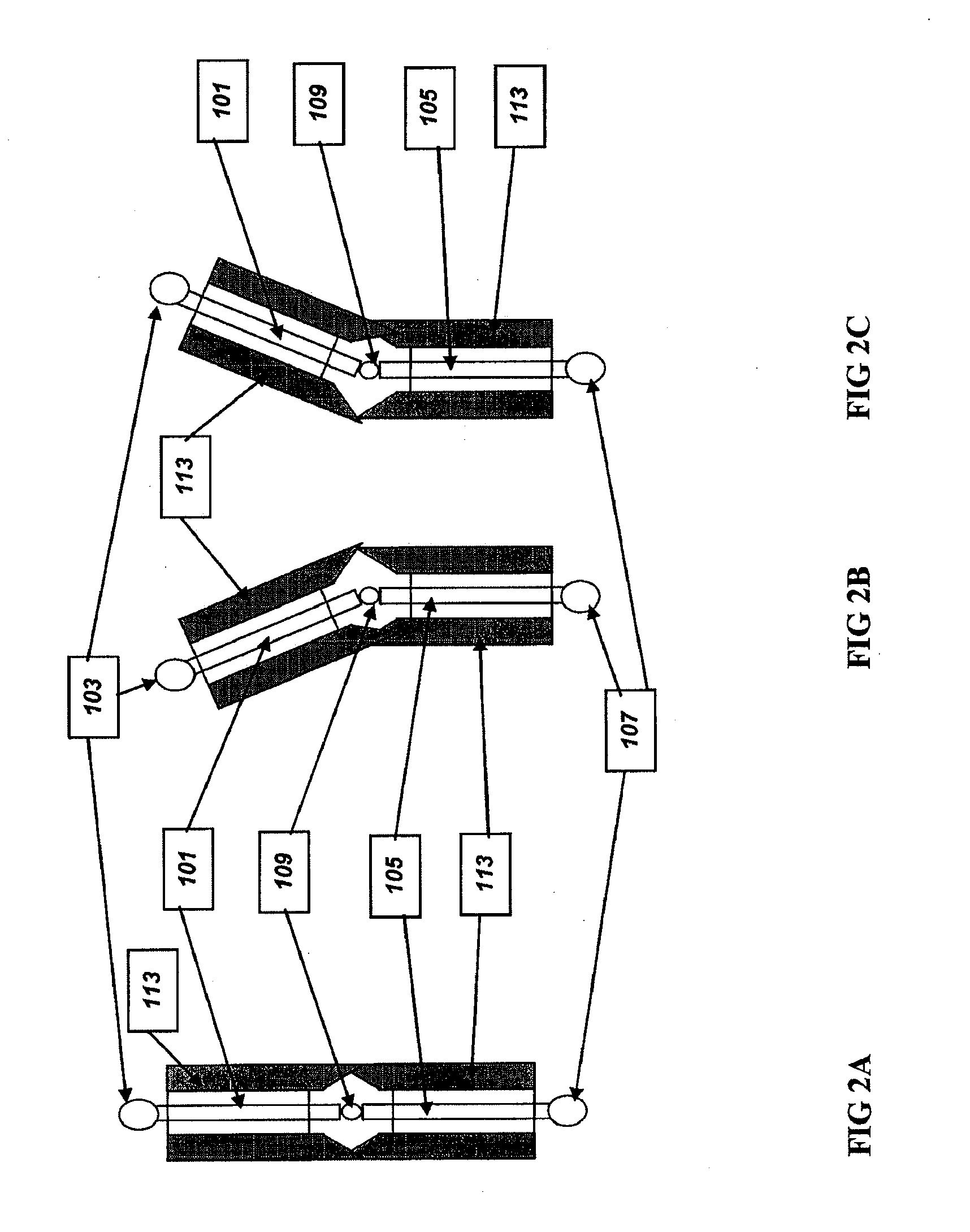
[0117]For a weight bearing joint like the neck or spine, there can be useful diagnostic data produced by affecting a comparison of weight bearing motion to non-weight bearing motion. For testing weight bearing motion, the present invention can be configured to be freestanding with both attachment arms occupying the line normal to the floor, and with one attachment arm attached to the patient's head with the other attached to the patient's shoulders. For testing non-weight bearing motion, the attachment arm attached to the shoulders in the previous example could be turned to be parallel to the floor, then attached to a patient on which the patient is lying. The other attachment arm could be attached to the patient's head (as in the previous configuration). Such a configuration would allow for the assessment of motion in a plane parallel to the floor (complete non-weight bearing), or in a plane perpendicular to the floor (partial weight bear...

example 2

Hypothetical Example 2

A Non-Weight Bearing Joint

[0118]For a non-weight bearing joint like the wrist, it would be possible to use a free-standing configuration of the motion control device, with both attachment arms parallel and occupying a plane parallel to the floor. One attachment mechanism could attach to the forearm, and the other to the hand. In this configuration, motion could be tested and compared between that which occupies a plane perpendicular to the floor (up and down wrist movement), and that which occupies a plane parallel to the floor (lateral bending wrist movement). Alternatively, comparisons could be made between active and passive motion on the part of the subject.


PUM


Abstract

Apparatuses are disclosed that is adapted and configured to cause and control joint motion of a patient. Apparatuses comprise, for example, a first motion member configured to engage a patient proximal to a target joint; a second motion member configured to engage a patient distal to a target joint; and a coupling member configured to connect the first motion member to the second motion member and further adapted to allow rotation of the first and second motion member around an axis, wherein the apparatus is engageable with a medical diagnostic device and further configured to capture patient specific data.

Description

CROSS-REFERENCE[0001]This application claims is a CONTINUATION of application Ser. No. 12 / 247,552 filed Oct. 8, 2008, entitled Devices, Systems and Methods for Measuring and Evaluating the Motion and Function of Joints, which claims the benefit of U.S. Provisional Application Nos. 60 / 978,907, filed Oct. 10, 2007, 60 / 981,922, filed Oct. 23, 2007, 61 / 015,149, filed Dec. 19, 2007, and 61 / 054,426, filed May 19, 2008, which applications are incorporated herein by reference.TECHNICAL FIELD[0002]The present invention relates to devices, systems and methods for measuring and evaluating the motion and pathologies of a joint structure in a subject. The invention also enables a determination of whether, and to what extent, muscles associated with a joint are impacting the joint's biomechanics. The invention enables the biomechanics of a joint to be evaluated with a precision of less than 5°, preferably less than 3°, and even more preferably less than 1°. The enhanced biomechanical assessment f...

Claims


Application Information

Patent Timeline
Patent Type & Authority Applications(United States)
IPC IPC(8): A61B5/103
CPCA61B5/1071A61B5/1121A61B5/4528A61B5/4519A61B5/1126A61B2562/17
Inventor DEITZ, ADAM
Owner AECC ENTERPRISES
Why Eureka
Try Eureka