Open pet/mri hybrid machine

a hybrid machine and open technology, applied in the field of open pet/mri hybrid machines, can solve the problems of difficult interventional treatment, inability of pet devices alone to acquire morphological information, etc., and achieve the effects of reducing subject stress, facilitating experiments, and imposing high psychological stress

Inactive Publication Date: 2012-06-14
NAT INST OF RADIOLOGICAL SCI
View PDF3 Cites 44 Cited by
  • Summary
  • Abstract
  • Description
  • Claims
  • Application Information

AI Technical Summary

Benefits of technology

[0022]Performing diagnosis on the head in a PET / MRI device with a long tunnel-like patient port imposes high psychological stress on the subject. According to the present invention, the simultaneous PET and MRI measurements of the open space can significantly reduce the stress on the subject.
[0023]Open-type devices provide doctors and operators easy access to the subject, allowing easy condition check. In particular, in the field of brain researches, responses to visual and other stimulations are sometimes inspected by PET or MRI. The presence of the open space facilitates experiments.
[0024]Moreover, the open space allows installation of a third diagnostic device and the like, whereby the same field of view can be simultaneously imaged in three or more modalities. Examples of the third diagnostic device include an ultrasonic device, a SPECT device, a fluorescent imager, an optical camera, and an optical CT device.

Problems solved by technology

In any case, however, the closed field of view imposes a lot of psychological stress on the subject as well as makes interventional treatment difficult.
The PET device, however, has had a problem that the PET device by itself is not capable of acquiring morphological information.

Method used


Image

  • Open pet/mri hybrid machine
  • Open pet/mri hybrid machine
  • Open pet/mri hybrid machine

Examples


first embodiment

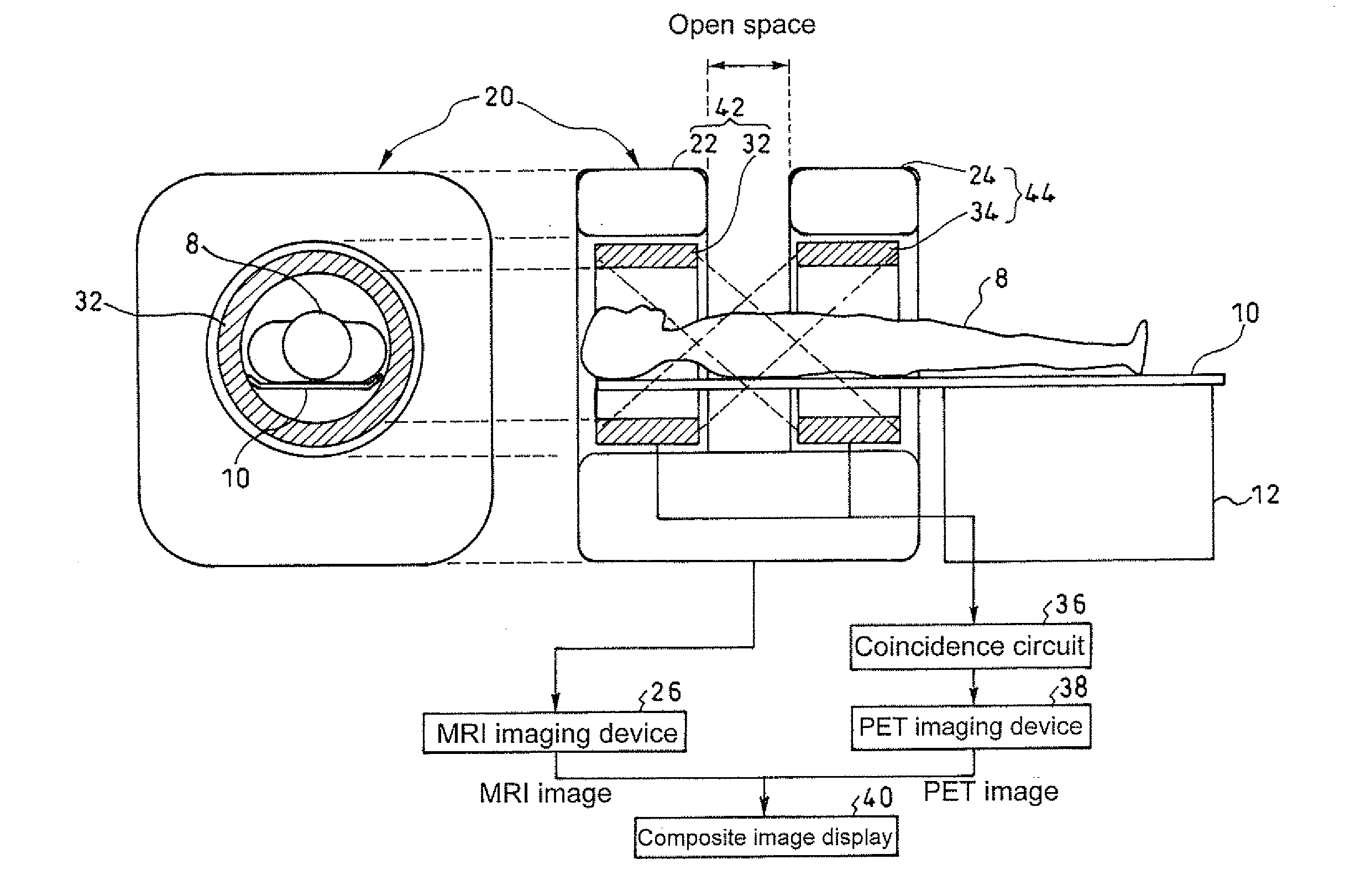
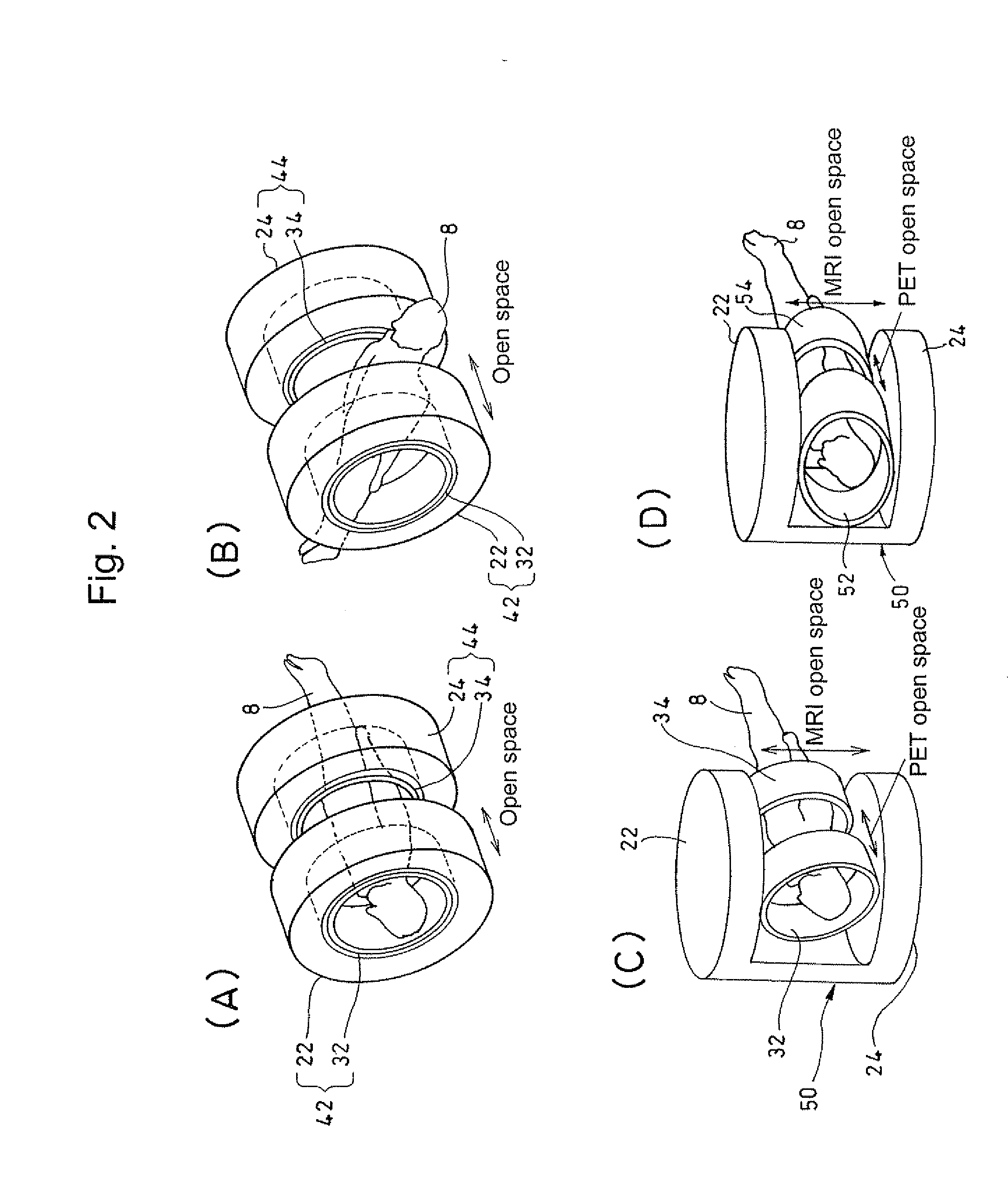
[0037]As shown in FIG. 1, the present invention includes an open MRI device 20 of so-called doubled doughnut type, having two magnets 22 and 24 which are arranged apart in the direction of the body axis of a subject 8. Antimagnetic PET detector rings 32 and 34 are arranged in patient ports on respective sides to constitute composite rings 42 and 44. The space between the two separated, opposed composite rings 42 and 44 is the overlapping open space of MRI and PET. In the diagram, 12 represents a pedestal of a bed 10, 26 represents an MRI imaging device, 36 represents a coincidence circuit for PET measurement, 38 represents a PET imaging device, and 40 represents a composite image display which displays a morphological image obtained by the MRI imaging device 26 and a functional image obtained by the PET imaging device 38 in a superposed manner.

[0038]With such a configuration, radiations are measured from lines of coincidence between the PET detector rings 32 and 34, whereby the same...

second embodiment

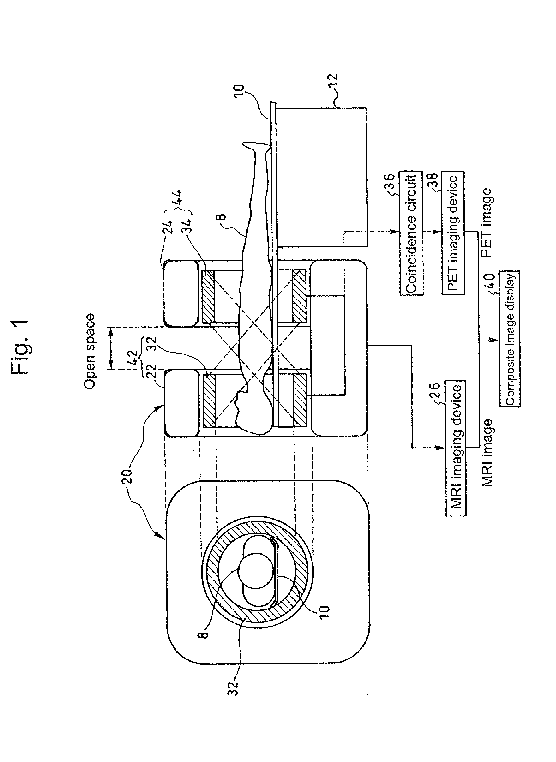
[0041]Next, the present invention will be described in detail with reference to FIG. 3.

[0042]In the present embodiment, an RF coil 60 for MRI is fixed to or movably arranged on the bed 10. The RF coil 60 and / or the bed 10 is / are moved in the direction of the body axis of the subject 8 so that the RE coil 60 enters the open space to make the PET field of view and the MRI field of view overlap each other. The RE coil typically has a circular cylindrical frame structure to surround the affected area. The RF coil usually has a lot of openings and will not interfere with access to the affected area.

[0043]According to the present embodiment, the RF coil 60 and / or the bed 10 can be moved in the body axis of the subject 8 to adjust the field of view of the MRI image in position.

[0044]FIG. 4 shows an example where an inspection tube 64 of a laparoscope device 62 is inserted into the open space as a working unit and laparoscopic inspection / treatment is administered to the subject 8 according ...


PUM


Abstract

A PET/MRI hybrid machine that combines a PET device with an MRI device, includes: an open PET device that has a PET field of view at least part of which is an open space accessible from outside; and an open MRI device that has an MRI field of view at least part of which is an open space accessible from outside, and the open space of the PET field of view and the open space of the MRI field of view are allowed to overlap each other at least in part. This makes it possible to simultaneously measure and image the same open space accessible from outside by the PET device and the MRI device, thereby acquiring functional information and morphological information at the same time.

Description

TECHNICAL FIELD[0001]The present invention relates to a PET / MRI hybrid machine that combines a PET device with an MRI device, and more particularly to an open PET / MRI hybrid machine that can simultaneously measure and image the same open space accessible from outside by the PET device and the MRI device, thereby acquiring biological functional information and morphological information at the same time.BACKGROUND ART[0002]PET is a method of administering a compound that is labeled with positron-emitting radionuclides and imaging the distribution of the compound in the body as a tomographic image. While X-ray CT or MRI based tomographic images provide morphological information, PET images are referred to as functional images which express biological functional information. For example, PET images are capable of highly sensitive tumor detection though not precise in position. To add accurate positional information to a PET image, it is needed to superpose an X-ray CT image or MRI image...

Claims


Application Information

Patent Timeline
Patent Type & Authority Applications(United States)
IPC IPC(8): A61B5/055
CPCG01R33/3806G01T1/1603G01R33/481G01R33/381
Inventor YAMAYA, TAIGAMURAYAMA, HIDEOOBATA, TAKAYUKIAOKI, ICHIO
Owner NAT INST OF RADIOLOGICAL SCI
Why Eureka
Try Eureka