Electronic endoscope system and image processing method

Active Publication Date: 2012-07-26
FUJIFILM CORP
View PDF11 Cites 20 Cited by
  • Summary
  • Abstract
  • Description
  • Claims
  • Application Information

AI Technical Summary

Benefits of technology

[0029]The system and the method of the present invention are effective to prevent image data of an extraneous or artificial object like a surgical tool from unnecessary image-processing like image enhancement when the artificial object is

Problems solved by technology

The surgical tools may however cause problems when protruded into the imaging field of the endoscope.
For example, some of the surgical tools may reflect the illumination light so much that the monitored endoscopic images suffer smears or flares, which will obstruct the inspection of the target site.
In addition to the above problem, when endoscopic image containing the surgical tool beside the target subject is digitally processed for example for the above-mentioned frequency enhancement, if pixels corresponding to the surgical tool are processed in the same way as other pixels of the image, the image of the surgical tool may often be modified

Method used


Image

  • Electronic endoscope system and image processing method
  • Electronic endoscope system and image processing method
  • Electronic endoscope system and image processing method

Examples


Embodiment Construction

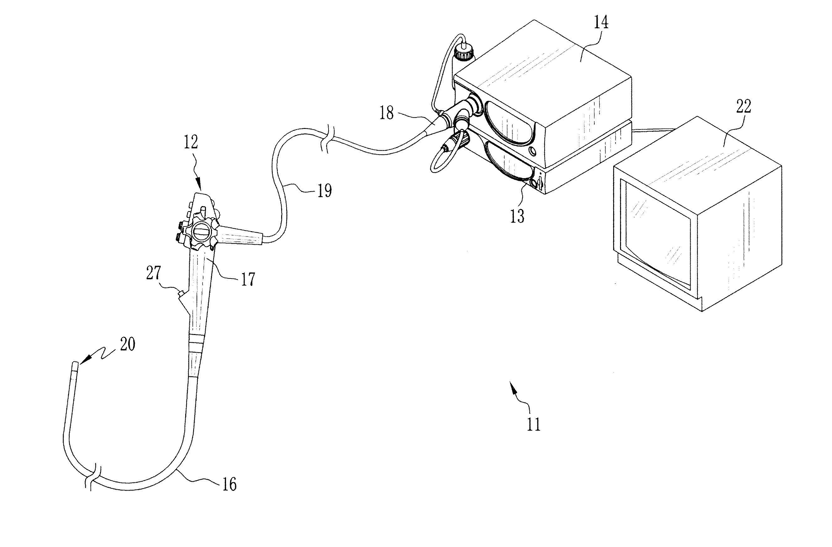
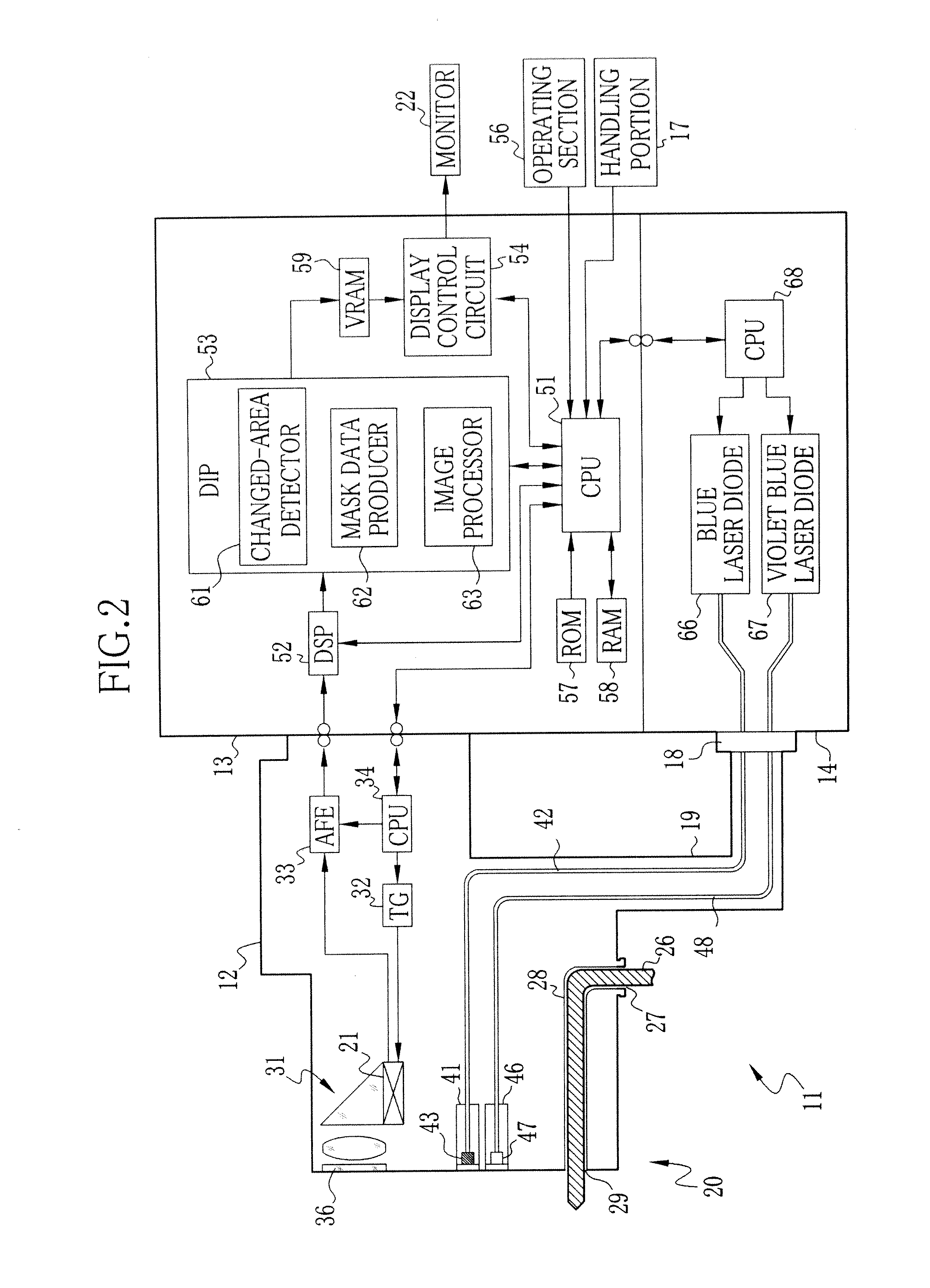
[0048]As shown in FIG. 1, an electronic endoscope system 11 includes an electronic endoscope 12, a processor unit 13, and a light source unit 14. The electronic endoscope 12 includes a flexible probing portion 16 to be inserted into the body cavity, a handling section 17 coupled to a proximal end of the probing portion 16, a connector 18 connected to the processor unit 13 and the light source unit 14, and an universal cord 19 connecting the handling section 17 and the connector 18. In a distal end 20 of the probing portion 16 is provided a CCD image sensor 21 (see FIG. 2) for imaging the interior of the body cavity.

[0049]The processor unit 13 is electrically coupled to the light source unit 14, and supervises the overall operation of the electronic endoscope system 11. Through a cable that is conducted through inside the universal cord 19 and the probing portion 16, the processor unit 13 controls power supply to the electronic endoscope 12 and also controls driving the CCD 21. The p...


PUM


Abstract

In an electronic endoscope system, a changed-area detector detects a changed area from an image captured by an endoscope, the changed area having different features from other area of the captured image. A mask data produce produces mask data based on the detected changed area, the mask data allocating an image processing parameter to each pixel of the captured image such that the changed area is processed in a different way from the other area. An image processor processes the captured image according to the mask data. Thus, an image area corresponding to an artificial object like a surgical tool may be detected as the changed area and excluded from unnecessary image processing.

Description

BACKGROUND OF THE INVENTION[0001]1. Field of the Invention[0002]The present invention relates to an electronic endoscope system that acquires images from inside of a test subject through an electronic endoscope. The present invention also relates to an image processing method for the electronic endoscope system.[0003]2. Description of the Related Art[0004]In the medical field, electronic endoscopes have been widely used for diagnoses, surgical procedures and treatments. Many of the electronic endoscopes conventionally project white light (hereinafter referred to as ordinary light) into a test subject body for imaging the interior of the subject body by an imaging device like CCD. Recently, for the sake of acquiring such images that show the details of particular tissues or biological components of the subject body, which are not clearly perceivable from endoscopic images acquired under the ordinary light, some of the electronic endoscopes also use rays of particular narrow wavelengt...

Claims


Application Information

Patent Timeline
IPC IPC(8): H04N7/18
CPCG06T7/0016G06T7/0081G06T2207/10016G06T2207/10064G06T2207/30101G06T2207/20012G06T2207/20132G06T2207/20148G06T2207/10068G06T7/11G06T7/136
Inventor KAKU, TOSHIHIKO
Owner FUJIFILM CORP
Why Eureka
Try Eureka