Method of generating 2D or 3D maps of MRI T1 and T2 relaxation times

Active Publication Date: 2012-09-06
ESAOTE
View PDF14 Cites 8 Cited by
  • Summary
  • Abstract
  • Description
  • Claims
  • Application Information

AI Technical Summary

Benefits of technology

[0017]Also, single-exponential relaxation is assumed for the longitudinal and transverse components of the magnetic moment associated with each voxel: inherent multi-exponential relaxation (on a microscopic scale) and Partial Volume effects, known as PVE (on a macroscopic scale) will genera

Problems solved by technology

Such great number of acquisitions has two apparent drawbacks, which might affect the quality of results and have an impact on everyday clinical practice: on the one hand the long duration of the protocol; on the other hand the considerable risk that any inadvertent movement of the patient might affect the quality of the individual images and perfect registration of different series (which is essential during combined post-processing of contrasts).
Thus using a TrueFISP sequence for determining T1

Method used


Image

  • Method of generating 2D or 3D maps of MRI T1 and T2 relaxation times
  • Method of generating 2D or 3D maps of MRI T1 and T2 relaxation times
  • Method of generating 2D or 3D maps of MRI T1 and T2 relaxation times

Examples


Embodiment Construction

[0041]Referring to a method according to the present invention, the T1 and T2 maps are obtained by acquiring 2D or 3D MRI signals using two sequences only.

[0042]The sequences that are used to generate relaxometry maps are steady-state 3D gradient-echo sequences. This will combine the typical advantages of 3D imaging sequences, i.e. speed, resolution and signal-to-noise ratio SNR with simple calibration of the system, which is a feature of the relaxometry method as disclosed herein.

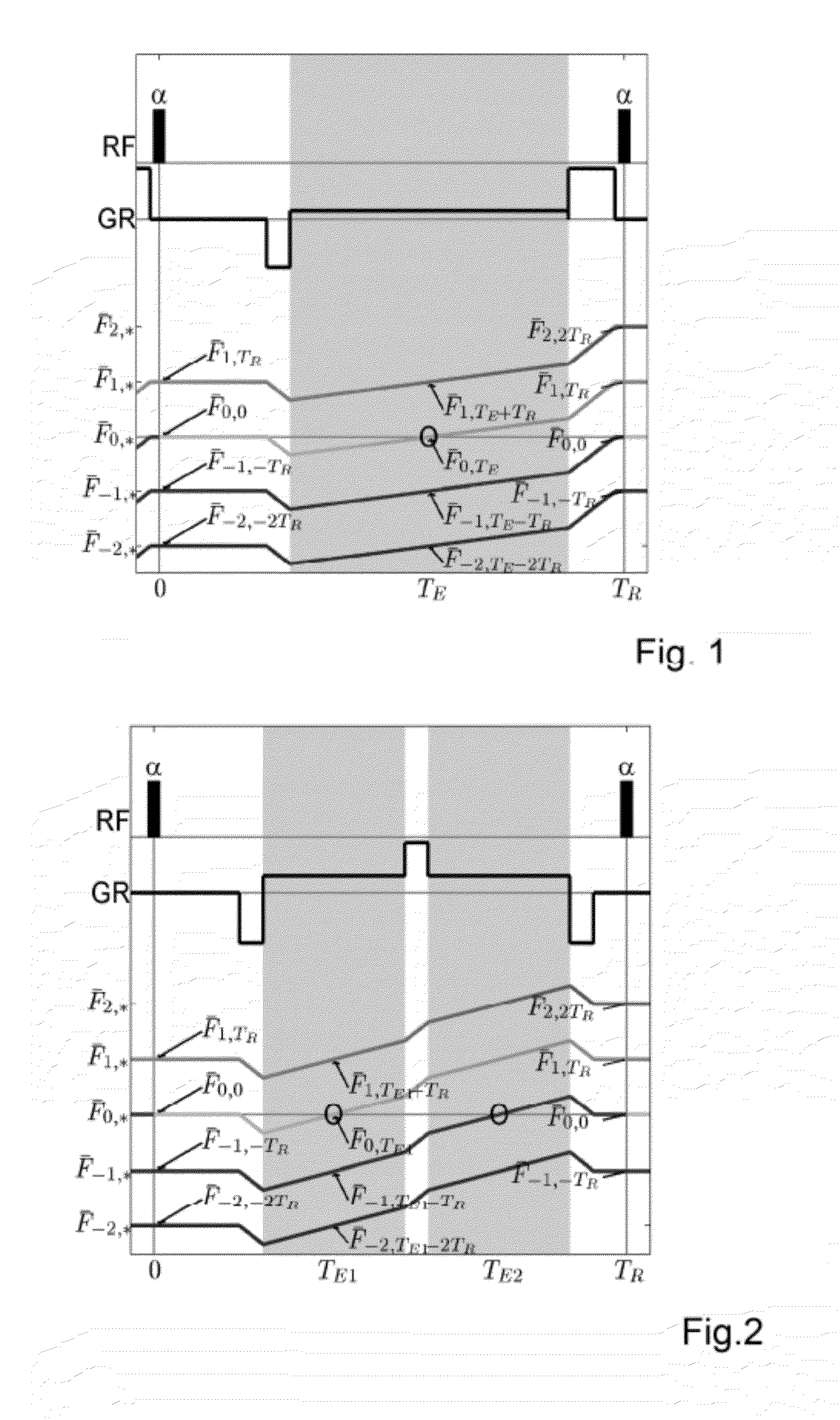
[0043]The first of the two sequences that are used in the method of the present invention is a so-called SSFP-FID sequence (see references above).

[0044]Assuming a SSFP-FID sequence (Steady-State Free-Precession Free Induction Decay—FIG. 1) with a repetition time TR1, an echo time TE0 and a flip angle α, the equation that describes the signal corresponding to 0-order unbalanced steady-state coherent magnetization is (Sobol W T & Gauntt D M, “On the Stationary States in Gradient Echo Imaging,” J. Magn. Reson...


PUM


Abstract

A method of generating 2D or 3D maps of MRI T1 and T2 relaxation times by acquiring 2D or 3D MRI gradient-echo images and extracting the T1 and T2 values from the images, wherein MRI images are acquired using a combination of gradient-echo sequences, including a first MRI image acquired using a SSFP-FID (Steady State Free Precession-Free Induction Decay) acquisition sequence; two further images acquired using a Dual-Echo SSFP acquisition sequence; and the T1 and T2 values are extracted for each image pixel or voxel from the corresponding MRI signals.

Description

FIELD OF THE INVENTION[0001]The present invention relates to a method for generating 2D or 3D maps of MRI T1 and T2 relaxation times by acquiring 2D or 3D MRI gradient-echo images and extracting the T1 and T2 values from said images.BACKGROUND OF THE INVENTION[0002]The terms 2D and 3D designate images of two-dimensional and three-dimensional maps respectively, as is generally used in the field of diagnostic magnetic resonance imaging (MRI).[0003]In the field of MRI, relaxation time mapping is a useful imaging instrument which is profitably used in the diagnosis of diseases affecting a variety of anatomic regions, from brain to limbs.[0004]At present, the most advanced protocol for 3D T1 and T2 relaxometry involves the acquisition of 4 sequences (Deoni et al. US2005 / 0256393). Such great number of acquisitions has two apparent drawbacks, which might affect the quality of results and have an impact on everyday clinical practice: on the one hand the long duration of the protocol; on the...

Claims


Application Information

Patent Timeline
IPC IPC(8): G01R33/50
CPCG01R33/50G01R33/448G01R33/5613
Inventor PALMA, GIUSEPPEGRECO, DANILOINNOCENTI, STEFANIAALFANO, BRUNO
Owner ESAOTE
Why Eureka
Try Eureka