Diagnostic Marker for Hepatocellular Carcinoma Comprising Anti-FASN Autoantibodies and a Diagnostic Composition for Hepatocellular Carcinoma Comprising Antigens Thereof
a technology for hepatocellular carcinoma and diagnostic composition, which is applied in the direction of fluorescence/phosphorescence, instruments, fused cells, etc., can solve the problems of weak specificity and sensitivity of hepatocellular carcinoma, evidence of hepatocellular carcinoma, and inability to become independent diagnostic tools, etc., to achieve high specificity and sensitivity, the effect of easy diagnosis
- Summary
- Abstract
- Description
- Claims
- Application Information
AI Technical Summary
Benefits of technology
Problems solved by technology
Method used
Image
Examples
example 1
Acquisition of Disease Mouse Model and Autoantibody-Producing Cells Using the Same
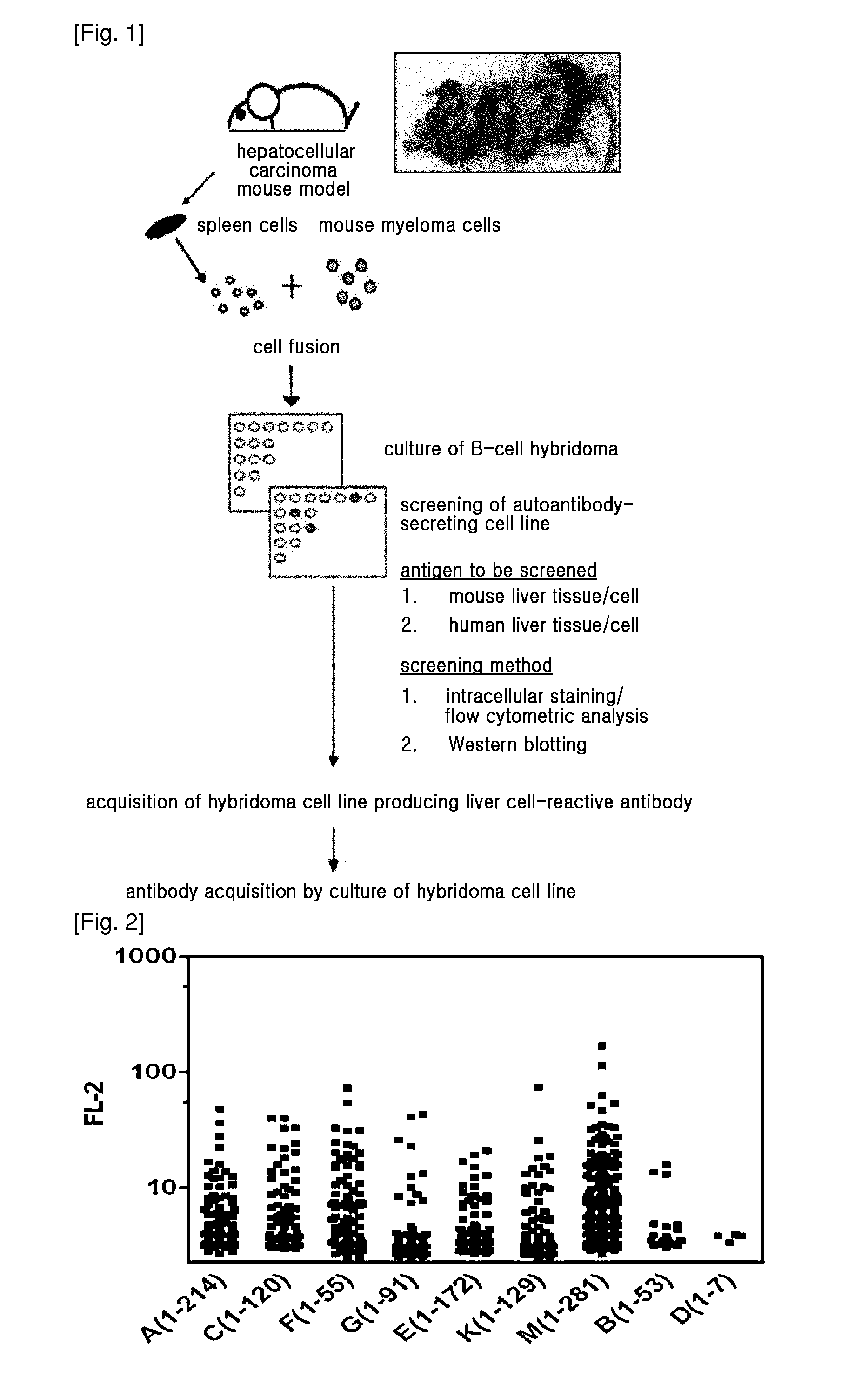
[0080]FIG. 1 is an overview of the method of obtaining hepatocellular carcinoma-associated autoantibody from hepatocellular carcinoma mouse model. H-ras 12V transgenic HCC mouse model, characterized by the occurrence of hepatocellular carcinoma at 8-10 months of age, was acquired, and splenocytes from the mouse models that developed hepatocellular carcinoma were fused with the mouse myeloma cells Sp2 / 0. The fused cells were primarily selected using HAT medium, and cells forming clones were separately cultured. B-cell hybridomas producing HCC-associated autoantibodies were only selected from the cell media, and maintained. The reactivity of the B-cell hybridoma medium to HCC cells was examined by the following method in Example 2.
example 2
Flow Cytometric Analysis for Detection of Autoantibody Generated by B-Cell Monoclone
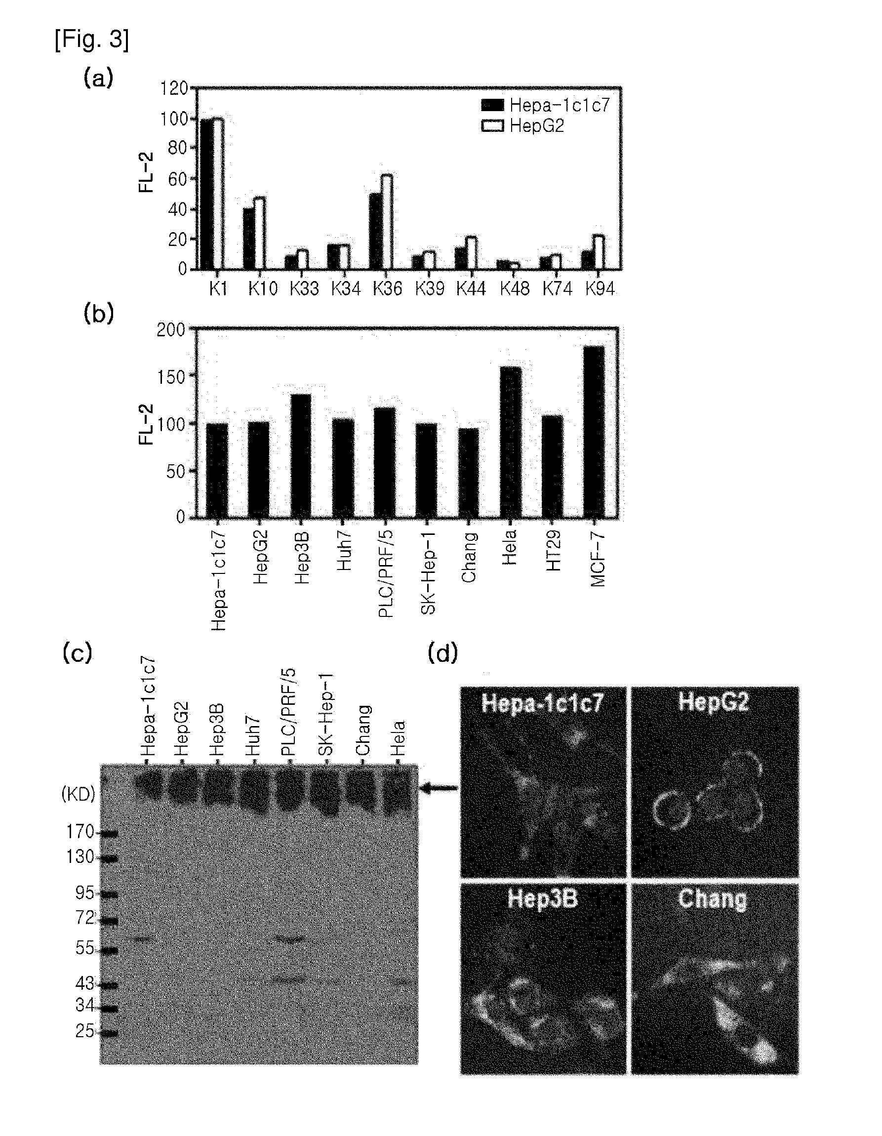
[0081]To Select Antibodies Reactive to Hepatocellular Carcinoma Cells from the Antibodies that were produced by the B-cell hybridomas obtained from cancer mouse model, intracellular staining of fixed and permeabilized cancer cell lines was performed, followed by flow cytometric analysis. 70-80% confluent HepG2 or Hepa-1c1c7 cells were treated with trypsin, and detached from a cell culture plate, and washed with PBS. Then, the cells were treated with a Cytofix / Cytoperm solution (BD) (100 μl per 2×10⁵ cells) at 4° C. for 20 min for fixation and permeabilization. After the incubation, 1 ml of Cytowash / Cytoperm solution (BD) was added, and vortexed, and then centrifuged at 1700 rpm for 5 min to obtain cell pellets. After washing the cells, 50 μl of primary antibody solution (B-cell hybridoma culture media or purified primary antibody solution) was added thereto, followed by incubation at 4° C. for 40 min...
example 3
Western Blotting Analysis for K1-Autoantigen
[0082]As a sample for protein analysis, the subject cells were collected and lysed in RIPA buffer (PBS containing 0.1% SDS, 0.1% sodium deoxycholate, 1.0% NP40, protease inhibitor cocktail (Roche)), or lysed in SDS-PAGE sample buffer and heated. Bradford assay was performed for protein quantification. The prepared protein sample was run on 8%-10% reduced SDS-PAGE, and transferred onto a PVDF membrane. Thereafter, the membrane was blocked with 5% skim milk (TBS), and treated with a primary antibody. The purified K1 antibody was used at a concentration of 10 μg / ml as a primary antibody. After treatment of primary antibody, the membrane was washed with TBST (0.02% Tween-20 containing TBS), and treated with secondary antibody (anti-mouse IgGAM-HRP). Then, the protein band corresponding to antibody reaction was confirmed by ECL. β-actin was used as a protein loading control for result comparison. As shown in FIG. 3c, it was found that the K1 an...
PUM
| Property | Measurement | Unit |
|---|---|---|
| Gloss | aaaaa | aaaaa |
| Gloss | aaaaa | aaaaa |
| Composition | aaaaa | aaaaa |
Abstract
Description
Claims
Application Information
 - R&D
- Intellectual Property
- Life Sciences
- Materials
- Tech Scout
- Unparalleled Data Quality
- Higher Quality Content
- 60% Fewer Hallucinations
Browse by: Latest US Patents, China's latest patents, Technical Efficacy Thesaurus, Application Domain, Technology Topic, Popular Technical Reports.
© 2025 PatSnap. All rights reserved.Legal|Privacy policy|Modern Slavery Act Transparency Statement|Sitemap|About US| Contact US: help@patsnap.com


