Use of microvesicles in analyzing kras mutations

a technology of kras mutation and microvesicles, which is applied in the field of medical diagnosis, prognosis, treatment efficacy, and patient monitoring, can solve the problems of insufficient sensitivity of the method, inconvenient use, and complicated methods,

Inactive Publication Date: 2013-01-31
THE GENERAL HOSPITAL CORP
View PDF4 Cites 15 Cited by
  • Summary
  • Abstract
  • Description
  • Claims
  • Application Information

AI Technical Summary

Problems solved by technology

The former method is invasive, complicated, possibly harmful to subjects, and not particularly sensitive.
The latter method inherently lacks sensitivity due to extremely low copy number of mutant cancer DNA in bodily fluid (Gormally et al., 2007).
Therefore, one challenge facing cancer diagnosis is to develop a diagnostic method that can detect tumor cells at different stages non-invasively, yet with high sensitivity and specificity.

Method used


Image

  • Use of microvesicles in analyzing kras mutations
  • Use of microvesicles in analyzing kras mutations
  • Use of microvesicles in analyzing kras mutations

Examples


example 1

[0155]Method of Analyzing Kras mutations using microvesicles isolated from serum and plasma samples.

[0156]Following proper protocols, serum and plasma samples from 12 colorectal cancer patients were obtained for the following analysis.

[0157]To isolate microvesicles, 0.6-2 milliliter serum was filtered through a 0.8 μm filter to remove any cell contamination. Microvesicles were then pelleted by ultracentrifugation at 110,000×g for 70 minutes.

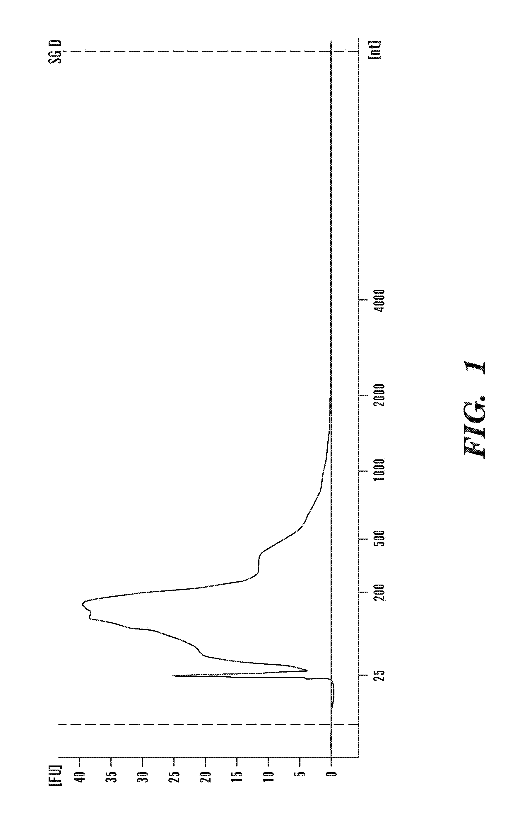
[0158]For the extraction of RNA from microvesicles, the pelleted microvesicles were incubated in an RNAse inhibitor solution for 20 minutes at room temperature. The RNase inhibitor can be obtained from various known vendors, e.g., SUPERase-In (Ambion Inc). Total RNA was then extracted from the RNAse-treated microvesicles using miRNeasy RNA extraction kit (Qiagen). Alternatively, various commercial RNA extraction kits such as the QIAamp RNA Blood Mini Kit from Qiagen, QIAamp viral RNA mini kit (Qiagen) and the MirVana RNA isolation kit from Ambion...

example 2

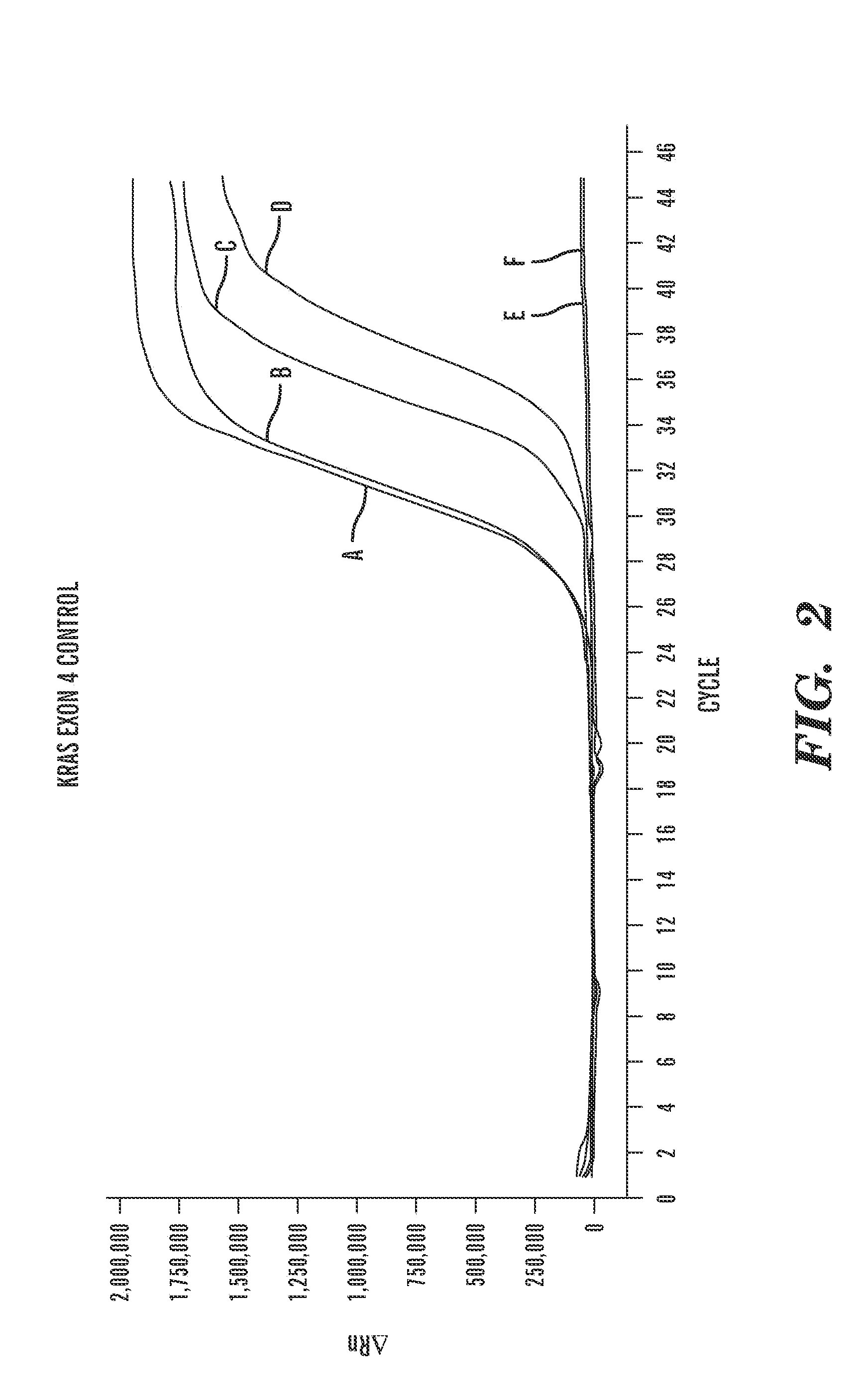
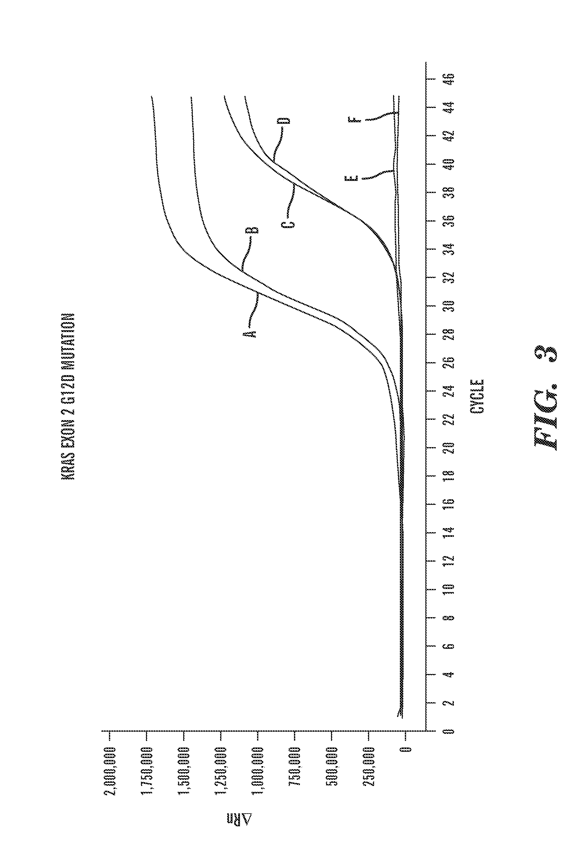
[0163]Positive and Negative Control Reactions For the Detection of Kras Exon 4 and Kras G12A Mutations.

[0164]Scorpion® Kras mutation detection PCR reactions to detect Kras Exon 4 were performed on positive and negative controls (FIG. 4(a)). Scorpion® Kras mutation detection PCR reactions to detect Kras G12A mutations were performed on positive and negative controls (FIG. 4(b)). As is shown in FIG. 4, the positive controls can be detected and the negative controls cannot.

example 3

[0165]DNase pre-treatment in method of analyzing Kras Exon 4 in RNA and DNA associated with microvesicles isolated from serum and plasma samples.

[0166]Following proper protocols, serum and plasma samples were obtained for the following analysis, from a patient diagnosed with colorectal cancer and having a Kras G12A mutation, confirmed by pathology evaluation of a biopsy.

[0167]Microvesicles were isolated as described previously in Example 1. Prior to isolation of nucleic acids from the microvesicles, a subset of the isolated microvesicles was pre-treated with DNase (Turbo™ DNase (Ambion®)) in order to eliminate or substantially eliminate any DNA located on the surface of the microvesicles or outside of the microvesicles. The remaining subset of microvesicles was left untreated. Samples of nucleic acid (both RNA and DNA) were obtained for analysis as described previously in Example 1. The RNA was reverse-transcribed into cDNA, as described in Example 1. The purified cDNA (FIG. 5(a)) a...


PUM

PropertyMeasurementUnit
diametersaaaaaaaaaa
diameteraaaaaaaaaa
diameteraaaaaaaaaa

Abstract

Microvesicles are small membrane vesicles that either shed or bud off eukarotic cells. Analysis of the nucleic acid content of microvesicles may be useful in detecting the presence or absence of genetic aberrations. This invention discloses novel methods of diagnosing, prognosing, monitoring, or treating a disease, such as cancer, or other medical condition in a subject involving analyzing one or more nucleic acids contained within an isolated microvesicle for the presence or absence of one or more Kras genetic aberrations.

Description

CROSS REFERENCE TO RELATED APPLICATIONS[0001]This Application claims the benefit of priority under 35 U.S.C. §119(e) of U.S. Provisional Application Ser. No. 61 / 241,020, filed Sep. 9, 2009, the contents of which are incorporated herein by reference in their entirety.FIELD OF THE INVENTION[0002]The present invention relates to the fields of medical diagnosis, and prognosis, patient monitoring, treatment efficacy, and molecular diagnostics based on the analysis of Kras nucleic acids extracted from microvesicles.BACKGROUND[0003]Molecular diagnostics, used to diagnose, monitor, treat, and evaluate diseases and other medical conditions, is becoming an increasingly important tool, particularly with the accumulating knowledge of the molecular mechanisms underlying various types of diseases and medical conditions. Molecular diagnostics is particularly valuable in the context of cancer, since our knowledge of the underlying genetic causes of cancers is rapidly expanding.[0004]Cancers arise t...

Claims


Application Information

Patent Timeline
Patent Type & Authority Applications(United States)
IPC IPC(8): C12Q1/68
CPCC12Q1/6858C12Q1/6886C12Q2600/106C12Q2527/125C12Q2600/156C12Q2600/118
Inventor SKOG, JOHAN KARL OLOVBREAKEFIELD, XANDRA O.
Owner THE GENERAL HOSPITAL CORP
Why Eureka
Try Eureka