Methods of detecting and treating cancers using autoantibodies

a cancer and autoantibody technology, applied in the field of autoantibody detection and treatment of cancer, can solve the problems of neoplasia escaping detection and treatment, complicated diagnosis and therapy, and insufficient diagnosis of many tumor types

Inactive Publication Date: 2013-03-14
THE ROCKEFELLER UNIV
View PDF0 Cites 12 Cited by
  • Summary
  • Abstract
  • Description
  • Claims
  • Application Information

AI Technical Summary

Benefits of technology

[0035]In certain embodiments, the autoantibodies are detected using an antibody detection reagent. In certain embodiments, the antibody detection reagent is Anti-IgG, Protein A, Protein G, an anti-human IgG, Fab(2) fragment of an anti-human IgG, Fab(1) of an anti-human IgG and a humanized mouse anti-human IgG, peptides that bind to the Fc region of human antibodies, and small molecules that bind IgGs.
[0036]In certain embodiments, the autoantibodies and sample are autologous. In other embodiments, the autoantibodies and sample are heterologous but are from one or more individuals of the same species.

Problems solved by technology

This variability complicates diagnosis and therapy.
The sensitivity and specificity of tumor detection in the sera is increased by testing for the presence of a panel of antibodies, rather than a single antibody (Kobold et al., 2010); however, this is still not sufficient for diagnosis in many tumor types.
The use of the broad spectra of antibodies generated against a neoplasia ensures that individual, or even bulk changes in the tumor, will not allow the neoplasia to escape detection and treatment.

Method used


Image

  • Methods of detecting and treating cancers using autoantibodies
  • Methods of detecting and treating cancers using autoantibodies
  • Methods of detecting and treating cancers using autoantibodies

Examples


example 1

Materials and Methods

[0163]1. Mouse Models:

[0164]The Alb-c-myc mouse models (Murakami et al., 1993) were a generous gift from Herman Stellar and both prostate models (Pb-_ENREF—23MYC (Ellwood-Yen et al., 2003) in FBV background and conditional Pten knockout (Pb-Cre X Ptenf / f) in C57 / B6 background (Trotman et al., 2003a, b)) were a generous gift from Charles Sawyers. Alb-myc and prostate sample controls were C57BL / 6J mice purchased from The Jackson Laboratory. All FVB / N-Tg(MMTVneu)202Mul / J mice, and the corresponding control FVB / NJ mice were purchased from The Jackson Laboratory. In addition six week old BALB / c mice (Jackson Labs) and CBySmn.CB17-Prkdcscid / J mice (Jackson Labs) were injected with a 4T1 cell line. One-hundred thousand cells were injected into a single mammary fat pad of each mouse, and mice were euthanized 14 days after injection. All experiments were approved by the Institutional Animal Care and Use Committee at The Rockefeller University.

[0165]2. Preparation of Tiss...

example 2

Alb-myc Model: Differentiating Abnormal Vs. Normal Tissue

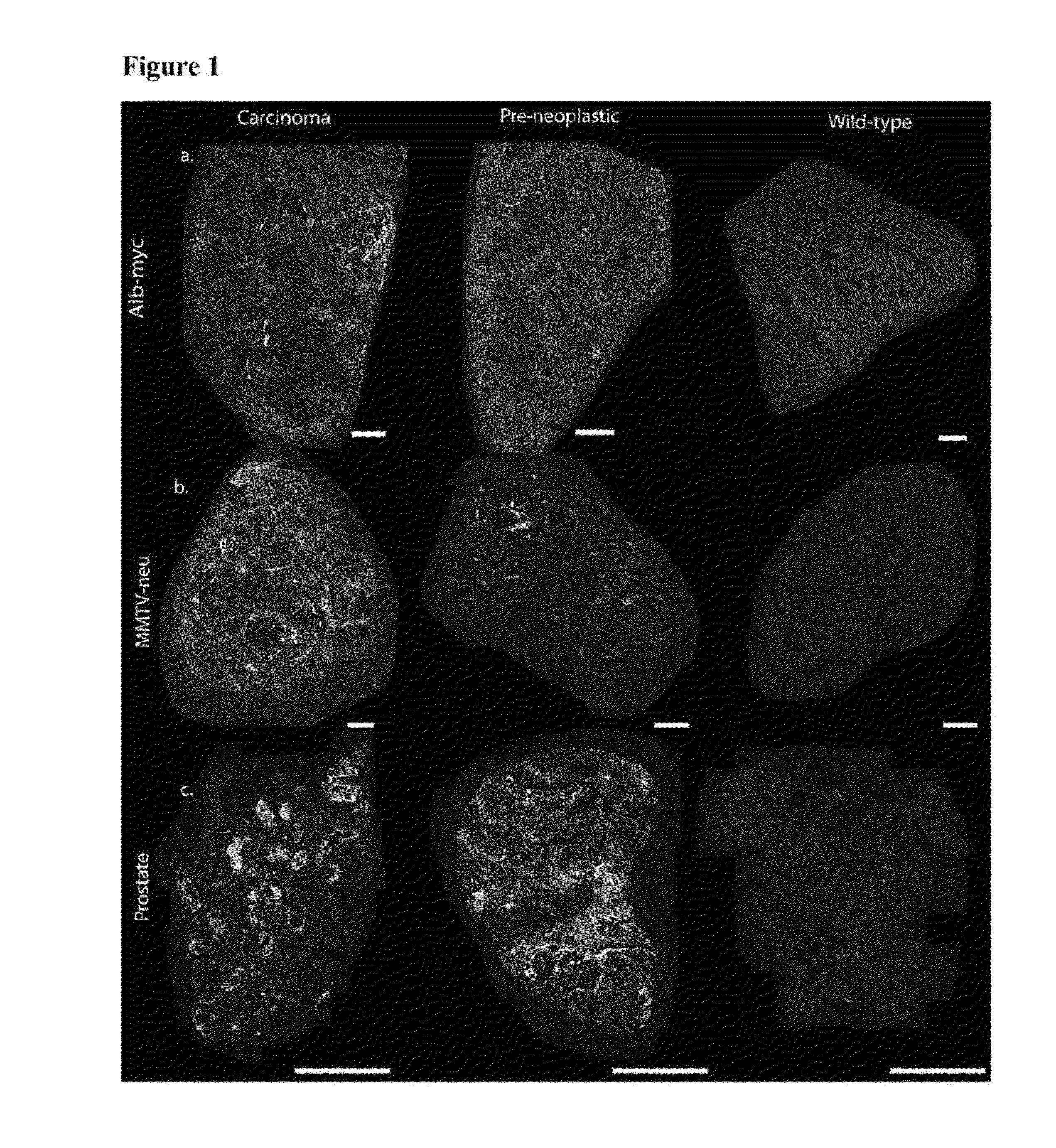
[0179]MYC oncogene overexpression is frequently seen in patients with hepatocellular carcinoma (Shachaf et al., 2004). Therefore, the presence of autoantibodies in an alb-myc mouse model of cancer was probed.

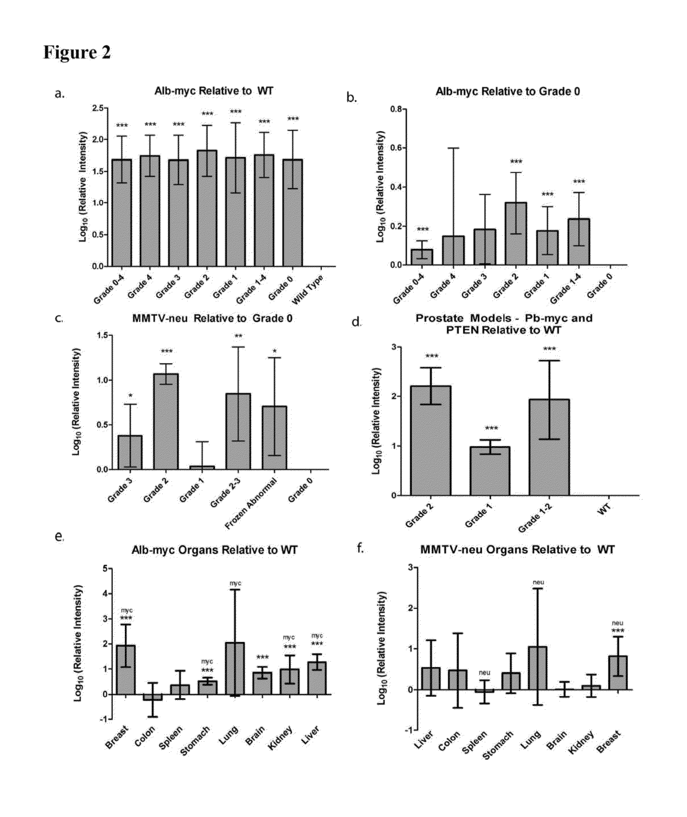
[0180]Liver tissue samples from this tumor model (Murakami et al., 1993) and from wild-type (“WT”) mice were paraffin embedded, mounted on the same slide, and probed with fluorescently tagged horse anti-mouse IgG antibody. In the alb-myc mice, anti-mouse IgG recognized autoantibodies throughout the tissue. Fluorescence was speckled with some areas considerably brighter than others. The anti-mouse IgG associated fluorescent intensity was approximately 50-fold higher in all liver tissue from alb-myc mice relative to tissue from WT mice (1.68±0.366 log 10-fold greater, pa, 2a).

[0181]Similar results were observed with goat anti-mouse IgG. In contrast, there was no difference observed between alb-myc and WT tissue when probed w...

example 3

MMTV-neu Model: Differentiating Abnormal Vs. Normal Tissue

[0185]Human breast tumors contain amplification of HER-2 / neu in 25-30% of patients (Slamon et al., 1989). Thus, the presence of endogenous antibodies in mammary tissue from virgin and multiparous mice expressing the un-activated neu oncogene, driven by a mouse mammary tumor virus (MMTV) promoter was probed (Guy et al., 1992).

[0186]Breast tissue was paraffin embedded and probed with fluorescently labeled anti-mouse IgG; adjacent sections were stained with H&E (n=10 MMTV-neu / WT pairs, 1-2 mammary glands taken from each mouse). Regions were histopathologically graded as either 0 (normal), 1 (hyperplasia without atypia / physiological hyperplasia), 2 (hyperplasia with atypia), or 3 (carcinoma). In the eighteen MMTV-neu tissue samples, six had grade 3 lesions, four had grade 2 lesions, and the remaining samples were grade 0. In the eighteen WT samples, five had grade 2 lesions, eleven had grade 1 regions, and two samples were unifor...


PUM

PropertyMeasurementUnit
concentrationsaaaaaaaaaa
temperatureaaaaaaaaaa
exposure timeaaaaaaaaaa

Abstract

This invention generally relates to a method of identifying pre-neoplastic or neoplastic tissue of a mammal by utilizing autoantibodies that detect the pre-neoplastic or neoplastic tissue. Also described herein are methods of killing pre-neoplastic or neoplastic tissue by either binding toxins to autoantibodies that detect the pre-neoplastic or neoplastic tissue or introducing toxin-conjugated molecules that can in turn recognize the autoantibodies already bound to the pre-neoplastic or neoplastic tissue.

Description

RELATED APPLICATIONS[0001]This application claims priority to U.S. provisional application Ser. No. 61 / 534,341, filed Sep. 13, 2011, which is herein incorporated by reference in its entirety.BACKGROUND OF THE INVENTION[0002]Cancer is a heterogeneous collection of diseases. Within a given cancer type, each individual tumor is defined by a distinct set of mutations resulting in different molecular profiles that could elicit an immune response. A neoplastic cell may have many mutations throughout its DNA (Lobe et al., 2003). Further, a particular tumor continues to mutate over time, which causes an evolving and diversifying phenotype. This variability complicates diagnosis and therapy.[0003]It has long been thought that the immune system suppresses developing tumors (Ehrlich 1909). This is consistent with the observation that immunosuppressed transplant recipients have higher rates of non-viral associated tumors than the general population (Birkeland 1995; Penn 1995; Penn 1996; Pam 199...

Claims


Application Information

Patent Timeline
Patent Type & Authority Applications(United States)
IPC IPC(8): G01N33/574G01N21/64A61K49/16A61K49/00A61K51/10
CPCG01N21/6428G01N21/6458G01N33/57434G01N33/5743G01N33/57415
Inventor SIMON, SANFORD M.
Owner THE ROCKEFELLER UNIV
Why Eureka
Try Eureka