
Contrast Agent Perfusion Adaptive Imaging System

a technology of adaptive imaging and contrast agent, which is applied in the field of adaptive imaging of contrast agent perfusion, can solve the problems of long contrast agent “fall off” period, limited image acquisition in the presence of contrast agent in known imaging system (e.g. ct (computed tomography) system), and patients may be over-radiated

Active Publication Date: 2013-03-14
SIEMENS HEALTHCARE GMBH
View PDF3 Cites 18 Cited by
  • Summary
  • Abstract
  • Description
  • Claims
  • Application Information

AI Technical Summary

Benefits of technology

This patent describes a system that monitors the movement of a contrast agent in a patient's body and adjusts the timing of image acquisition and post-processing methods based on the specific needs of the patient and the type of contrast agent used. The system analyzes data about the patient and the type of contrast agent to select the best acquisition protocol and post-processing method for a specific patient scan. The technical effect of this patent is to improve the accuracy and efficiency of medical imaging procedures by optimizing the timing of image acquisition and post-processing methods based on patient characteristics and the type of contrast agent used.

Problems solved by technology

The acquisition of images in the presence of contrast agent in known imaging systems (e.g. a CT (computed tomography) system) is limited by either acquisition time or number of image acquisition cycles.
A common reoccurring issue is that the time to peak or enhancement of contrast agent is different for each patient and, scanning acquisitions need to begin before contrast agent is present in an image.
In some patients, where their contrast agent “peaks” earlier than average, patients may be over-radiated due to acquisition of data beyond a necessary time point.
In other patients, especially patients with poor / low cardiac output, their contrast agent peaks substantially later, resulting in a longer contrast agent “fall off” period, that gets “cut off”—not imaged.
When the “tail” or fall off is not imaged, the calculated estimate for Cerebral Blood Volume (CBV) may be inaccurate, for example, causing problems or inaccuracies with diagnosis, and perhaps treatment.
This interval allows collection of enough time points for these patients with later peaks, but results in over-radiation for some patients and reduced temporal resolution for other patients.

Method used


Image

  • Contrast Agent Perfusion Adaptive Imaging System
  • Contrast Agent Perfusion Adaptive Imaging System
  • Contrast Agent Perfusion Adaptive Imaging System

Examples


Embodiment Construction

Definition

[0011]Hounsfield unit (HU)—indicates X-ray radiation absorption and attenuation of CT scanner radiation on a scale comprising a linear transformation of an original linear attenuation coefficient measurement into one in which the radiodensity of distilled water at standard pressure and temperature (STP) is defined as zero Hounsfield units (HU), while the radiodensity of air at STP is defined as −1000 HU. In a voxel with average linear attenuation coefficient, the corresponding HU value is therefore given by:

HU=1000×μX-μwaterμwater

Where μwater is the linear attenuation coefficient of water.

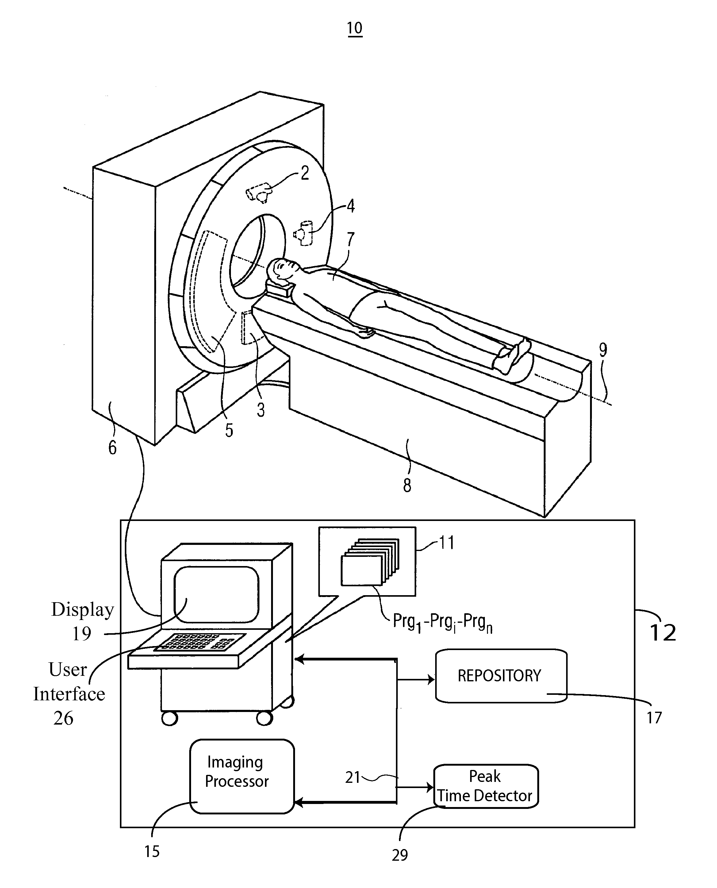
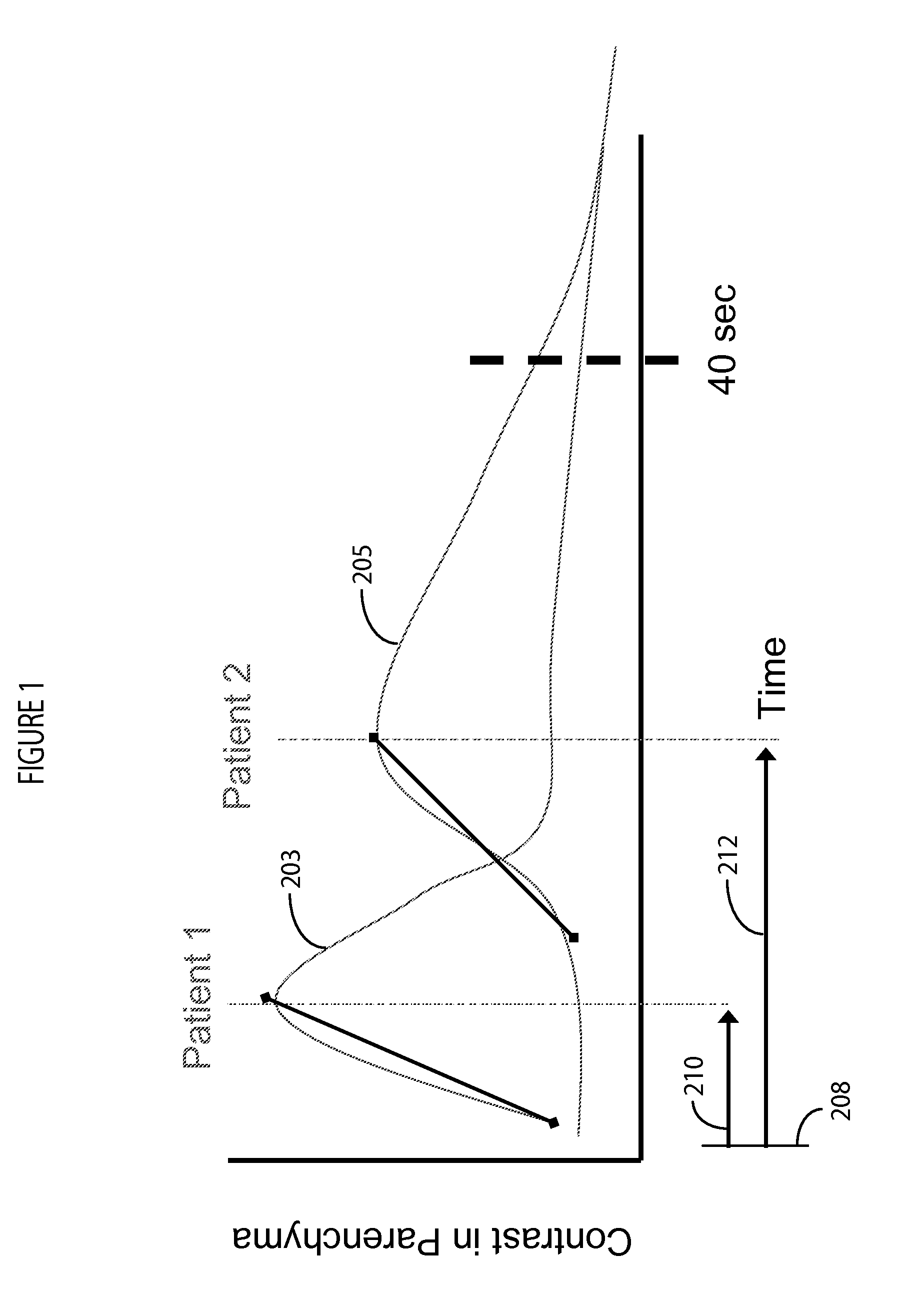
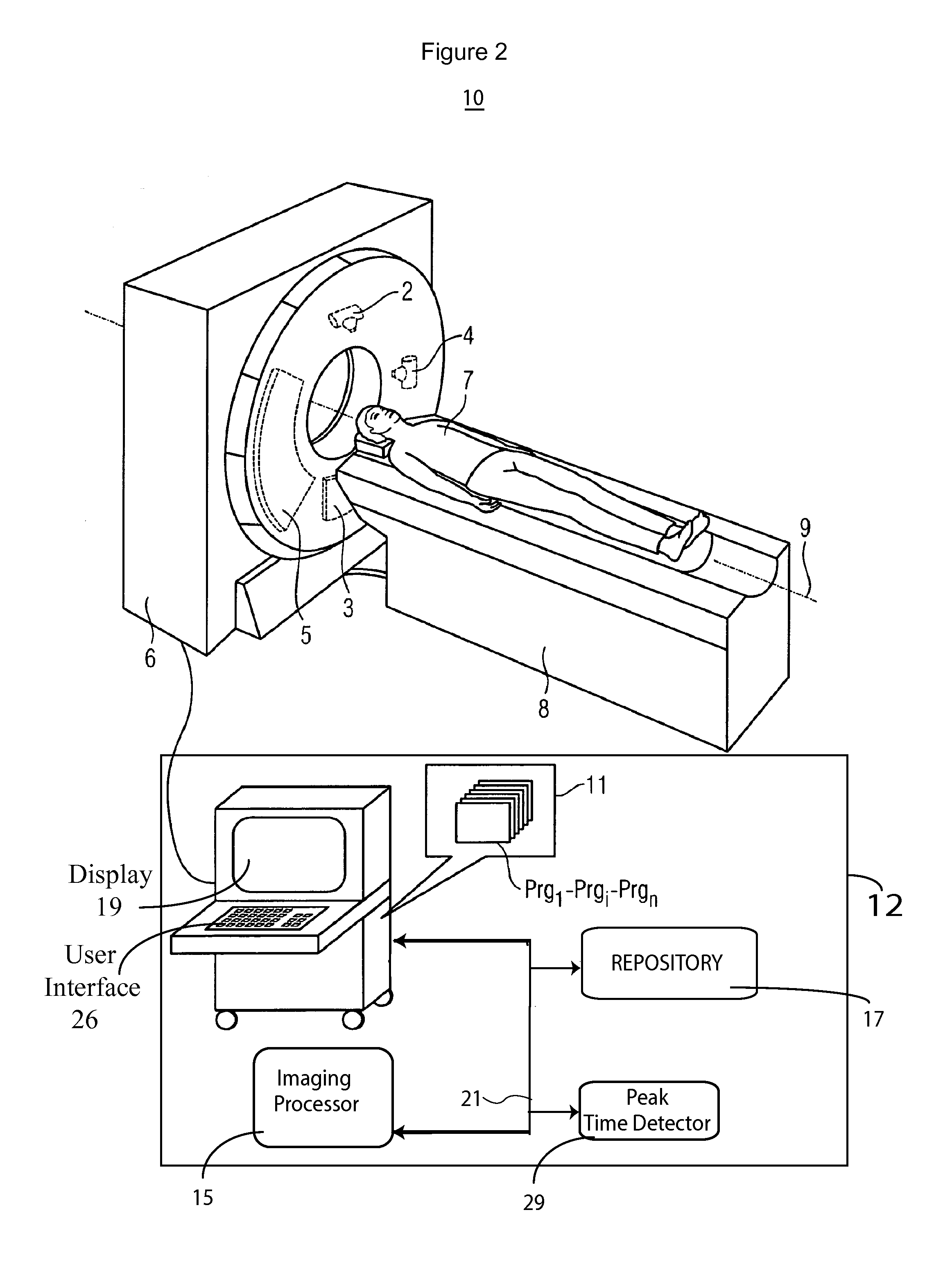
[0012]A system according to invention principles monitors a contrast bolus, and detects a contrast agent peak in the parenchyma by determining the peak in real-time (time-to-peak). The system adaptively adjusts an image acquisition rate and period in response to patient specific contrast agent timing (time-to-peak). The acquisition rate in one embodiment is selected by a user if there is ...


PUM


Abstract

An imaging system selects a medical imaging protocol using a repository of information associating multiple ranges of contrast agent peak time with corresponding different imaging protocols. An imaging protocol comprises a method for acquiring images using an imaging system and using data identifying at least one of (a) an imaging rate within an imaging scan cycle and (b) an interval between imaging scans. A contrast agent peak time comprises a time a contrast agent concentration substantially reaches a peak value in an anatomical region of interest of a patient relative to a time of start of contrast agent injection. A contrast agent peak time detector detects a contrast agent peak time. An imaging processor adaptively selects an imaging protocol from the imaging protocols in response to a comparison of a detected contrast agent peak time with at least one of the plurality of ranges.

Description

[0001]This is a non-provisional application of provisional application Ser. No. 61 / 532,587 filed Sep. 9, 2011, by K. L. R. Grant et al.FIELD OF THE INVENTION[0002]This invention concerns a system for automatically adaptively selecting a medical image acquisition protocol usable by a Computed Tomography (CT), X-ray or other imaging system based on time duration it takes injected contrast agent to reach a peak concentration.BACKGROUND OF THE INVENTION[0003]The acquisition of images in the presence of contrast agent in known imaging systems (e.g. a CT (computed tomography) system) is limited by either acquisition time or number of image acquisition cycles. A common reoccurring issue is that the time to peak or enhancement of contrast agent is different for each patient and, scanning acquisitions need to begin before contrast agent is present in an image. In a majority of patients, a single, stable scan protocol is sufficient to properly gather necessary image data. In some patients, wh...

Claims


Application Information

Patent Timeline
IPC IPC(8): A61B6/00A61B6/03
CPCA61B6/032A61B6/481A61B6/5217A61B6/507A61B6/486G16H50/30
Inventor GRANT, KATHARINE LYNN ROWLEYSCHMIDT, BERNHARD
Owner SIEMENS HEALTHCARE GMBH
Learn More