Medical image processing device, method and program

a medical image and image processing technology, applied in image enhancement, instruments, healthcare informatics, etc., can solve the problems of inability to accurately extract the endocardium in the image of interest correspondingly to the variation of shape, and the inability to achieve the desired endocardium shape, etc., to achieve accurate extraction of the endocardium

Inactive Publication Date: 2013-04-04
FUJIFILM CORP
View PDF8 Cites 16 Cited by
  • Summary
  • Abstract
  • Description
  • Claims
  • Application Information

AI Technical Summary

Benefits of technology

[0008]In view of the above-described circumstances, the present invention is directed to providing a medical image processing device, a medical image processing method and a medical image processing program which allow accurate detection of the endocardium of the left ventricle from 3D image data representing the left ventricle.
[0011]For example, in a case where the identification of the convex and concave points and the deformation of the contour are two-dimensionally carried out, the convex and concave points identifying unit may identify the convex points forming the convex hull and the concave points, which are the sample points other than the convex points, by finding the convex hull from the sample points on the contour of the extracted blood region in each of pieces of 2D image data representing cross sections perpendicular to an axis passing through the base and the apex of the heart set in the left ventricle; and the endocardium determining unit may obtain, in each of the pieces of 2D image data, a deformed contour of the blood region deformed by moving at least a part of the concave points outward from the blood region based on a positional relationship among each concave point and the convex points located on both sides of the concave point, and may determine the endocardium based on the deformed contour.
[0015]The endocardium determining unit may make the degree of the movement of the concave point smaller as an angle formed between straight lines respectively connecting an arbitrary point set in a central area between the blood region with convex points located around the concave point is larger.
[0019]According to the medical image processing device, method and program of the invention, in order to extract the endocardium of the left ventricle from 3D image data representing the left ventricle, a blood region in the left ventricle is extracted from the image data. Then, a convex hull is found from sample points on a contour of the extracted blood region to identify convex points forming the convex hull and concave points, which are sample points other than the convex points. Then, the contour of the blood region is deformed by moving at least a part of the concave points outward from the blood region by an amount determined based on a positional relationship among each concave point and the convex points located around the concave point, and a deformed contour of the blood region is determined to be the endocardium. Therefore, as shown in FIG. 2, the deformed contour of the blood region, which is deformed to contain the papillary muscle regions located at the concave portion thereof, is determined to be the endocardium, thereby accurately extracting the endocardium of the left ventricle, which faces the blood region and the papillary muscles, correspondingly to variation of the shape due to individual differences. In this manner, the present invention can achieve more accurate extraction of the endocardium than that by the above-described conventional method, which extracts the endocardium by limiting the shape using a standard model.

Problems solved by technology

With the above-described conventional method, however, the shape of the endocardium to be extracted is limited by the standard model.
Therefore, although the result of the extraction of the endocardium does not deviate from a proper shape of the endocardium, the endocardium in the image of interest may not accurately be extracted correspondingly to variation of the shape due to individual differences.
For example, in a case where a convex portion or concave portion is locally present in the endocardium, some standard model cannot express such a shape of the endocardium in the image of interest, and even when an amount of shift for each feature point is correctly calculated, the shape may converge at a local solution (which is not an optimal solution), resulting in unsuccessful acquisition of a desired shape of the endocardium.

Method used


Image

  • Medical image processing device, method and program
  • Medical image processing device, method and program
  • Medical image processing device, method and program

Examples


Embodiment Construction

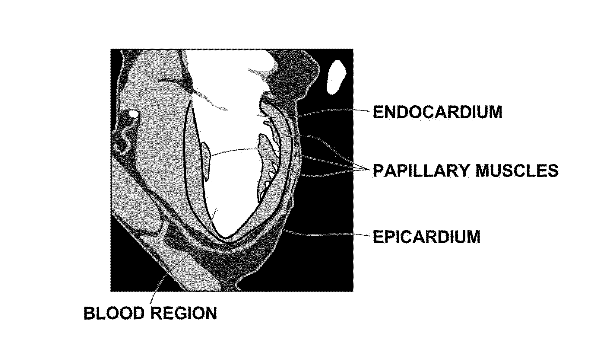
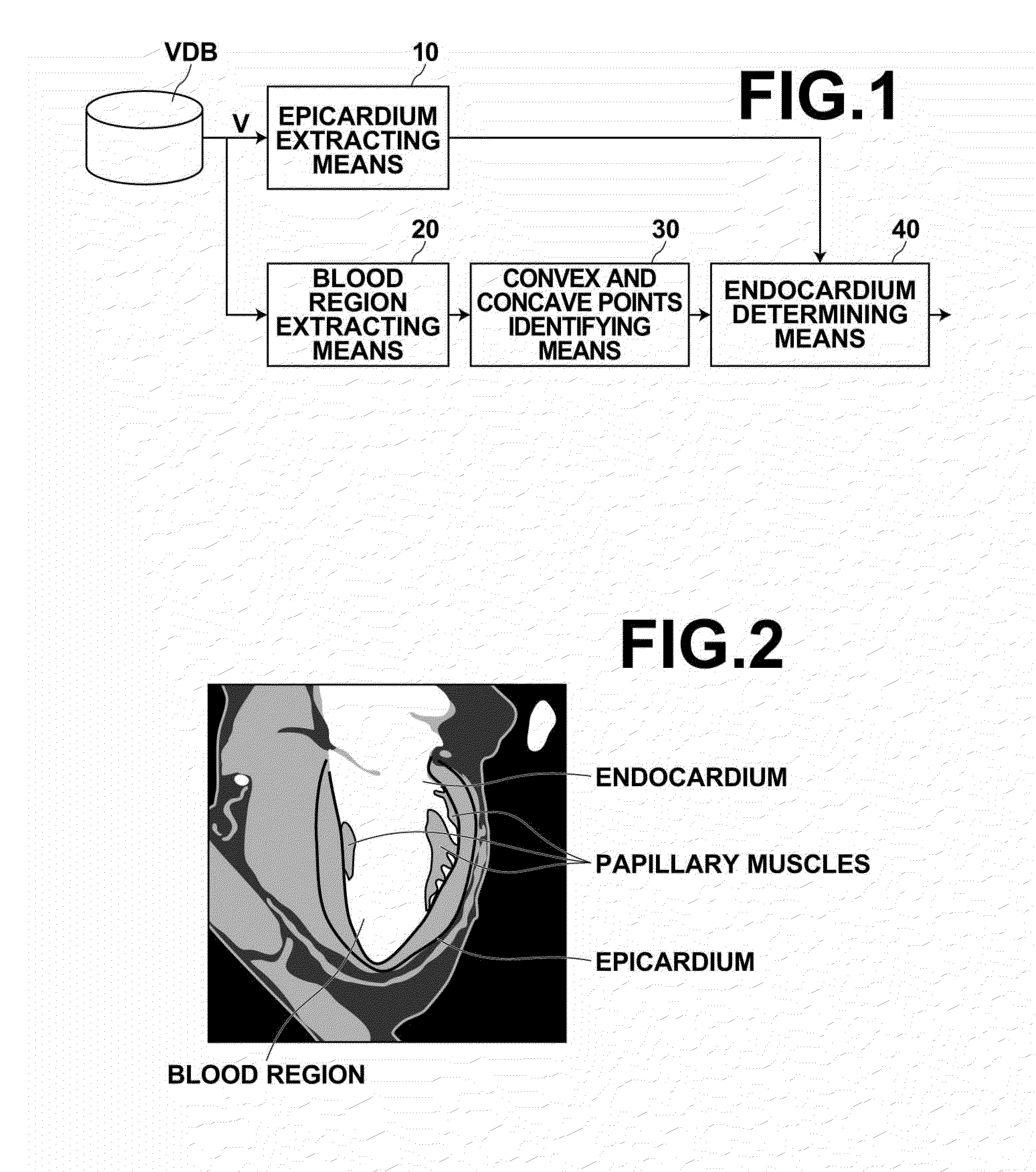
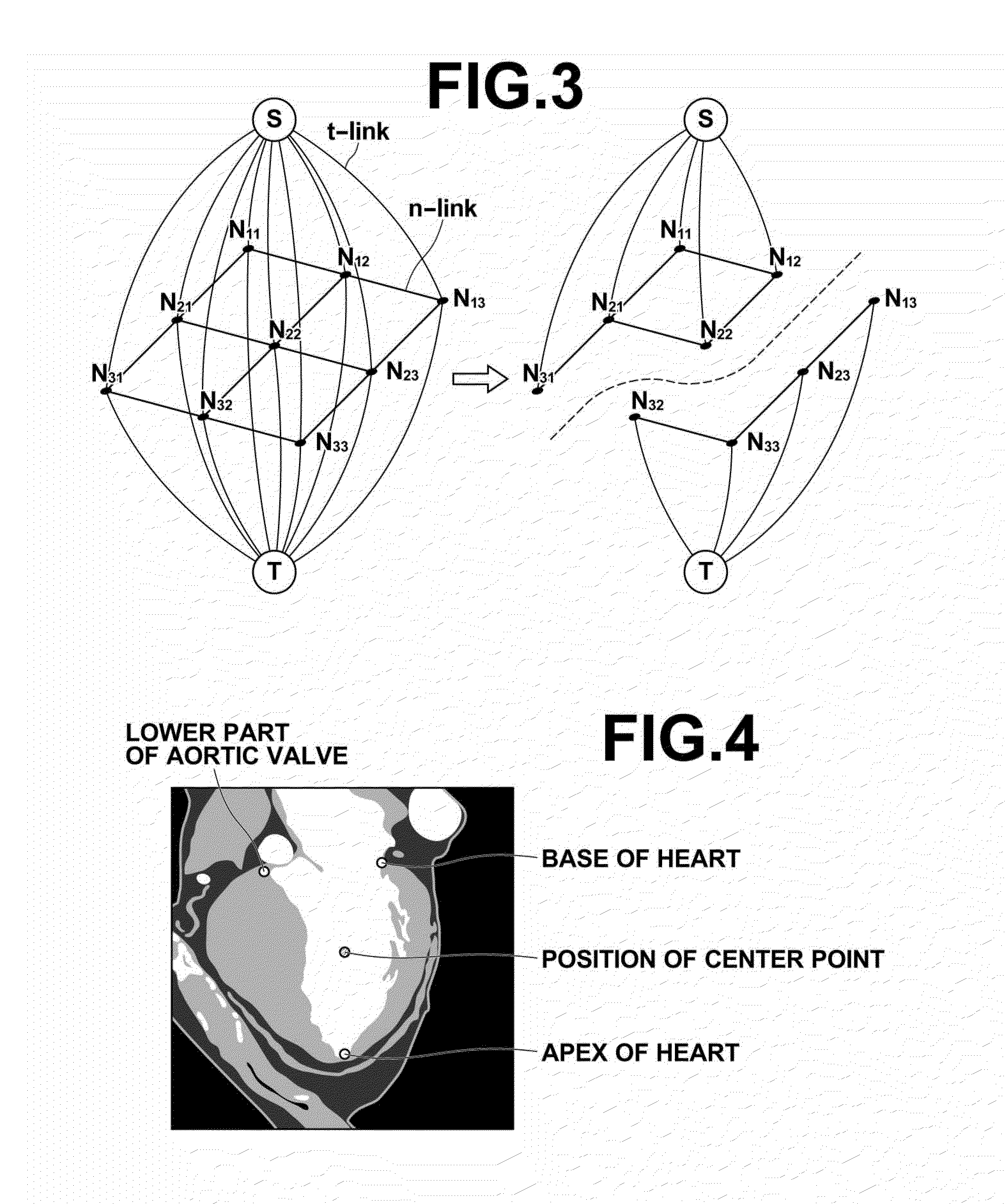
[0033]Hereinafter, an embodiment of a medical image processing device, a medical image processing method and a medical image processing program of the present invention will be described in detail with reference to the drawings. FIG. 1 is a diagram illustrating a schematic configuration of a medical image processing device 1 according to an embodiment of the invention. The configuration of the medical image processing device 1 as shown in FIG. 1 is implemented by a computer executing a medical image processing program, which is read in an auxiliary storage device of the computer. The medical image processing program is distributed with being stored in a storage medium, such as a CD-ROM, or via a network, such as the Internet, to be installed on a computer. The medical image processing device 1 shown in FIG. 1 extracts the endocardium of the left ventricle from 3D image data representing the left ventricle. The medical image processing device 1 includes an epicardium extracting means...


PUM


Abstract

A medical image processing device for extracting an endocardium of a left ventricle from 3D image data representing the left ventricle is provided. The device includes: a blood region extracting unit to extract a blood region in the left ventricle from the image data; a convex and concave points identifying unit to find a convex hull from sample points on a contour of the extracted blood region and identify convex points forming the convex hull and concave points, which are sample points other than the convex points; and an endocardium determining unit to deform the contour of the blood region by moving at least a part of the concave points outward from the blood region by an amount determined based on a positional relationship among each concave point and the convex points located around the concave point and determine a deformed contour of the blood region to be the endocardium.

Description

BACKGROUND OF THE INVENTION[0001]1. Field of the Invention[0002]The present invention relates to a medical image processing device, a medical image processing method and a medical image processing program for extracting the endocardium of the left ventricle from 3D image data representing the left ventricle.[0003]2. Description of the Related Art[0004]Along with the spread of the multislice CT technology at medical treatment sites, medical image analyzing technologies are rapidly developing. For example, with respect to the heart region, it is now possible to obtain a plurality of 3D images during a single beat of the heart. With this development, functions of the heart, such as ejection rate, end-diastolic volume, end-systolic volume, single cardiac output, cardiac output and cardiac muscle weight, are analyzed by analyzing a plurality of pieces of 3D image data obtained by imaging the heart.[0005]In order to achieve such analysis of the cardiac functions based on image information...

Claims


Application Information

Patent Timeline
Patent Type & Authority Applications(United States)
IPC IPC(8): G06K9/46
CPCG06T7/0083G06T7/0089G06T2207/10072G06T7/0012G06T2207/30048G06K2009/366G06F19/321G06T2207/20164G06T7/12G06T7/149G16H30/20G06V10/248
Inventor NAKAMURA, KEIGO
Owner FUJIFILM CORP
Why Eureka
Try Eureka