

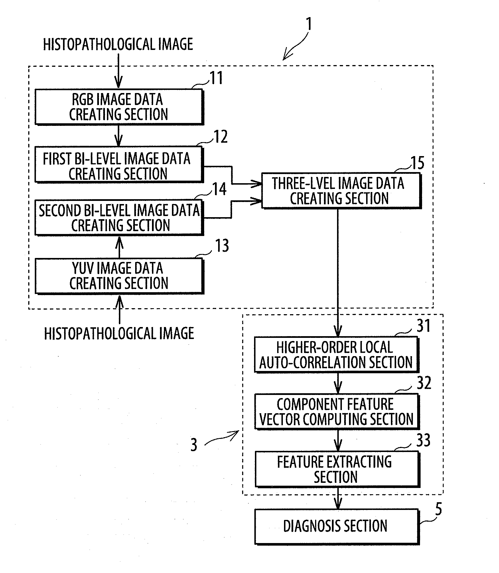
Region segmented image data creating system and feature extracting system for histopathological images
a technology of image data and histopathological images, which is applied in image enhancement, image analysis, instruments, etc., can solve the problems of no cure for the shortage of pathologists and heavy and achieve the effect of reducing the burden on the pathologis
- Summary
- Abstract
- Description
- Claims
- Application Information
AI Technical Summary
Benefits of technology
Problems solved by technology
Method used
Image

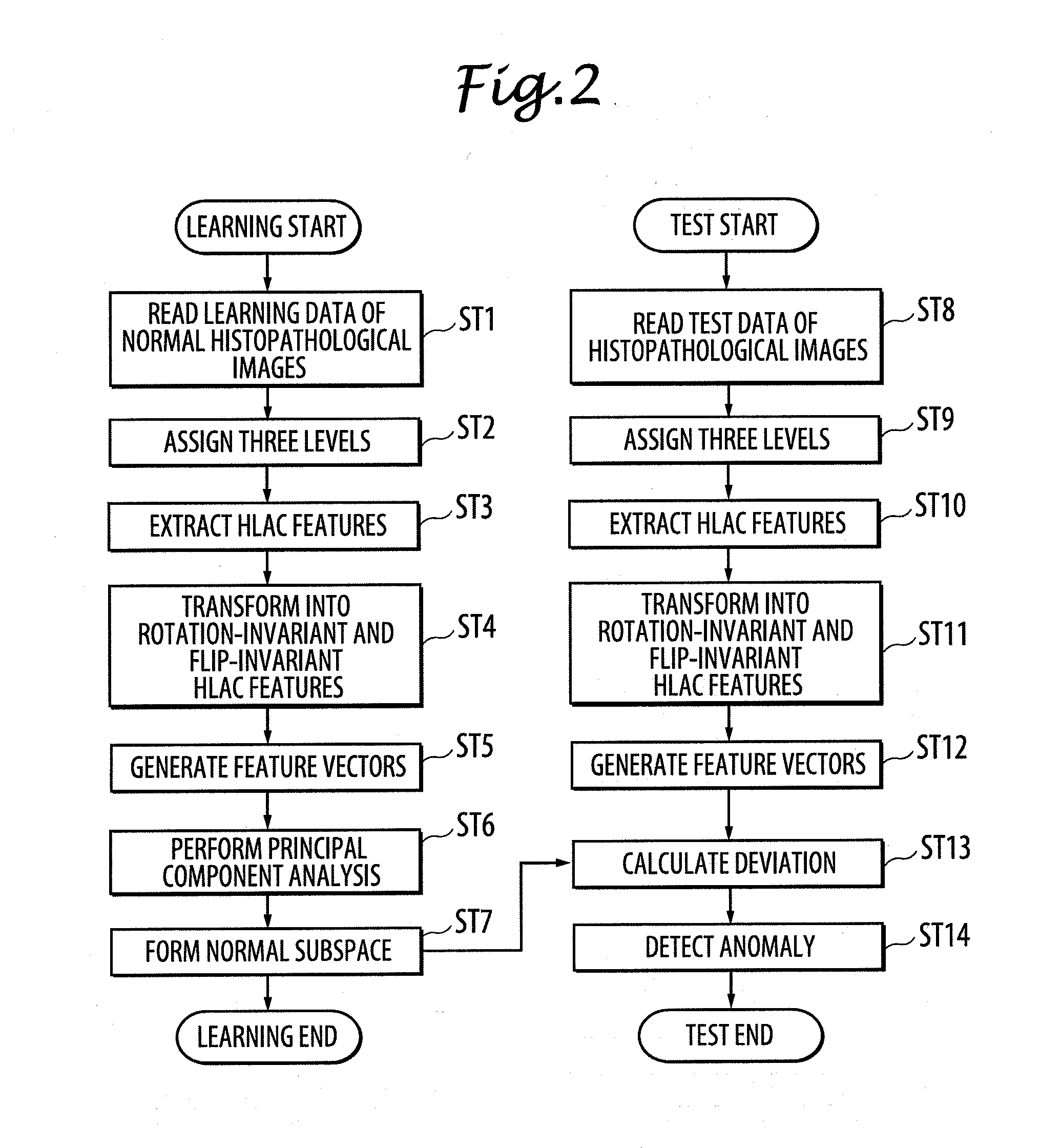
Examples
experiment 1
[0108] Verification of Proposed Assignment of Three Levels
experiment 2
[0109] Verification of Proposed Rotation Invariance and Flip Invariance
[0110][Test Data for Experiments]
[0111]In the conducted experiments, histopathological images of the biopsy taken from patients were used. The biopsy samples were confirmed by the pathologists that some of them were clearly non-cancerous or normal and the others were clearly cancerous in order to verify that the cancerous histopathological image can properly be detected as anomaly through learning of non-cancerous or normal histopathological images.
[0112]FIG. 14 shows datasets used in the verification experiments. 250 samples, which were diagnosed as non-cancerous or normal by the pathologists, were used as the learning data. In addition to the learning data, 50 non-cancerous samples and 24 cancerous samples were used as test data. The histopathological images used in the experiments were taken by microscopy of 20 times power and stored in jpeg format of 1280×960 pixels as the non-cancerous image shown in FIG. 15...
verification experiment 1
[0116][ Verification of Effectiveness of Assignment of Three Levels]
[0117]To verify the effectiveness of the proposed three-level image technique used in the present embodiment, three techniques were used in comparison experiment as shown in FIG. 16. In this experiment, rotation and flip features were not transformed. The results from the accumulated contribution rates C of 0.999, 0.9999, and 0.99999 were compared with each other in the three techniques to find the best condition.
[0118]FIG. 17 shows an original image and gray scaled region segmented images of the respective techniques for visual comparison. FIG. 17A shows an original image. In the image of the proposed technique, the pixel values of nucleus, cytoplasm, and background were set to 255, 127, and 0 for display. FIG. 17B shows a gray scaled image which is the closest to the original image and the tissue structure may visually be discriminated clearly. FIG. 17C shows a bi-level image in which most of the cytoplasm regions...
PUM
 Abstract
Description
Claims
Application Information