Adaptive visualization for direct physician use

Active Publication Date: 2013-05-16
AI VISUALIZE INC
View PDF9 Cites 38 Cited by
  • Summary
  • Abstract
  • Description
  • Claims
  • Application Information

AI Technical Summary

Benefits of technology

[0017]It is another object of the present invention to provide a method that enables physicians to control views generated from a three dimensional image of an anatomical structure in real time during diagnosis instead of relying on scan technologists to generate those views.
[0018]A further object of the inven

Problems solved by technology

The current techniques emanate from having to improve scan technicians' ability from having to sketch the contours slice by slice using pointing devices such as a mouse or trackball, which they note that this is a very time-consuming and the results may suffer from intra-observer or inter-observer variability.
These segmentation techniques are both computer and human intensive that a lab technician is given the responsibility to pre-segment a scan.
Also, due to the time and cost aspects, segmentation has become an exception r

Method used


Image

  • Adaptive visualization for direct physician use
  • Adaptive visualization for direct physician use
  • Adaptive visualization for direct physician use

Examples


Embodiment Construction

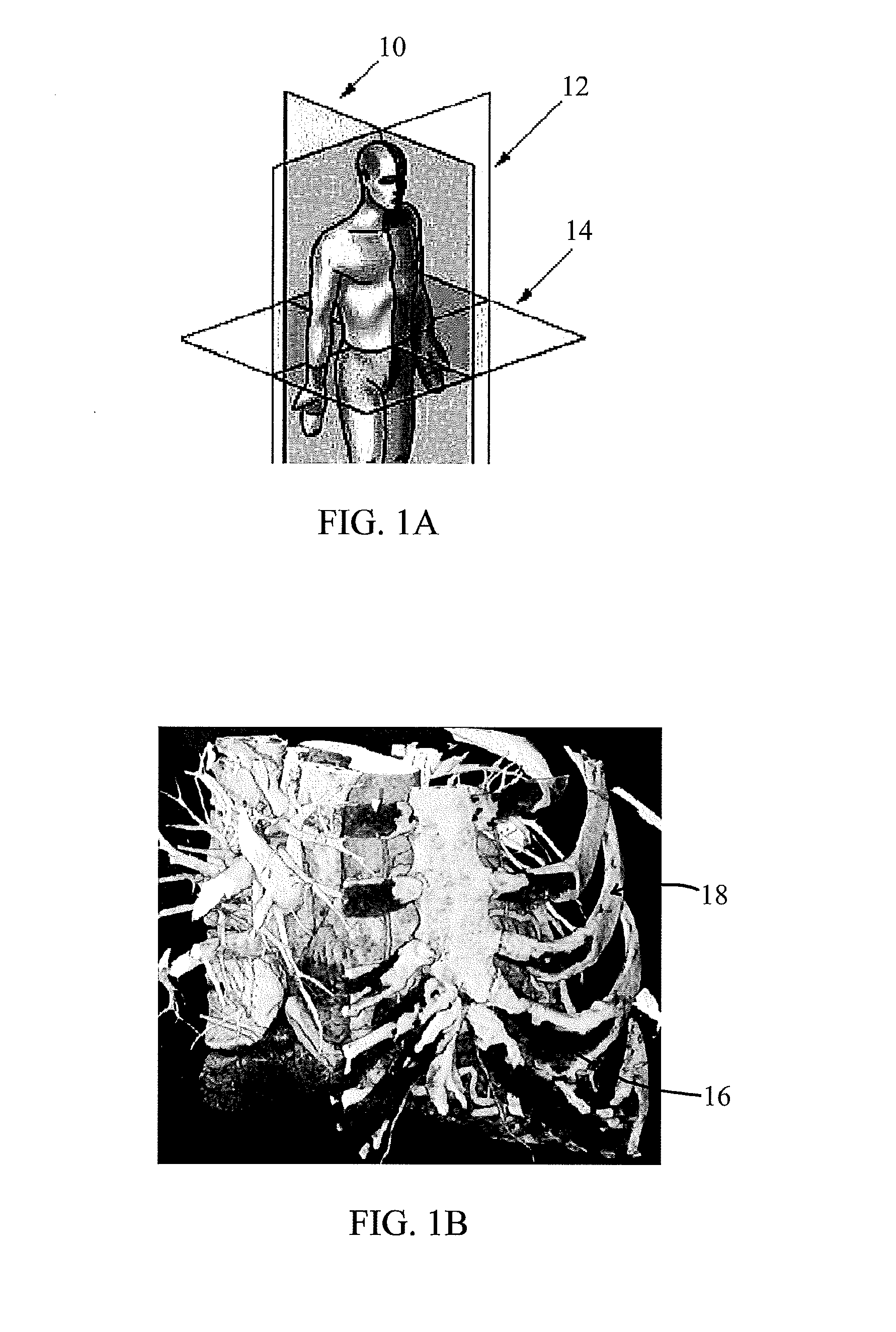
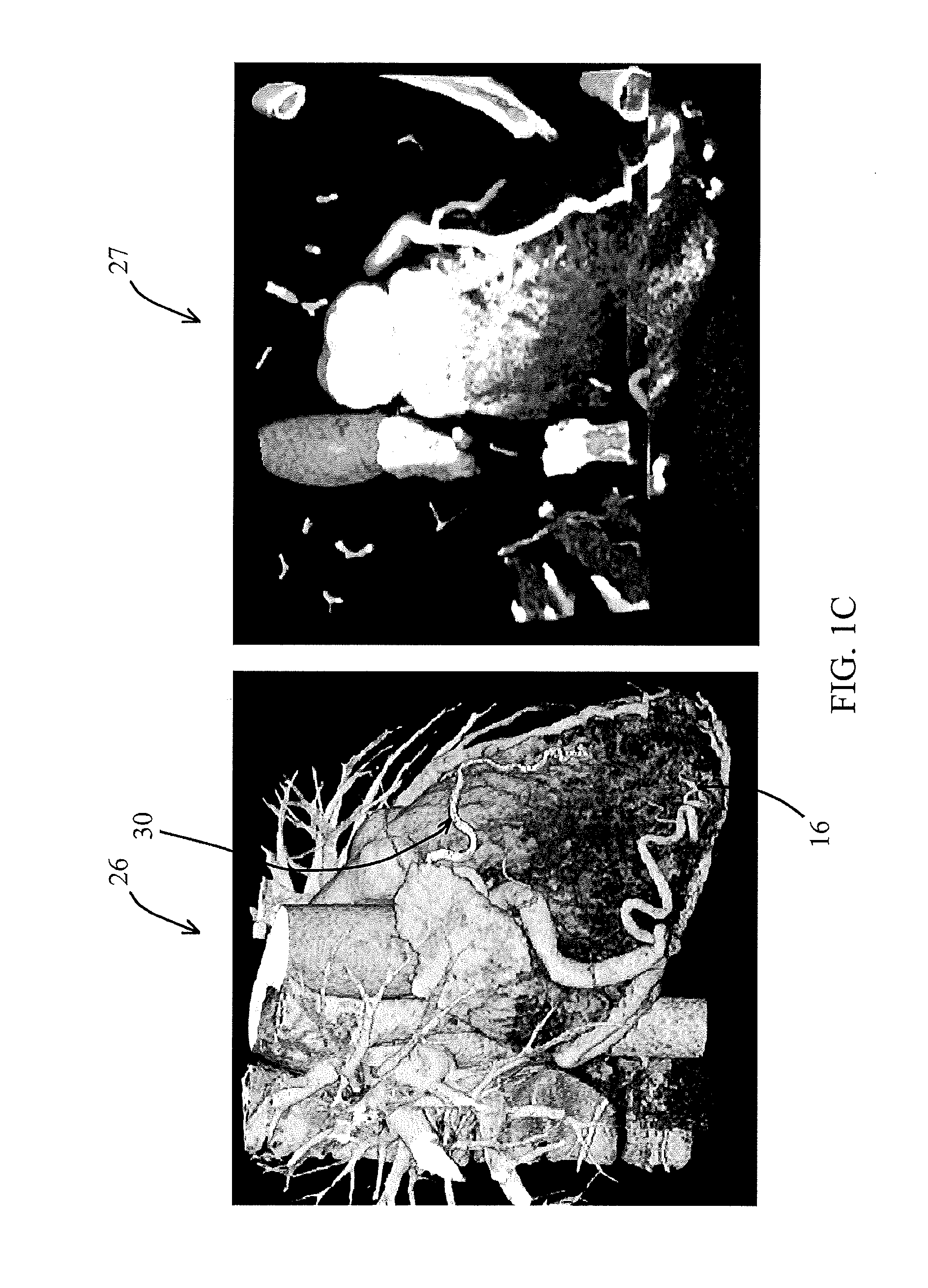
[0061]In describing the embodiment of the present invention, reference will be made herein to FIGS. 1-14 of the drawings in which like numerals refer to like features of the invention.

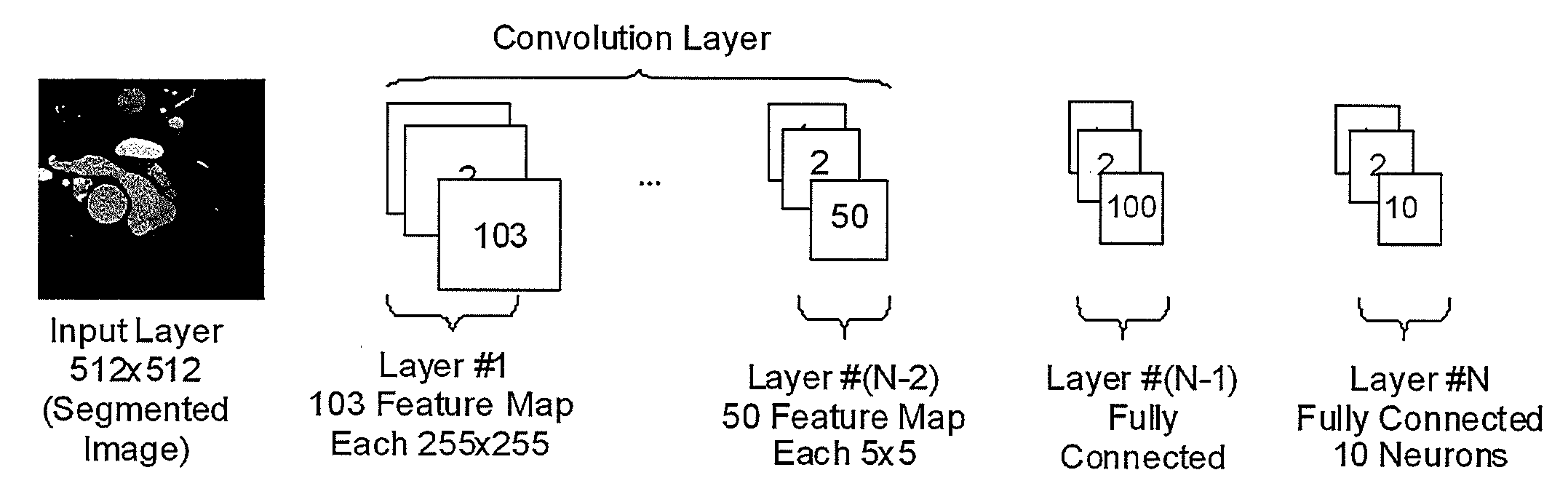
[0062]Medical image segmentation seeks to separate important anatomy from the image background. However, such methods are performed by a scan technician and are time consuming, and error prone. The present invention is directed to an on-demand and adaptive 3D intelligent visualization application that the physician can use to improve diagnosis and patient care without a technician's help.

[0063]Medical image segmentation seeks to change the representation of a medical scan to make it more amenable to interpretation by a physician. The extreme variability within the image requires that a scan technician be employed to separate important anatomical structures from the image background. Methods are time consuming and error prone and can lead to diagnostic inaccuracy. A solution to this problem is a semi-au...


PUM


Abstract

A method of modifying a three dimensional (3D) volume visualization image of an anatomical structure in real time to separate desired portions thereof. The method includes providing a two dimensional (2D) image slice of a 3D volume visualization image of an anatomical structure, identifying portions of the anatomical structure of interest, and providing a prototype image of desired portions of the anatomical structure. The method then includes using an evolver to evolve parameters of an algorithm that employs a transfer function to map optical properties to intensity values coinciding with the portions of the anatomical structure of interest to generate an image that sufficiently matches the prototype image. If the parameters match the prototype image, the method then includes applying the transfer function to additional 2D image slices of the 3D volume visualization image to generate a modified 3D volume visualization image of the anatomical structure. The method includes using a pattern recognizer to assist the evolver, to classify whether a view is normal or abnormal, and to extract the characteristic of an abnormality if and when detected.

Description

TECHNICAL FIELD[0001]The present invention relates to computer processing of three dimensional medical images and, in particular, to a method and system for modifying a three dimensional (3D) volume visualization image of an anatomical structure in real time to delineate desired portions thereof.BACKGROUND ART[0002]A visual medical scan such as computed tomography (CT) or magnetic resonance imaging (MRI) is acquired thru cross sections (FIG. 1A), for example CT scan has an axial cross sections acquisition. Sagittal 10, Coronal 12 and Oblique 14 sections and 3D visualizations may be generated automatically and on demand. However, the automatic 3D image may have the rib cage blocking the heart from being visible (FIG. 1B). A segmentation task would involve removing the bone from this view. According to standard practice, segmentations are prepared by scanner technologist and saved as snapshots for radiologists to view and use for diagnosis (FIG. 1D).). For instance, these series of im...

Claims


Application Information

Patent Timeline
IPC IPC(8): G06T7/00
CPCA61B5/055A61B6/032A61B6/466G06T15/08G06N3/086G06T2207/20084G06T2207/30004G06T7/0014G06T7/0081G06T2207/10072G06T2207/20081G06T2207/20104G06T7/11G06T7/143G06V10/454G06V2201/031
Inventor KOVALAN, KOVEYBRAILKO, NICKOLAY
Owner AI VISUALIZE INC
Why Eureka
Try Eureka