
Methods and systems for enhanced tomographic imaging

a tomographic imaging and enhanced technology, applied in the field of methods and systems for enhanced tomographic imaging, can solve the problems of limited scan time for acquiring image data, patient immobility, inadequate signal-to-noise ratio (snr) at the region of interest,

Inactive Publication Date: 2013-05-30
GENERAL ELECTRIC CO
View PDF2 Cites 30 Cited by
  • Summary
  • Abstract
  • Description
  • Claims
  • Application Information

AI Technical Summary

Benefits of technology

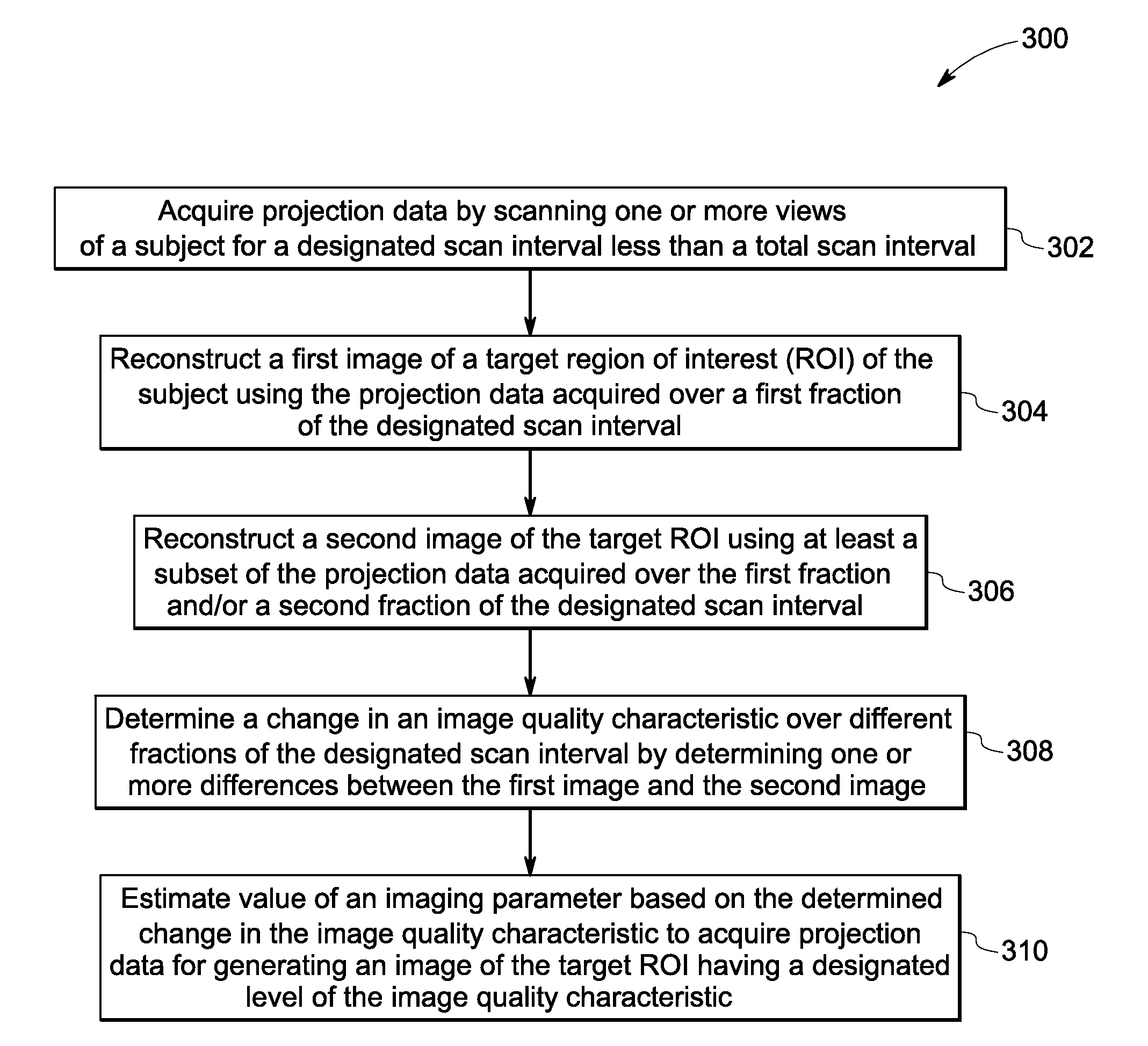
The present patent describes a method for tomographic imaging that involves acquiring projection data from a subject and reconstructing images of a target region of interest using the acquired data. The method includes determining the image quality characteristic of the target region of interest over a designated scan interval and estimating the value of an imaging parameter based on the change in image quality characteristic over the designated scan interval to acquire the projection data for generating an image of the target region of interest with a desired level of image quality. The method can also involve using synthetic projection of the target region of interest and non-transitory computer readable media and nuclear medicine imaging systems for implementing the method. The technical effects of the present patent include improved image quality and efficiency in tomographic imaging.

Problems solved by technology

However, the total scan time for acquiring the image data is limited by the decay of a radioactive isotope used in imaging and by the inability of the patients to remain immobile for extended durations.
Further, patient size, attenuation, physiology, injected dose and spatial distribution of the detected radiation events affect image quality, often resulting in inadequate signal-to-noise ratio (SNR) at the region of interest (ROI).
Use of a fixed scan time or detection of a fixed number of coincidence events, thus, does not guarantee acquisition of sufficient data for reconstructing a PET or SPECT image of the ROI at a desired SNR.
The imaging systems, however, may ignore “noise” sources introduced by processing steps such as scatter correction and interpolation, thus leading to inaccuracies during image reconstruction.

Method used


Image

  • Methods and systems for enhanced tomographic imaging
  • Methods and systems for enhanced tomographic imaging
  • Methods and systems for enhanced tomographic imaging

Examples


Embodiment Construction

[0013]The following description presents exemplary systems and methods for enhanced tomographic imaging. Particularly, embodiments illustrated hereinafter disclose imaging systems and methods that aim to estimate uncertainty in a reconstructed image using a “bootstrap” approach, and use the estimated uncertainty to optimize image data acquisition for reconstructing images of a targeted region of interest (ROI) with a desired spatial resolution.

[0014]In the bootstrap approach, a single data set is used to determine a statistical distribution of an estimated statistic θ, for example, a pixel value in a reconstructed image. To that end, multiple bootstrap replicates are generated from the original data set by randomly drawing samples from the original data set. Each bootstrap replicate is then treated as an independent measurement from which θ can be determined Particularly, a resulting variance in θ determined using the bootstrap replicates generated from a fraction of the original da...


PUM


Abstract

Nuclear imaging systems, non-transitory computer readable media and methods for tomographic imaging are presented. Projection data is acquired by scanning one or more views of a subject for a designated scan interval less than a total scan interval. A first image of a target region of interest (ROI) is reconstructed using projection data acquired over a first fraction of the designated scan interval. A second target ROI image is reconstructed using at least a subset of projection data acquired over the first and / or a second fraction. A change in an image quality characteristic over the first and the second fractions is determined by determining one or more differences between the first and the second images. A value of an imaging parameter is estimated based on the change to acquire projection data for generating a target ROI image having at least a predetermined level of the image quality characteristic.

Description

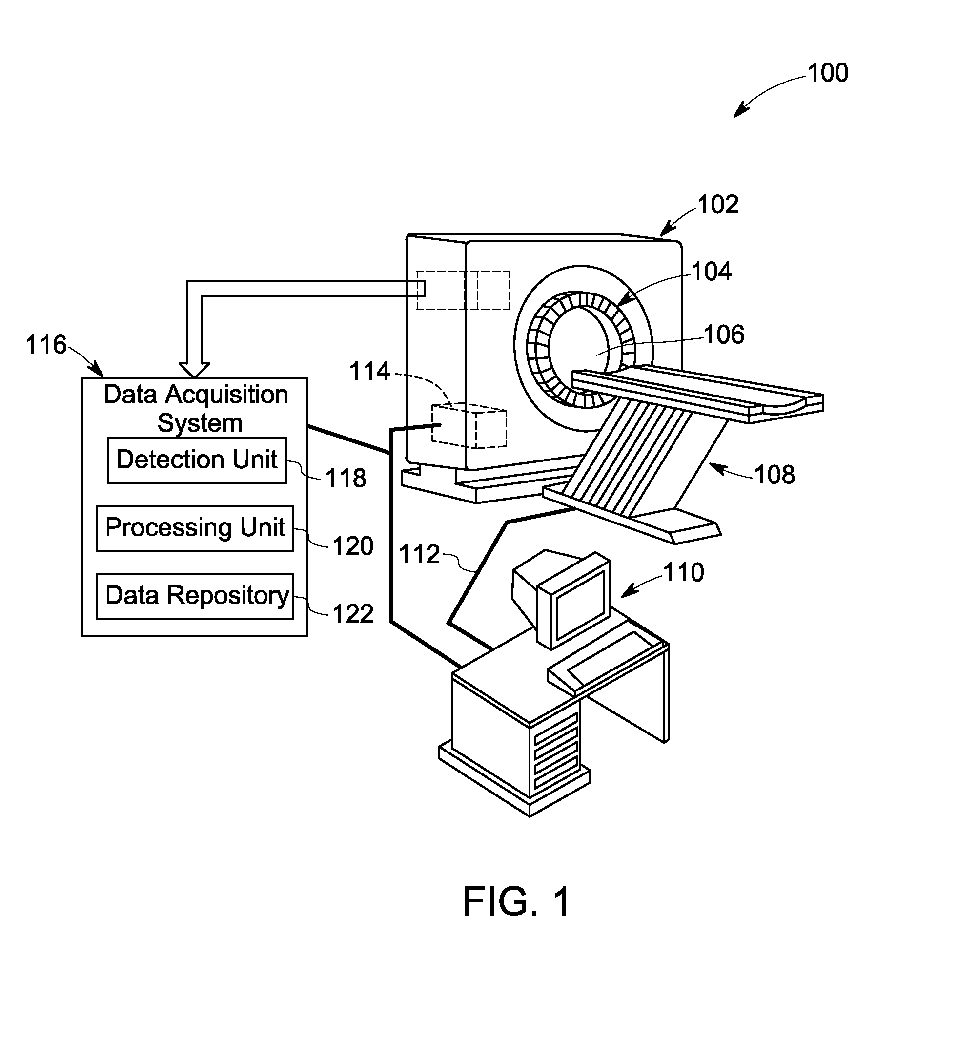
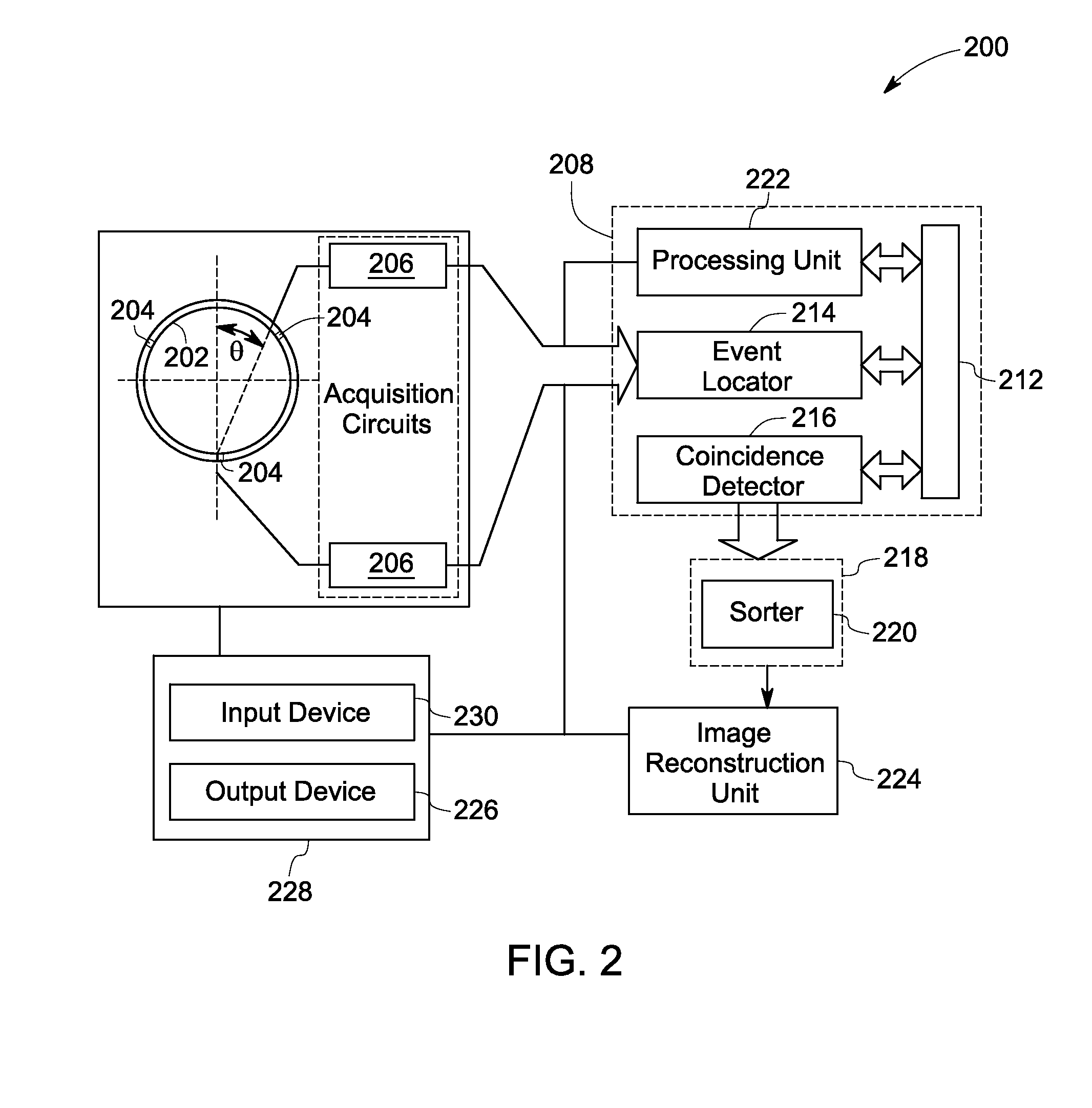
BACKGROUND[0001]Non-invasive imaging techniques are widely used in security screening, quality control, and medical diagnostic systems. Particularly, in medical imaging, non-invasive imaging techniques such as multi-energy imaging allow for unobtrusive, convenient and fast imaging of underlying tissues and organs. To that end, radiographic imaging systems such as nuclear medicine (NM) gamma cameras, computed tomography (CT) systems, single photon emission CT (SPECT) systems and positron emission tomography (PET) systems generate images that illustrate various biological processes and functions for medical diagnoses and treatment.[0002]PET systems, for example, generate images that represent a distribution of positron-emitting nuclides within a patient's body. Typically, a positron-electron interaction results in annihilation, thus converting entire mass of the positron-electron pair into two 511 kilo-electron volt (keV) photons emitted in opposite directions along a line of response...

Claims


Application Information

Patent Timeline
IPC IPC(8): G06K9/00
CPCG06T2211/436G06T11/005
Inventor JANSEN, FLORIBERTUS HEUKENSFELDTMANJESHWAR, RAVINDRA MOHAN
Owner GENERAL ELECTRIC CO
Learn More