
Rapid membrane isolation method for animal and plant samples

a technology of animal and plant samples and membrane isolation, which is applied in the field of rapid membrane isolation method for animal and plant samples, can solve the problems of affecting the use of glass homogenizers and tissue blenders, and requiring more than one hour to complete the procedur

Active Publication Date: 2013-08-22
INVENT BIOTECH
View PDF1 Cites 3 Cited by
  • Summary
  • Abstract
  • Description
  • Claims
  • Application Information

AI Technical Summary

Benefits of technology

The patent describes a method for quickly and easily isolating membranes and organelles from animal or plant samples for various applications such as protein electrophoresis and enzyme assays. The method is simple, fast, and can yield consistent results with both single and multiple samples. It also uses a filter cartridge in a spin column format, which is a new and innovative technique for membrane and organelle isolation.

Problems solved by technology

The procedures are tedious and time consuming; a typical procedure takes more than one hour to complete.
The use of glass homogenizer and tissue blenders is generally less user friendly for multiple samples and are time consuming Thorough cleaning of the homogenizer is required for each sample and there is a possibility of sample cross-contamination.
Currently available commercial kits for membrane and organelle isolation are relatively tedious and time consuming.
These procedures usually take more than one hour to complete.

Method used


Image

  • Rapid membrane isolation method for animal and plant samples
  • Rapid membrane isolation method for animal and plant samples
  • Rapid membrane isolation method for animal and plant samples

Examples


example 2

Isolation of Crude Membranes from Mouse Tissues

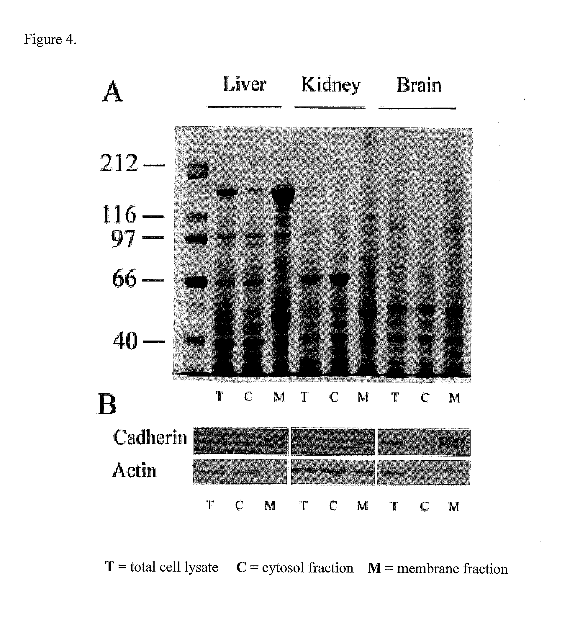
[0039]Fresh or frozen mouse tissue (20-40 mg) was placed in a filter cartridge. Two hundred microliters of cold hypotonic buffer (10 mM Tris-HCl, pH 7.5) was added to the tissue and the tissue was homogenized for 30 seconds with a flat end plastic rod. After homogenization 300 ul Tri-HCl buffer was added to the filter and the following steps were performed sequentially:[0040]1.Centrifuged the filter cartridge at 14,000 rpm in a microcentrifuge at 4° C. for 30 seconds.[0041]2. Discarded the filter and resuspended the pellet in collection tube by vortexing. Centrifuged the tube at 3000 rpm for 1 min to remove most nuclei, un-ruptured cells and tissue debris. Carefully removed the supernatant without disturbing the pellet. Transferred the supemantant to a 1.5 ml microfuge tube and centrifuged the tube at 14-16,000 rpm at 4° C. for 15 min. The pellet contained isolated crude membranes. FIG. 4 shows the results of proteins associated with me...

example 3

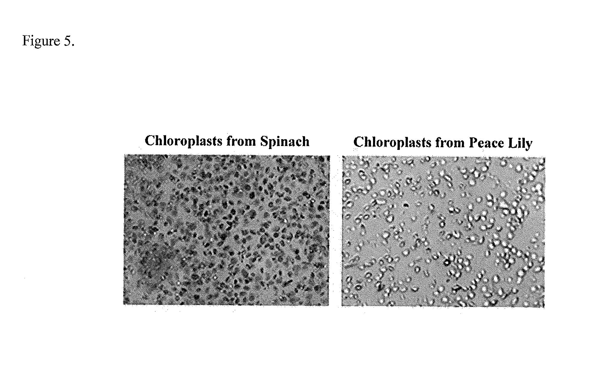
Isolation of Chloroplasts from Plant Cells

[0042]The following can be used for isolation of intact chloroplasts from 5-100 mg fresh plant tissue samples (leaves, seeds and soft stems etc.). If smaller or larger amounts of starting materials are used, the amount of buffers used may be adjusted proportionately.

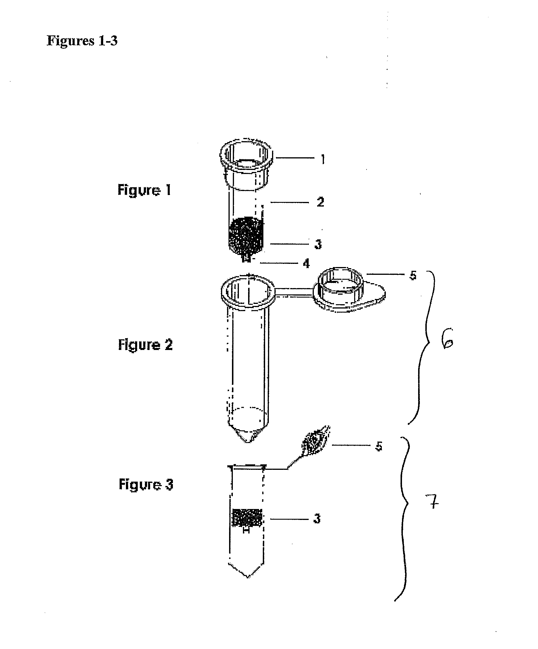
1. Pre-chill buffers and the filter cartridge in collection tube on ice.

2. Approximately 5-100 mg fresh plant tissue were placed in the filter. Plant leaves were folded or rolled and inserted into the filter. The leaf was punched in the filter repeatedly with a pipette tip about 60 times to reduce the volume (for tissues less than 50 mg punching with the tip is not necessary). Seeds and soft stems were cut with a sharp blade into smaller pieces and placed in the filter cartridge(s).

3. Cold homogenization buffer (200 μl, 1×PBS with 20% glass bead, Crystal Mark Inc. Glendale, Calif.) was added to the filter (shake the bottle vigorously for a few seconds prior to pipetting). The tis...


PUM


Abstract

Disclosed herein are methods for quickly obtaining crude membranes from cells, including animal and plant cells. The methods include incubating cells in a buffer and forcing the cells through a filter that causes rupture of the cells, and then separating the resulting crude membranes from most cytosolic proteins.

Description

CROSS-REFERENCE TO RELATED APPLICATIONS[0001]This application claims the benefit of U.S. Provisional Application Ser. No. 61 / 600,116, filed Feb. 17, 2012, which is incorporated by reference herein.BACKGROUND[0002]Isolation of crude membranes from animal and plant cells and / or tissues is the first step for further purification and characterization of membrane and organelle proteins. Over 70% of drug targets and biomarkers are found on plasma membranes, therefore isolation and purification of membrane proteins is one of the most commonly employed procedures in biomedical research. Traditional membrane and organelle isolation protocols often involve homogenizing cultured cells and / or tissues in a hypotonic buffer or a buffer containing detergents such as Triton-X 114. The cells and / or tissues are homogenized with a glass homogenizer (such as Dounce homogenizer), sonication or a tissue blender to break up cells and / or tissues. Insoluble membrane and organelles are separated from nuclei ...

Claims


Application Information

Patent Timeline
IPC IPC(8): C12N5/00
CPCC12N5/0006C12M47/06
Inventor YAN, LINLI, QUANZHI
Owner INVENT BIOTECH
Learn More