
Kinase and phosphatase activity measurement using fluorescent polarization

Inactive Publication Date: 2007-02-01
LIFE TECH CORP
View PDF2 Cites 3 Cited by
  • Summary
  • Abstract
  • Description
  • Claims
  • Application Information

AI Technical Summary

Benefits of technology

[0009] We have developed a simpler, more direct method for measuring kinase activity using fluorescence polarization assays. Fluorescence polarization is a versatile laboratory technique for measuring equilibrium binding, nucleic acid hybridization, and enzymatic activity. Fluorescence polarization assays are homogeneous in that they do not require a separation step such as centrifugation, filtration, chromatography, precipitation or electrophoresis. Assays are done in real time, directly in solution and do not require an immobilized phase. Polarization values can be measured repeatedly and after the addition or reagents since measuring the polarization is rapid and does not destroy the sample. Generally, this technique can be used to measure polarization values of fluorophores from low picomolar to micromolar levels.
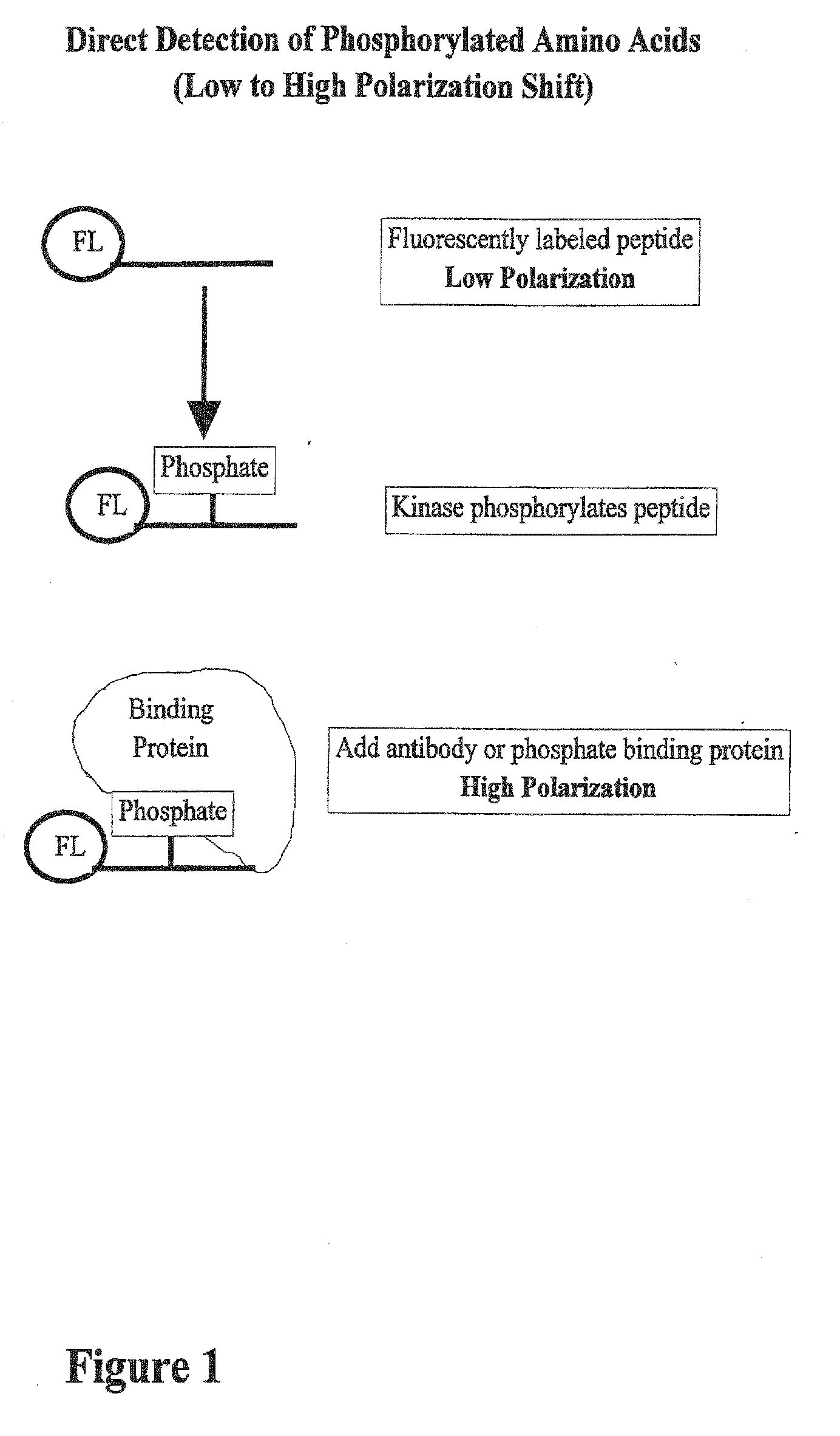
[0014] The rotational relaxation time is small (≈1 nanosecond) for small molecules (e.g. fluorescein) and large (≈100 nanoseconds) for large molecules (e.g. immunoglobulins) (Jolley, 1981). If viscosity and temperature are held constant, rotational relaxation time, and therefore polarization, are directly related to the molecular volume. Changes in molecular volume may be due to interactions with other molecules, dissociation, polymerization, degradation, hybridization, or conformational changes of the fluorescently labeled molecule. For example, fluorescence polarization has been used to measure enzymatic cleavage of large fluorescein labeled polymers by proteases, DNases, and RNases. It also has been used to measure equilibrium binding for protein / protein interations, antibody / antigen binding, and protein / DNA binding. In this invention, we sow that fluorescence polarization is a simple and economical way to measure protein kinase activity.

Problems solved by technology

A mutated or missing protein can change a regulated pathway to be constitutively ‘on’ or ‘off’, disrupting the steady state balance of biological control.
Using radiation provides for very sensitive assays but creates biological hazards and expensive disposal costs.
These assays can be sensitive but are often labor intensive, expensive, and enzyme kinetic information can be limited because of the immobilized component.
Often these assays are semi-quantitative because it is difficult or impossible to measure the amount of immobilized substrate.
A significant drawback is that two molecules are labeled in this method and the chemicals that are required are not widely available.

Method used


Image

  • Kinase and phosphatase activity measurement using fluorescent polarization
  • Kinase and phosphatase activity measurement using fluorescent polarization
  • Kinase and phosphatase activity measurement using fluorescent polarization

Examples


example 1

Fluorescein Amine Labeling Protocol for Labeling Peptides

[0043] The pp60c-src C-terminal phosphoregulatory peptide (BIOMOL; Plymouth Meeting, Pa.) was fluorescently labeled according to the instructions included with the fluorescein amine labeling kit (PanVera Corporation; Madison, Wis.). Briefly, 50 mg of the peptide was labeled at 37° C. for one hour in a 50 mL reaction containing 5 mL 10× coupling buffer (1 M KPO4, pH 7.0) and 5 mL 20 mM fluorescein. The reaction was then quenched with 5 mL 1 M Tris-HCl (pH 8.0) and incubated at room temperature for 30 minutes. Fluorescein-labeled products were then separated by thin layer chromatography. Using silica TLC plates, 2 mL of the reaction products were spotted onto the origin. The products were then separated for 6 hours using 4:1:1 n-butanol:acetic acid:water (v / v) as the solvent. The plate was then dried and photographed, and fluorescein-labeled peptides were scraped off the plate and eluted into 200 mL 50 mM Tris-HCl (pH 8.0).

[0...

example 2

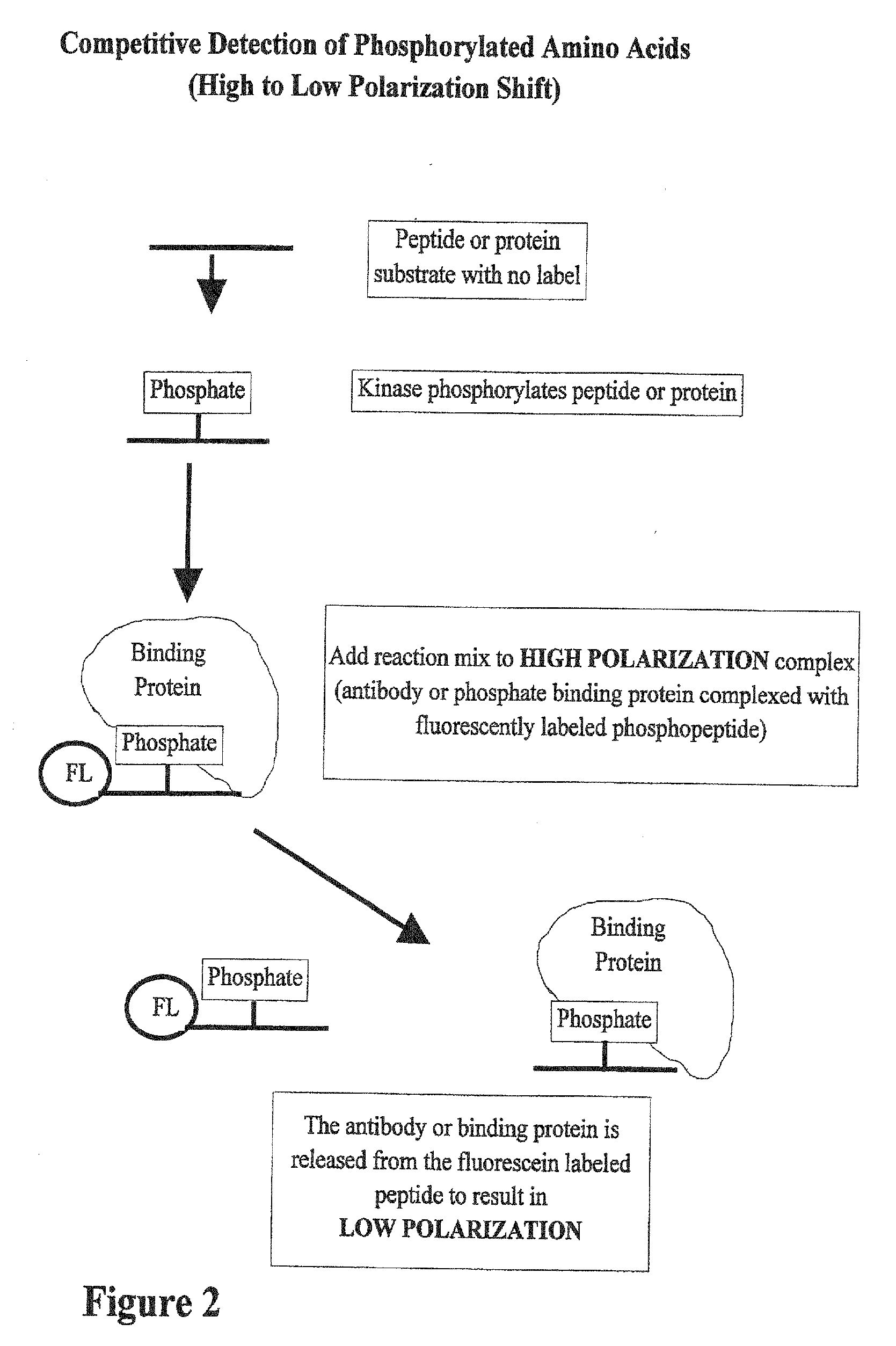
[0045] In FIG. 2, the substrate for the kinase can be any peptide or protein and size is not a limitation. Also, there is no limitation on the concentration of the peptide or protein. Once the kinase reaction has occurred, a high polarization complex is the added to the reaction. This complex contains a fluorescently labeled phosphorylated peptide bound to protein. When the reaction and high polarization complex are mixed, the phosphorylated amino acids produced in the reaction compete for binding to the proteins in a high polarization mix. As the binding proteins are released from the fluorescently labeled peptide, the polarization value of the peptide goes down. In this case the shift in polarization is from a high polarization complex to a low polarization free phosphopeptide. In a simplified version of this assay, both the kinase reaction and the high polarization complex are mixed together at the beginning of the reaction. As the reaction proceeds, the polarization value goes d...

example 3

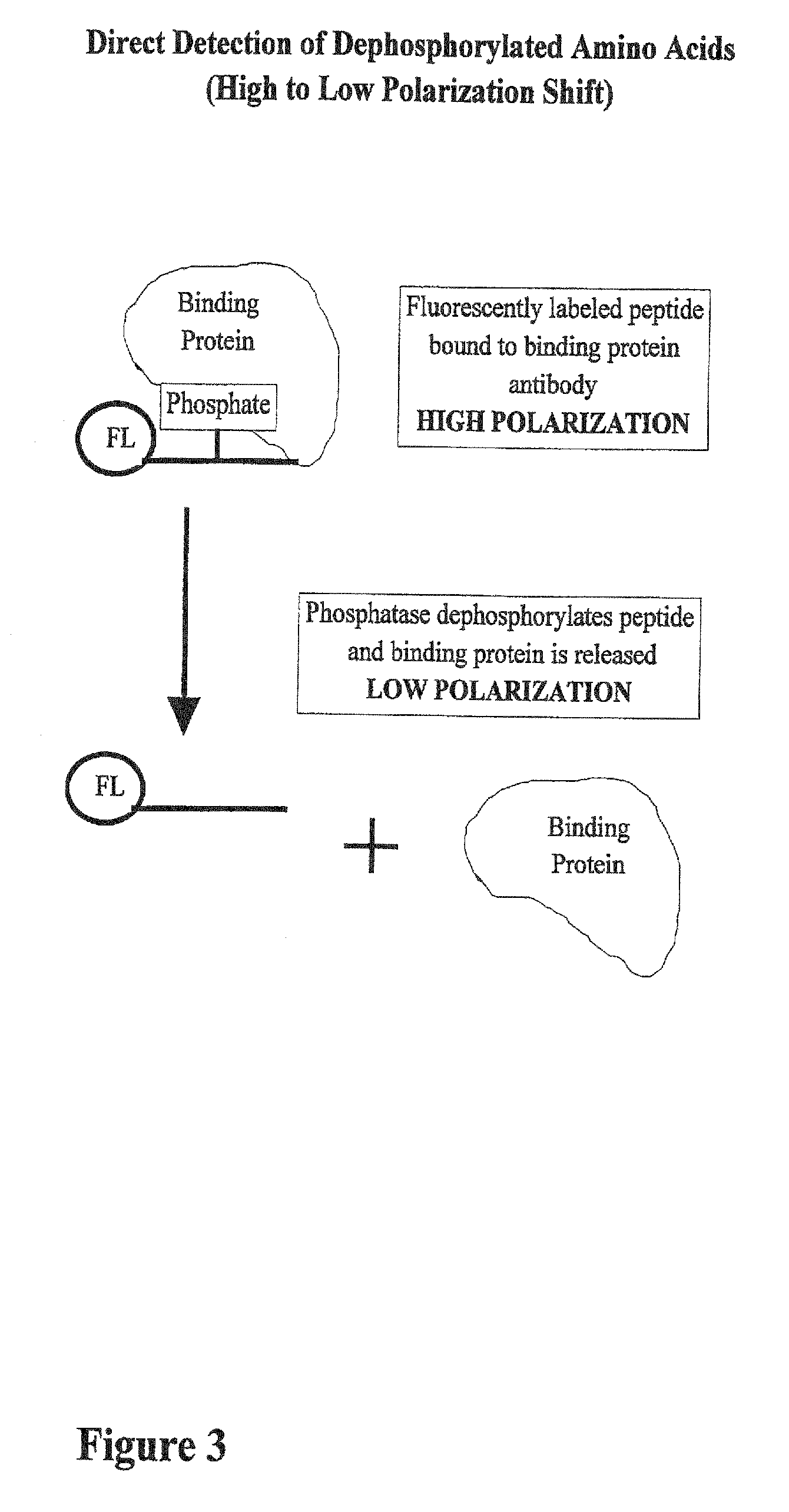
[0046] In FIG. 3, the reaction begins with a high polarization mixture, which is a binding protein attached to a fluorescently labeled phosphopeptide. A phoshatase, an enzyme that removes phosphate groups from other molecules, is added to the reaction and the polarization value goes down. Phosphatase enzymes are important in cellular regulation because they perform the opposite role of kinases.


PUM


Abstract

A method for quantitating enzyme activity that causes phosphorylation or dephosphorylation of a peptide or protein substrate is provided. The method utilizes the differences in fluorescence polarization of a phosphorylated amino acid-containing fluorescence-emitting reporter molecule free in solution as compared to being bound to a binding molecule.

Description

CROSS-REFERENCE TO RELATED APPLICATIONS [0001] This is a continuation application of application Ser. No. 10 / 133,528, filed Apr. 26, 2002, which is a divisional of application Ser. No. 09 / 464,142, filed Dec. 16, 1999, now abandoned, which is a divisional of application Ser. No. 08 / 959,367, filed Oct. 28, 1997, now abandoned, which claims the benefit of Provisional Application No. 60 / 029,831, filed Oct. 28, 1996, all of which are incorporated herein by reference in their entirety.FIELD [0002] The field of the invention is the detection of phosphorylated amino acids using a fluorescence polarization (anisotropy) competition assay. In particular, the process of the invention detects and measures protein kinase activities ad monitors phosphatases. In another application, the assay can be used to quantitatively measure phosphorylated amino acids in extracts. BACKGROUND OF THE INVENTION [0003] Kinases are enzymes that catalyze the transfer of a phosphate molecule from a nucleotide triphos...

Claims


Application Information

Patent Timeline
IPC IPC(8): G01N33/53G01N33/542G01N33/573G01N33/68
CPCC12Q1/485G01N33/542G01N33/6842G01N33/68G01N33/573
Inventor BURKE, THOMAS J.BOLGER, RANDALL E.SCHALL, REBECCAPARKER, GREGORY
Owner LIFE TECH CORP
Learn More