
Methods, Systems and Computer Program Products for Non-Invasive Determination of Blood Flow Distribution Using Speckle Imaging Techniques and Hemodynamic Modeling

a technology of speckle imaging and hemodynamic modeling, applied in the direction of angiography, instruments, diagnostic recording/measure, etc., can solve the problems of limited conventional methods for measuring blood flow and perfusion, high cost of ultrasound detection, and hybrid operating room used for coronary angiography, etc., to achieve accurate determination of blood flow speed

Active Publication Date: 2013-08-29
EAST CAROLINA UNIVERISTY
View PDF7 Cites 22 Cited by
  • Summary
  • Abstract
  • Description
  • Claims
  • Application Information

AI Technical Summary

Benefits of technology

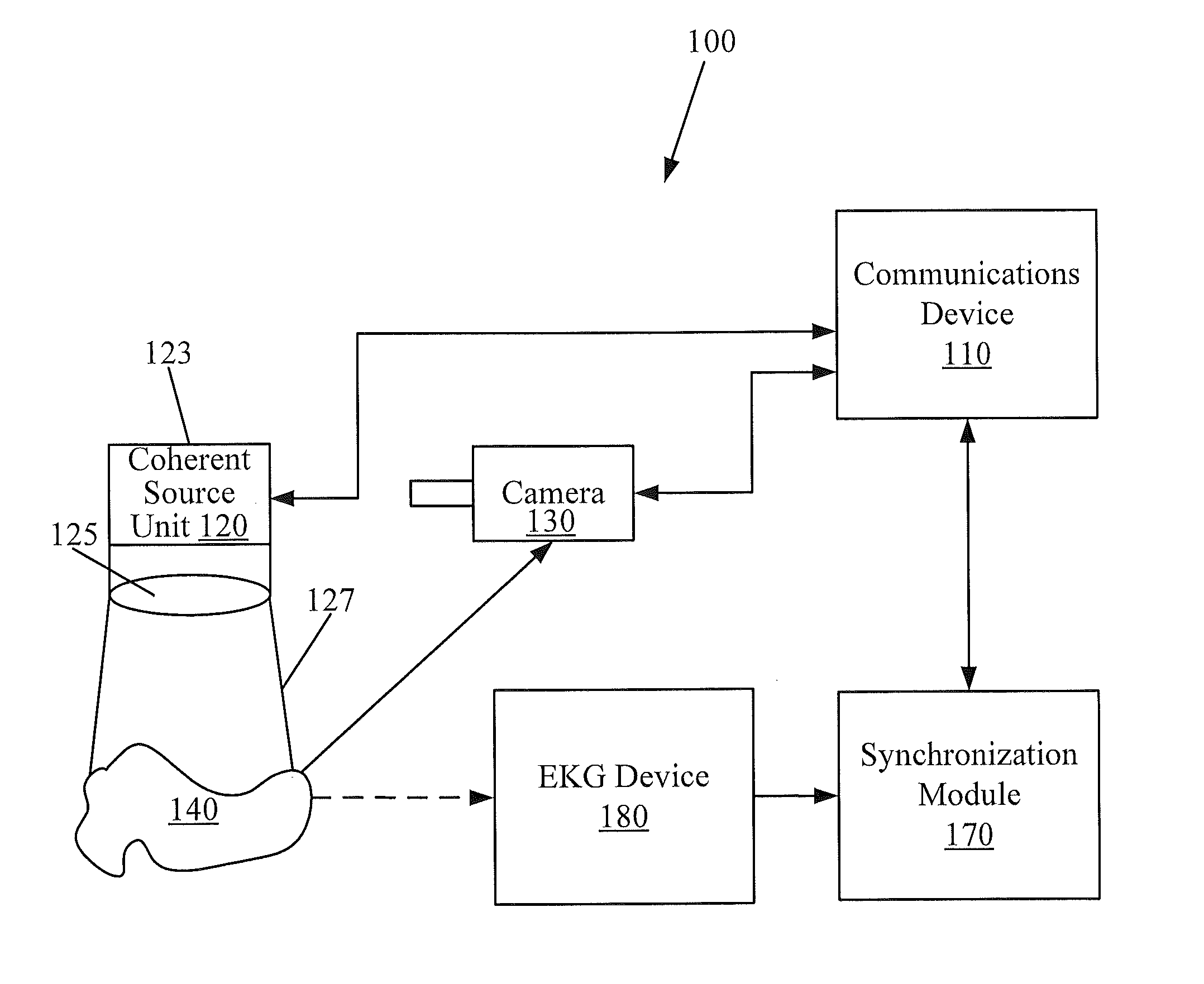
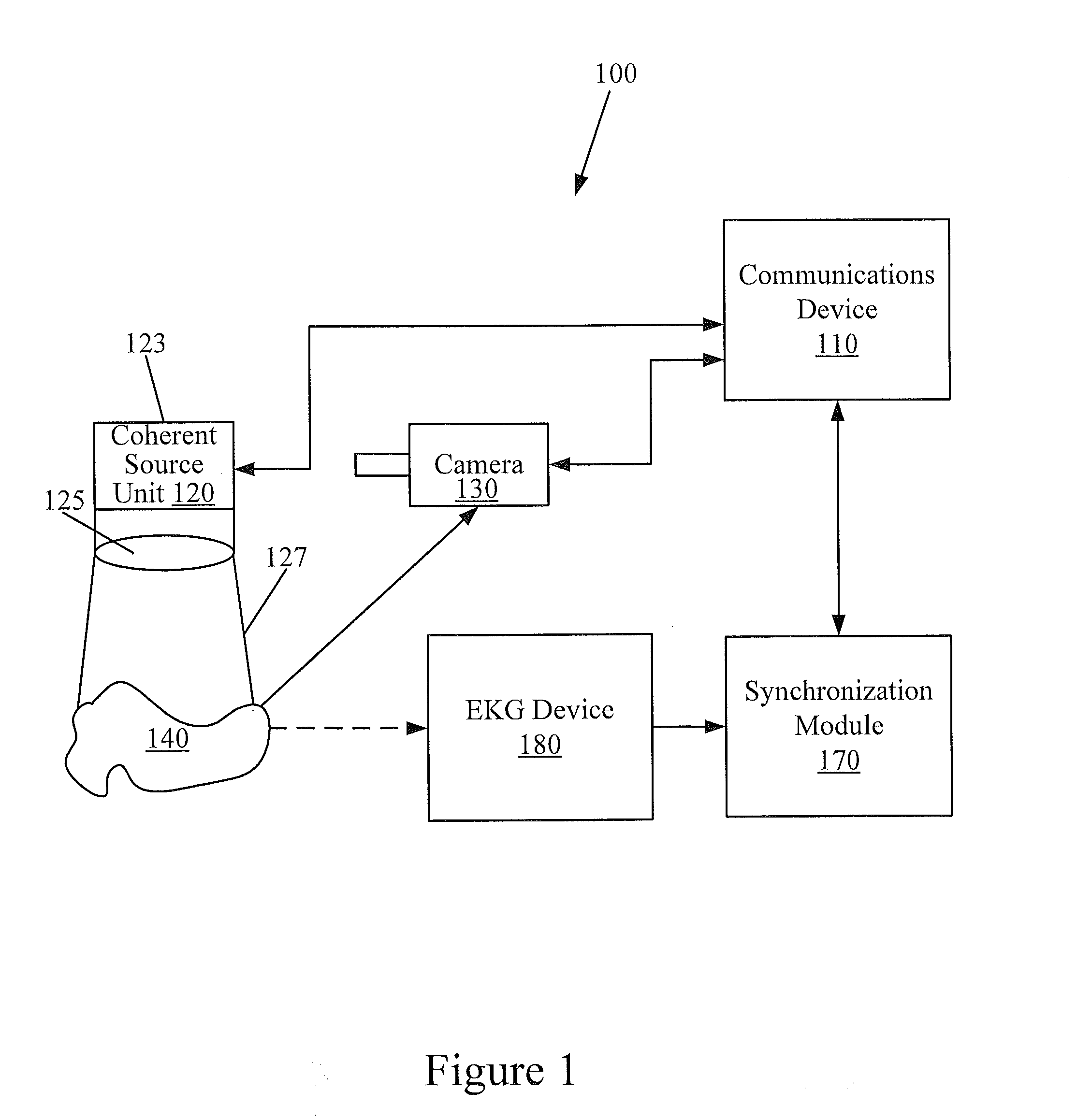
The patent text describes a device that uses coherent light to determine blood flow speed in main vessels and how well the blood is flowing through the tissues. The device uses a laser that emits light with a specific wavelength, which is able to penetrate deep into tissues. The device is designed to provide accurate results for determining blood flow and perfusion distribution.

Problems solved by technology

Conventional methods for measurement of blood flow and perfusion are limited, despite the benefit these measurements would bring to the clinical evaluation of the quality of the revascularization procedure.
However, ultrasound detection typically requires physical contact between the graft vessel and a probe.
Furthermore, ultrasound detection typically relies on proper placement of the probe around the vessel to obtain accurate measurement of flow speed and can be unreliable, measurement to measurement.
Furthermore, hybrid operating rooms used for coronary angiography can be relatively expensive, making this method unavailable to many patients undergoing CABG.

Method used


Image

  • Methods, Systems and Computer Program Products for Non-Invasive Determination of Blood Flow Distribution Using Speckle Imaging Techniques and Hemodynamic Modeling
  • Methods, Systems and Computer Program Products for Non-Invasive Determination of Blood Flow Distribution Using Speckle Imaging Techniques and Hemodynamic Modeling
  • Methods, Systems and Computer Program Products for Non-Invasive Determination of Blood Flow Distribution Using Speckle Imaging Techniques and Hemodynamic Modeling

Examples


examples

[0113]Referring to FIG. 5, a digital photograph of a prototype system 500 to detect flow speed using laser speckle contrast imaging (LSCI) technology in accordance with some embodiments of the present inventive concept will be discussed. As illustrated in FIG. 5, the system includes a communication device 510, such as a laptop computer, a laser unit 520 including a laser generator and a focusing lens, a camera 530, a flow generator 580, flow liquid 590 and a flow target 585. Table 1 set out below summarizes the actual equipment / devices used in this experiment.

TABLE 1Devices used in Experiment 1NotesCCD camera (530)Lumenera Lm075Laser (520)633 nm in wavelength, 1 mW in powerLiquid used in flow (590)20% Intralipid 1 to 4 ratio mixed withwater plus fruit colorComputer / CommunicationsLaptop PCDevice (510)

[0114]As discussed above, the faster the camera 530, the smaller the fixed time period has to be to obtain an adequate number of speckle images to provide a meaningful result. Thus, the ...


PUM


Abstract

A non-invasive method for measuring blood flow in principal vessels of a heart of a subject is provided. The method includes illuminating a region of interest in the heart with a coherent light source, wherein the coherent light source has a wavelength of from about 600 nm to about 1100 nm; sequentially acquiring at least two speckle images of the region of interest in the heart during a fixed time period, wherein sequentially acquiring the at least two speckle images comprises acquiring the at least two speckle images in synchronization with motion of the heart of the subject; and electronically processing the at least two acquired speckle images based on the temporal variation of the pixel intensities in the at least two acquired speckle images to generate a laser speckle contrast imaging (LSCI) image and determine spatial distribution of blood flow rate in the principal vessels and quantify perfusion distribution in tissue in the region of interest in the heart from the LSCI image.

Description

CLAIM OF PRIORITY[0001]The present application claims priority from U.S. Provisional Application No. 61 / 431,161 (Attorney Docket No. 5218-200PR), filed Jan. 10, 2011 and U.S. Provisional Application No. 61 / 476,854 (Attorney Docket No. 5218-200PR2), filed Apr. 19, 2011, the disclosures of which are hereby incorporated herein by reference as if set forth in their entirety.RESERVATION OF COPYRIGHT[0002]A portion of the disclosure of this patent document contains material which is subject to copyright protection. The copyright owner, East Carolina University of Greenville, N.C., has no objection to the reproduction by anyone of the patent document or the patent disclosure, as it appears in the Patent and Trademark Office patent file or records, but otherwise reserves all copyright rights whatsoever.FIELD[0003]The present inventive concept relates generally to determination of blood flow distribution and, more particularly, to the use of speckle imaging techniques for non-invasive determ...

Claims


Application Information

Patent Timeline
Patent Type & Authority Applications(United States)
IPC IPC(8): G06T7/00
CPCA61B5/0059A61B5/02007G06T7/0012G02B27/48A61B5/0261
Inventor FERGUSON, JR., THOMAS BRUCEHU, XIN-HUACHEN, CHENG
Owner EAST CAROLINA UNIVERISTY
Learn More