Ultrasound imaging system with patient-specific settings

an ultrasound imaging system and patient-specific technology, applied in ultrasonic/sonic/infrasonic image/data processing, applications, tomography, etc., can solve the problems of time-consuming and error-prone manual changes of parameters for every acquisition

Inactive Publication Date: 2013-09-26
KONINKLIJKE PHILIPS ELECTRONICS NV
View PDF7 Cites 17 Cited by
  • Summary
  • Abstract
  • Description
  • Claims
  • Application Information

AI Technical Summary

Benefits of technology

[0005]It is an object of the present invention to provide an ultrasound imaging system that ensures that in subsequent examinations of the same patient ultrasound images are acquired under conditions such that the images can be compared and can be used to monitor disease progression.

Problems solved by technology

Manually changing the parameters for every acquisition is time consuming and error-prone.

Method used


Image

  • Ultrasound imaging system with patient-specific settings
  • Ultrasound imaging system with patient-specific settings
  • Ultrasound imaging system with patient-specific settings

Examples


Embodiment Construction

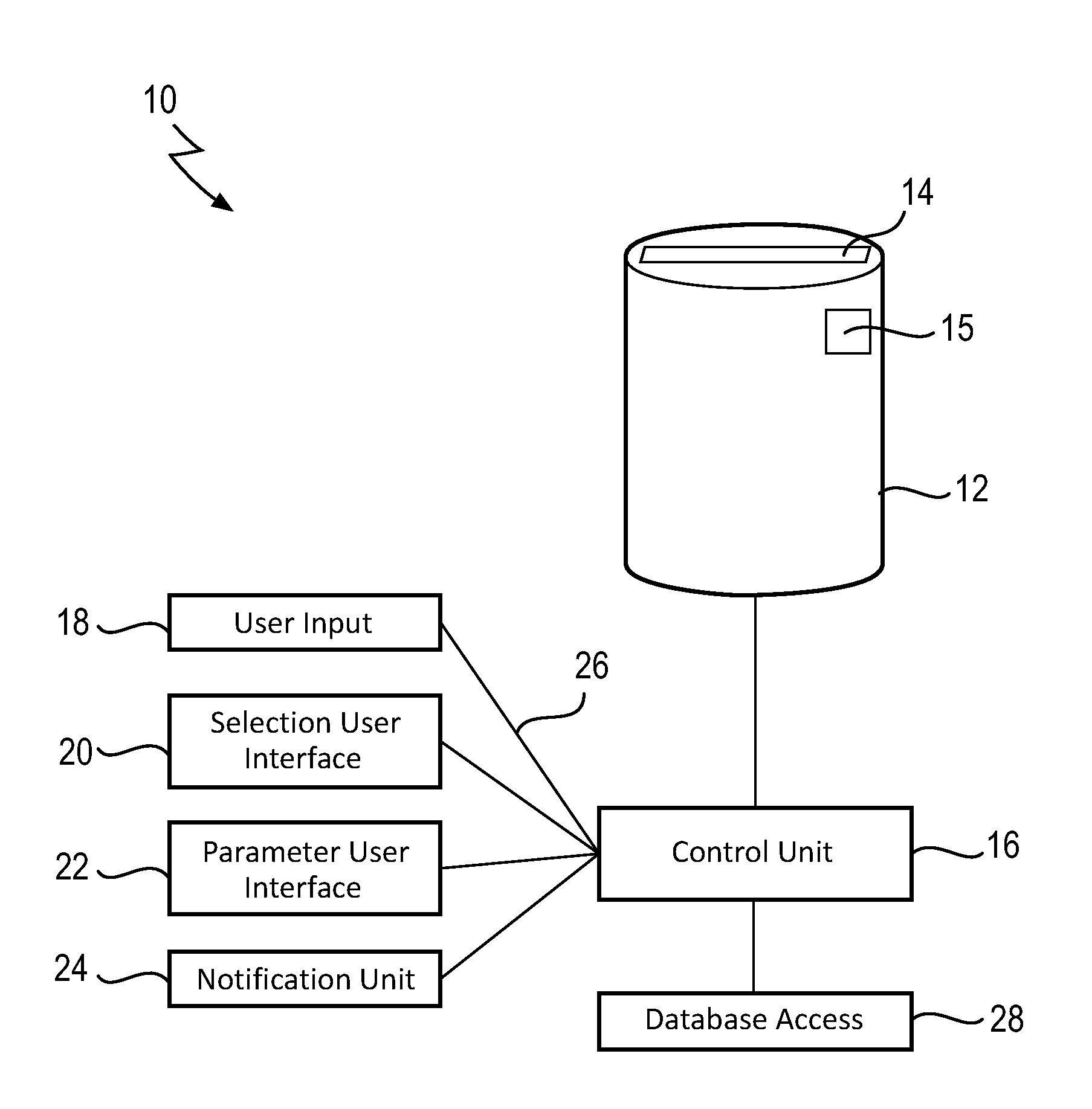
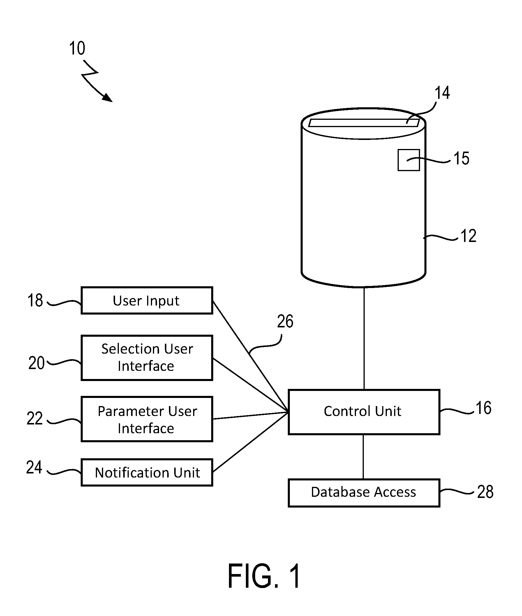
[0031]FIG. 1 shows a schematic block diagram of an ultrasound imaging system 10 according to the present invention. The image acquisition unit 12 comprises an exchangeable transducer unit 14 and a transducer sensor unit 15 which is capable of recognizing different exchangeable transducer units 14.

[0032]The control unit 16 is connected to a number of user interface components: the user input 18, the selection user interface 20, the parameter user interface 22, and the notification unit 24. Based on the patient identifier 26 that is entered in the user input 18, the control unit 16 is adapted to retrieve a set of acquisition parameters that corresponds to this patient. In this embodiment, the user input 18 directly takes a patient identifier as input, e.g. a number that is unique for this patient. In other embodiments, the user input 18 could also comprise input fields for patient name, gender, and birth date, such that the user input 18 can look up the corresponding unique patient. O...


PUM


Abstract

The present invention relates to an ultrasound imaging system (10) and a corresponding method which enable that in subsequent examinations of the same patient ultrasound images are acquired under conditions such that the images can be compared and can be used to monitor disease progression. The proposed ultrasound imaging system (10) comprises an image acquisition unit (12) configured to acquire an ultrasound image based on a set of acquisition parameters, a user input (18) for entering (S10) a patient identifier (26), a database access (28) configured to access a database of sets of acquisition parameters, wherein the sets of acquisition parameters are associated with patient identifiers (26), and a control unit (16) configured to automatically retrieve (S12) a set of acquisition parameters that is associated with said patient identifier (26) based on an entered patient identifier (26) and control the image acquisition unit (12) to acquire (S22) an ultrasound image based on the retrieved set of acquisition parameters.

Description

FIELD OF THE INVENTION[0001]The present invention relates to a system and method for acquiring ultrasound images. The invention also relates to a computer program for implementing said method.BACKGROUND OF THE INVENTION[0002]Ultrasound imaging is commonly used to diagnose many different diseases and the progression of these diseases. In order to best adapt the image acquisition to the specific disease, the organ under investigation, and different characteristics of the patient, there are numerous acquisition parameters that need to be set appropriately. These parameters are related to the transmission and reception of the ultrasound signals, the processing of the measured signals, image reconstruction, image display and image storage. They include such operating parameters as the depth of an image, the location of the transmit focus, the number of focal zones, whether to use the B mode or color Doppler mode, whether harmonic or fundamental frequencies are to be used for imaging, ima...

Claims


Application Information

Patent Timeline
Patent Type & Authority Applications(United States)
IPC IPC(8): A61B8/08A61B8/13A61B8/00
CPCA61B8/468A61B8/5292A61B8/54A61B8/467G06F19/3406A61B8/085A61B8/13A61B8/585G16H40/63
Inventor GAUTHIER, THOMAS PATRICE JEAN ARSENE
Owner KONINKLIJKE PHILIPS ELECTRONICS NV
Why Eureka
Try Eureka