
Method and apparatus for generating a signal indicative of motion of a subject in a magnetic resonance apparatus

a magnetic resonance and signal technology, applied in the field of generation of a signal indicative of motion of a subject in a magnetic resonance (mr) apparatus, can solve the problems of under-treatment of the target tissue, unwanted collateral damage to healthy or critical surrounding anatomical structures, and requiring compromise with respect to certain parameters

Inactive Publication Date: 2013-10-17
SIEMENS AG
View PDF7 Cites 13 Cited by
  • Summary
  • Abstract
  • Description
  • Claims
  • Application Information

AI Technical Summary

Benefits of technology

This patent is about a method to take into account the movement of a person during an MR-guided intervention procedure. The method ensures that the therapy is effective at the desired time, depending on the person's movement. The patent also describes an MR apparatus to implement this method. The technical effect of this invention is to improve the accuracy and effectiveness of MR-guided intervention procedures.

Problems solved by technology

If the tissue motion management during HIFU sonication is not accurate, this may result in under-treatment of the target tissue, and / or unwanted collateral damage to healthy or critical surrounding anatomical structures.
In principle, this motion monitoring can be achieved by analyzing the magnetic resonance images themselves, but this requires compromises with respect to certain parameters (temporal resolution v. signal-to-noise ratio (SNR)), or image contrast.
This procedure, however, can only manage liver deformations caused by the periodic breathing cycle, and is not capable of handling the non-rigid liver deformations that are caused by intestinal activity or muscle relaxation, as noted by von Siebenthal et al. in “4D MR Imaging of Respiratory Organ Motion and its Variability,” Phys. Med. Biol., Vol. 52, No. 6 (2007) pgs.
This setup is not suitable for clinical application, because the external ultrasound imaging probe cannot emit ultrasound parallel to the axis of respiratory motion.
Moreover, the local motion in liver is spatially dependent, so as one-dimensional projection would not be sufficient.
By contrast to images obtained with ultrasound, external images of the subject do not provide tomographic information from the interior of the body.
This technology has been used for the correction of fMRI data, but has not been used in the context of image-guided therapy.

Method used


Image

  • Method and apparatus for generating a signal indicative of motion of a subject in a magnetic resonance apparatus
  • Method and apparatus for generating a signal indicative of motion of a subject in a magnetic resonance apparatus
  • Method and apparatus for generating a signal indicative of motion of a subject in a magnetic resonance apparatus

Examples


Embodiment Construction

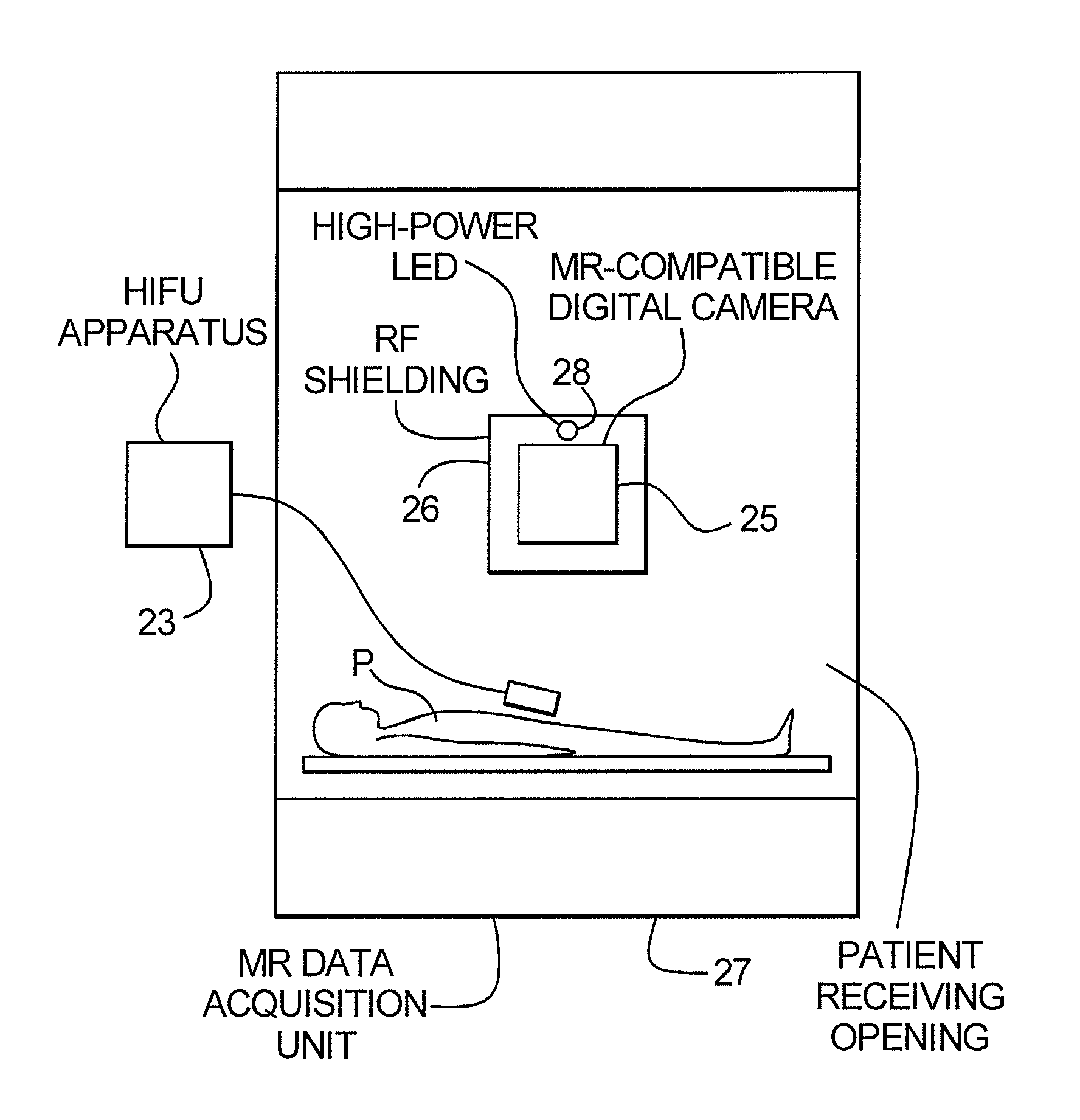
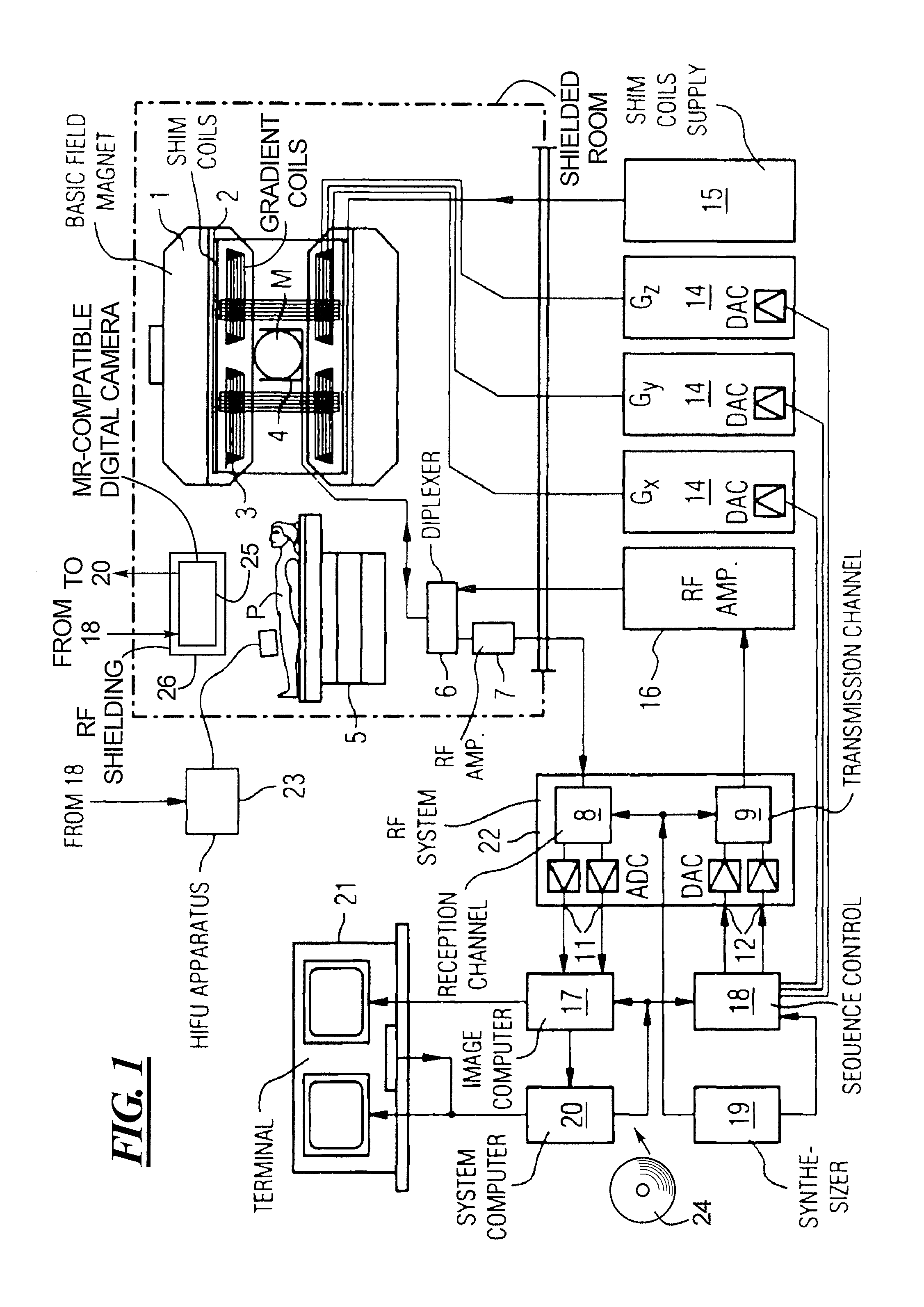
[0027]FIG. 1 is a schematic illustration of a magnetic resonance tomography apparatus operable according to the present invention. The structure of the magnetic resonance tomography apparatus corresponds to the structure of a conventional tomography apparatus, with the differences described below. A basic field magnet 1 generates a temporally constant, strong magnetic field for the polarization or alignment of the nuclear spins in the examination region of a subject such as, for example, a part of a patient P to be examined. The high homogeneity of the basic magnetic field required for the magnetic resonance measurement is defined in a spherical measurement volume M into which the parts of the patient P to be examined are introduced. For satisfying the homogeneity requirements and, in particular, for eliminating time-invariable influences, shim plates of ferromagnetic material are attached at suitable locations. Time-variable influences are eliminated by shim coils 2 that are driven...


PUM


Abstract

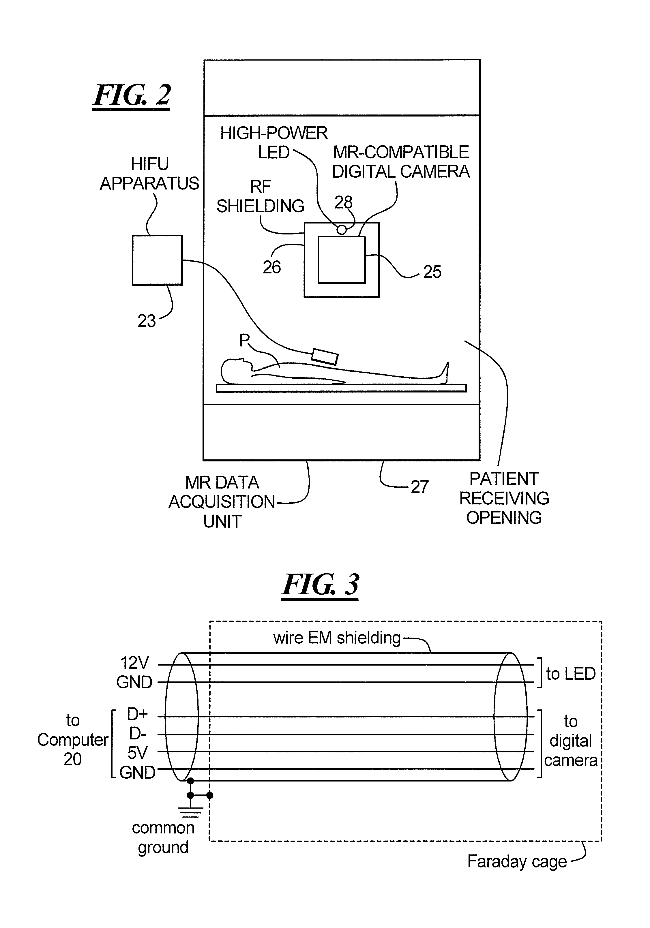
In a method and magnetic resonance (MR) apparatus for implementing an MR-guided procedure, an MR-compatible digital camera is placed in the patient receiving opening of the MR data acquisition unit that is operated to acquire MR data for reconstructing images that are used to guide the MR-guided intervention. The digital camera is operated to obtain digital images of the exterior of the patient, from which motion of the patient is detectable. The images are analyzed in a processor to identify therefrom the motion of the patient and the result of the analysis is represented as a processor output that is used to control the timing, with respect to the motion of the examination subject, of the occurrence of at least one event in the MR-guided procedure. One important application is respiratory gating / triggering of HIFU sonication for the treatment of moving organs.

Description

BACKGROUND OF THE INVENTION[0001]1. Field of the Invention[0002]The present invention concerns the generation of a signal indicative of motion of a subject in a magnetic resonance (MR) apparatus, and in particular to the generation of such a signal for triggering an event in an MR-guided procedure.[0003]2. Description of the Prior Art[0004]Many types of procedures are implemented by interaction with an examination subject while the examination subject is located in a magnetic resonance apparatus, namely while the subject is located inside the data acquisition unit of such a magnetic resonance apparatus. One example of such a procedure is treatment with high intensity focused ultrasound (HIFU).[0005]The principle of HIFU treatment is to concentrate a high acoustic intensity within a focal spot having a size of a few millimeters, in order to produce sharply localized mechanical or thermal effects. This treatment is implemented using an external source of energy, and a propagating ultr...

Claims


Application Information

Patent Timeline
Patent Type & Authority Applications(United States)
IPC IPC(8): A61B5/055
CPCA61N7/02A61N2007/0086A61B2090/0436A61B2090/3612A61B90/30A61B2090/309A61B90/361A61B34/10A61B2090/374
Inventor AUBOIROUX, VINCENTVIALLON, MAGALIEPETRUSCA, LORENASALOMIR, RARES
Owner SIEMENS AG
Learn More