Automated doppler velocimetry using a low-cost transducer

a transducer and automatic technology, applied in the field of volume examination, can solve the problems of limiting the availability and access of ultrasound by specialists, and the cost of both devices is relatively high, and achieve the effect of low cos

Active Publication Date: 2013-10-17
KONINKLIJKE PHILIPS ELECTRONICS NV
View PDF4 Cites 8 Cited by
  • Summary
  • Abstract
  • Description
  • Claims
  • Application Information

AI Technical Summary

Benefits of technology

[0010]In accordance with the present invention, an ultrasound device is designed for carrying out a series of steps automatically and without need for user intervention. These steps include examining, using ultrasound, a volume for bo

Problems solved by technology

In emerging market countries such as India, the shortage of specialists limits the avail

Method used


Image

  • Automated doppler velocimetry using a low-cost transducer
  • Automated doppler velocimetry using a low-cost transducer
  • Automated doppler velocimetry using a low-cost transducer

Examples


Embodiment Construction

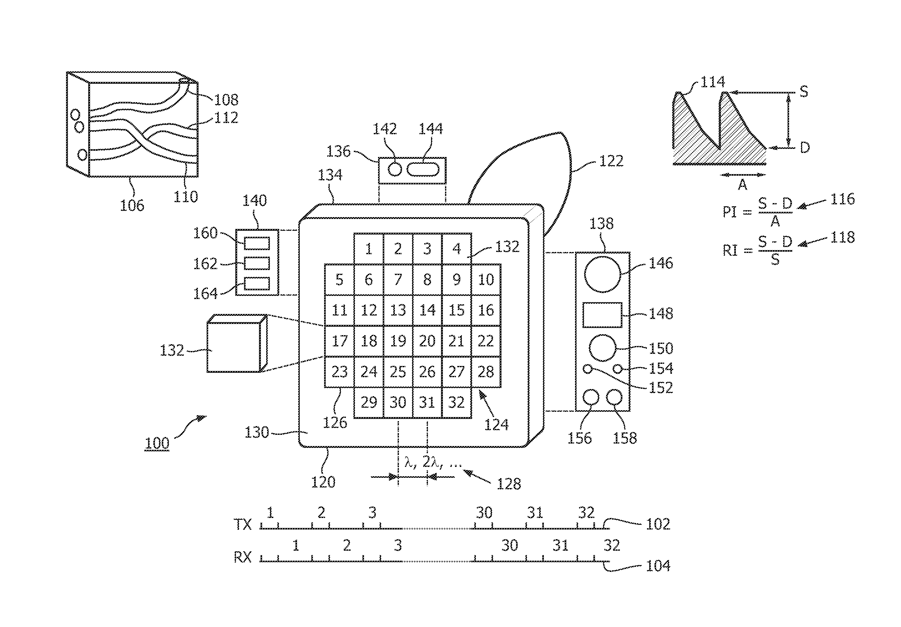
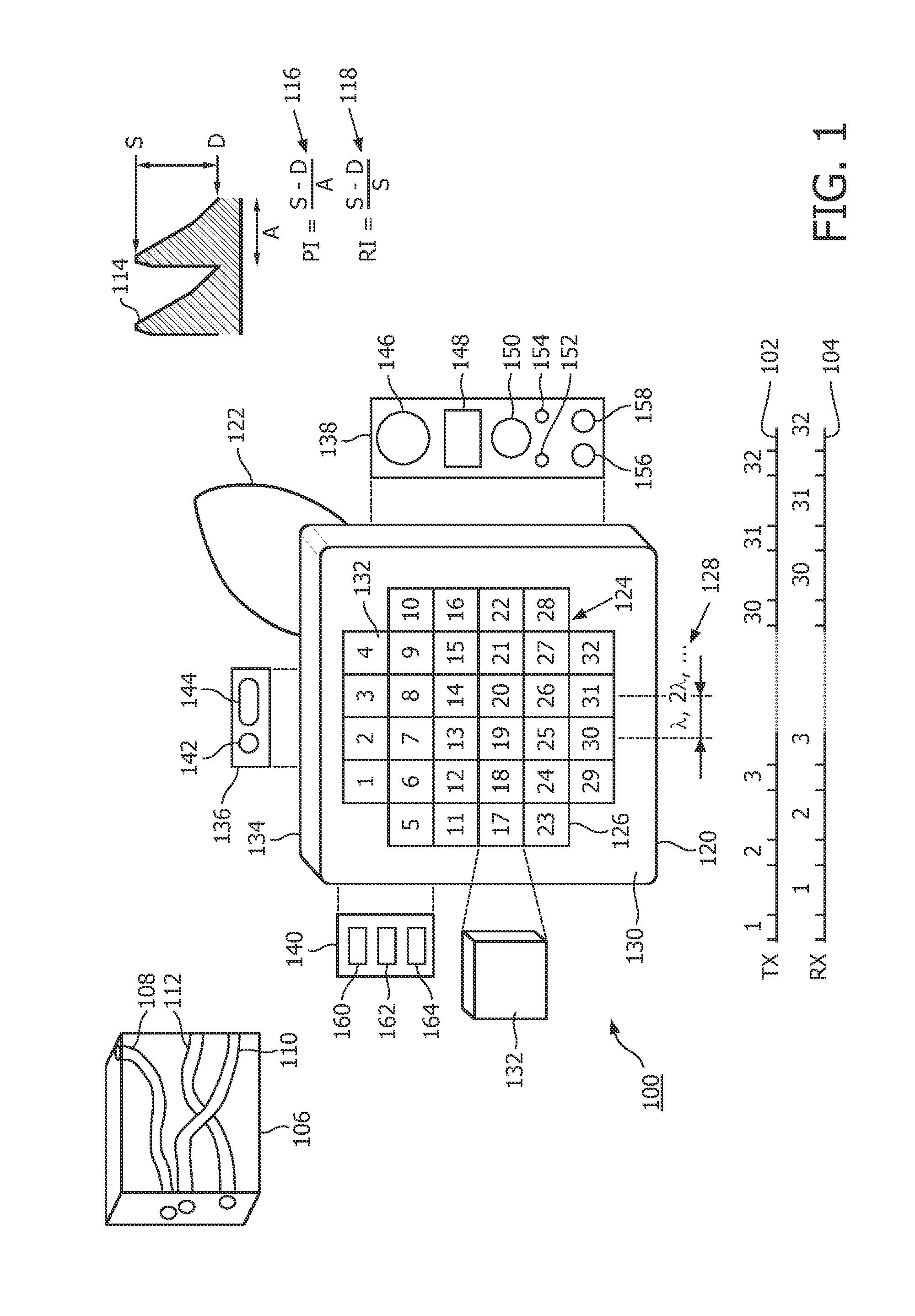
[0034]FIG. 1 depicts, by way of illustrative and non-limitative example, an ultrasound probe 100, transmit / receive timing diagrams 102, 104, and a volume or “volume of interest”106 containing blood vessels 108, 110, 112. Further depicted are a blood-flow, or “spectral Doppler ultrasound”, waveform 114 and respective clinical Doppler indices 116, 118.

[0035]The probe 100 is implementable as an automatic, handheld, stand-alone, self-contained, ultrasound examination device. It has a transducer housing 120 and a handle 122.

[0036]Within the transducer housing 120, a non-phased, two-dimensional transducer 124 is comprised of transducer elements 126, the number of elements being determined by the scan volume and anatomy.

[0037]As seen in FIG. 1 by way example, the number of elements 126 is 32. Thus, with an element size of 10 mm, an approximately 6 cm×6 cm volume is covered. Flush with a front surface 130 of the housing 120, are ultrasound-receiving faces 132 of the transducer elements 126,...


PUM


Abstract

An automatic, stand-alone, hand-held ultrasound blood-vessel examination device (100) requires a reduced number of transducer elements (126) and presents a simplified user interface (136-140), without the need for displaying an image of any of the vessels. The probe, in one embodiment, acquires and examines a volume of interest (106), searches for a target vessel, tests the vessel for normality of blood flow, and reports the diagnosis, all automatically and without need for user intervention. In another embodiment, the probe finds a body vessel (108-112) in a volume, and extracts a clinical Doppler parameter, all automatically and without need for user intervention.

Description

FIELD OF THE INVENTION[0001]The present invention relates to examining a volume of interest for a body vessel and, more particularly, to using ultrasound for the examining.BACKGROUND OF THE INVENTION[0002]Assessing the well being of fetus is a very important clinical practice in pregnancy care. Currently, the most prevalent ways for doctors to assess fetal well-being are the analysis of fetal heart rate using a cardio-tocograph (CTG) and the assessment of maternal and fetal blood vessel flows using ultrasound Doppler. Ultrasound Doppler waveform analysis of specific blood flows of fetus and mother is part of established medical practice, and a standard recommendation in various clinical guidelines for the diagnosis and assessment of high-risk pregnancies (type-2 diabetes, hypertension or pre-eclampsia in mother and IUGR—intra-uterine growth restriction of fetus). One of the main aims of routine antenatal care is to identify the “at risk” fetus in order to clinically intervene, there...

Claims


Application Information

Patent Timeline
IPC IPC(8): A61B8/08A61B8/00A61B8/06
CPCA61B8/5223A61B8/06A61B8/488A61B8/4427A61B8/4444A61B5/489A61B8/467A61B8/54A61B8/585A61B8/0891A61B8/483A61B8/466
Inventor ANAND, AJAYPETRUZZELLO, JOHNSISODIA, RAJENDRA SINGHGUPTA, LALITVAJINEPALLI, PALLAVIFIRTION, CELINE
Owner KONINKLIJKE PHILIPS ELECTRONICS NV
Why Eureka
Try Eureka