Component Frame Enhancement for Spatial Compounding in Ultrasound Imaging

a component frame and spatial compounding technology, applied in the field of spatial compounding, can solve the problems of not being able to pick up the target, not being able to achieve the best suited preservation of angled specular targets, and not performing average performan

Active Publication Date: 2013-11-07
SIEMENS MEDICAL SOLUTIONS USA INC
View PDF29 Cites 14 Cited by
  • Summary
  • Abstract
  • Description
  • Claims
  • Application Information

AI Technical Summary

Benefits of technology

The patent describes a method, computer-readable media, and system for improving ultrasound imaging by combining data from different spatial responses. This allows for more accurate detection and segmentation of anatomical structures and soft tissue. The system includes a beamformer for acquiring data, a processor for detecting variance and segmenting the data, and a display for displaying the resulting compound image. The technical effect of this invention is improved accuracy and precision in ultrasound imaging.

Problems solved by technology

Averaging performs well for speckle variance reduction, but is not best suited for preserving the angled specular targets.
Other components steered differently may not pick up the target as well, or may miss the target all together.
Averaging all the component frames may diminish the level of such matched structures.
However, maximum compounding provides poor speckle reduction and is ridden with a host of other problems, such as motion artifacts and increased clutter noise.

Method used


Image

  • Component Frame Enhancement for Spatial Compounding in Ultrasound Imaging
  • Component Frame Enhancement for Spatial Compounding in Ultrasound Imaging
  • Component Frame Enhancement for Spatial Compounding in Ultrasound Imaging

Examples


Embodiment Construction

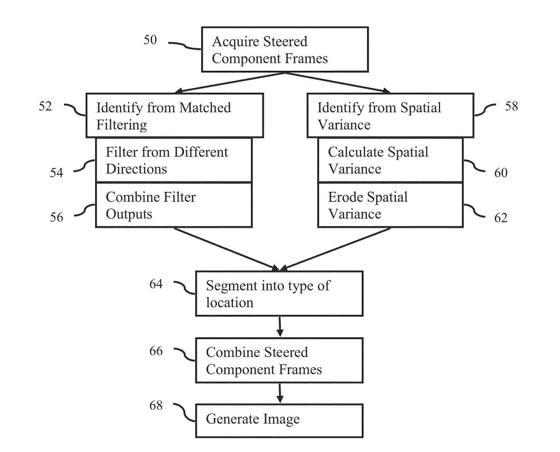
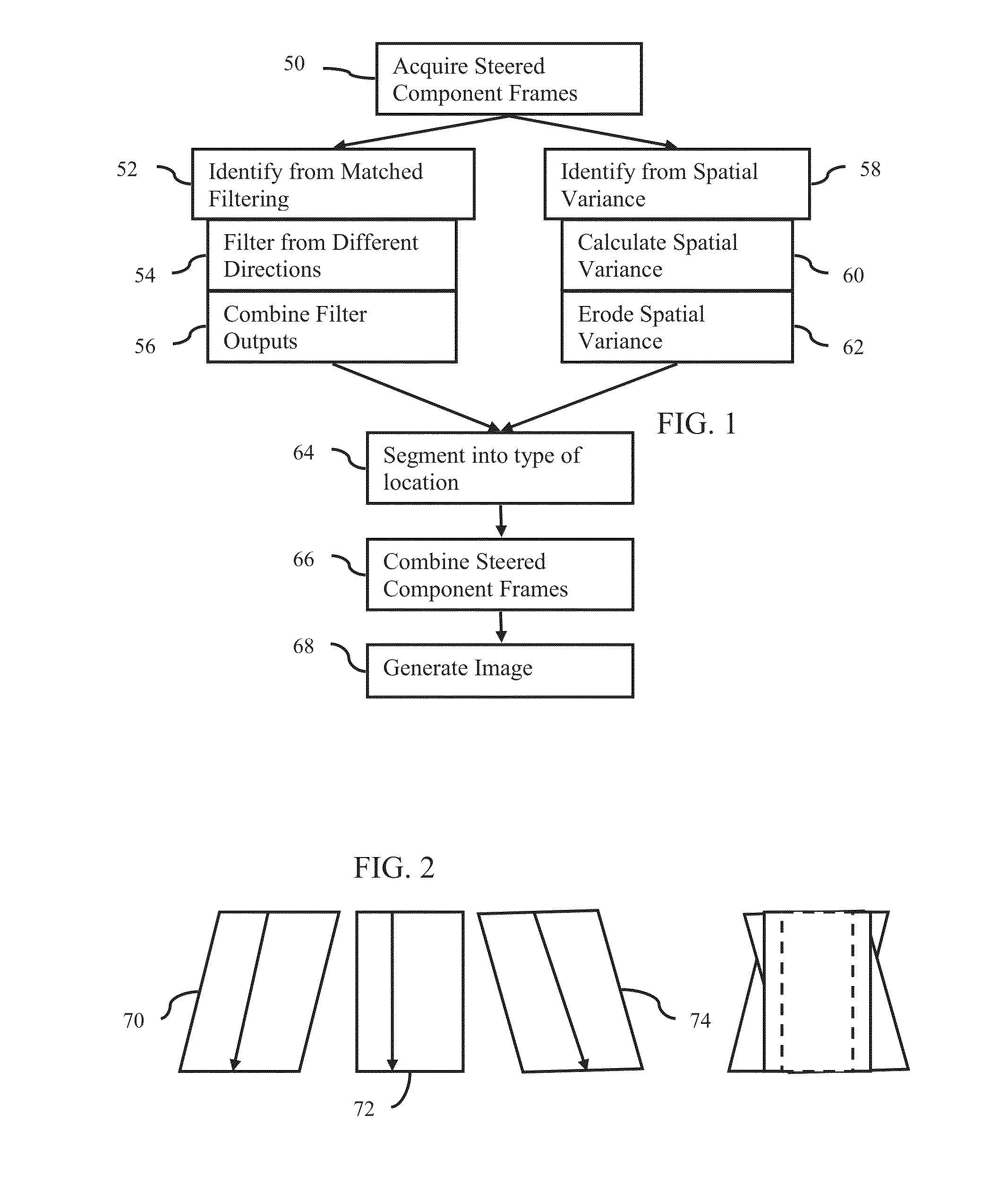
[0018]No one combining method is best suited for the entire spatially compounded image. Instead, the image is segmented into soft tissue (speckle), noise, and anatomic structures or other combination of sources. Compounding appropriate to each segment is applied. An optimal method of combining the steered ultrasound images by location is used.

[0019]For ultrasound imaging, the component frames are pre-processed for segmentation. Most image processing techniques perform segmentation based on detailed analysis of the image data. Such an image analysis stage is computationally intensive as well as time-consuming. Rather than image analysis, matched filtering and / or variance with erosion processing is used. These types of processing may be well suited for use in a real time ultrasound system.

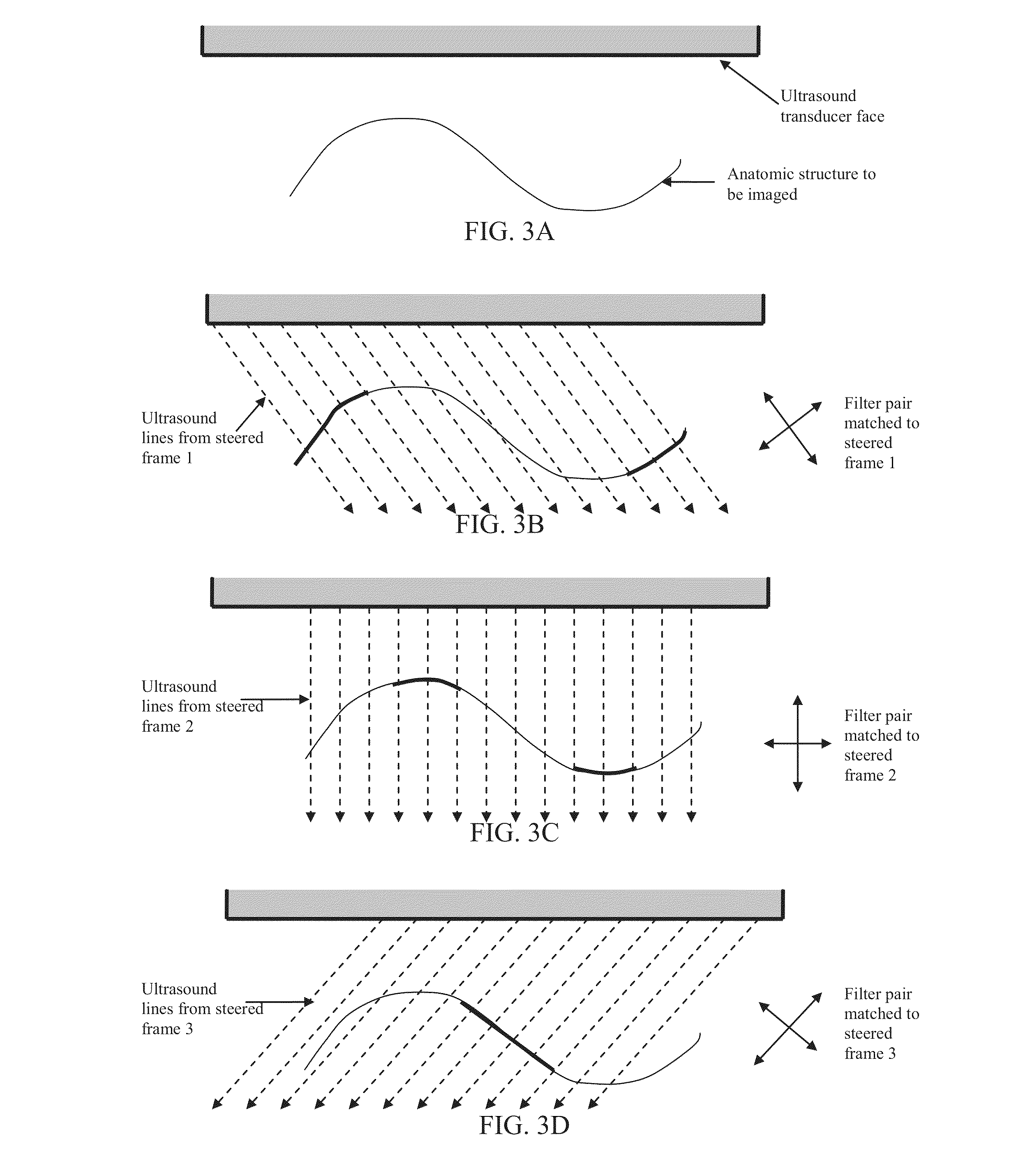
[0020]Prior knowledge of the system state, such as the beam directions, is used to set up matched filters. Matched or directional filtering may identify and enhance structures best imaged by individu...


PUM


Abstract

Steered spatial compounding is provided in ultrasound imaging. Component frames of data associated with different spatial response are processed to identify different sources of signals. Matched filtering and / or spatial variance with erosion distinguish between sources of signals, such as distinguishing between (1) anatomical structure and (2) soft tissue and / or noise. The locations in the patient are segmented by the source of signals and different compounding is applied for the different sources.

Description

BACKGROUND[0001]The present invention relates to spatial compounding. Steered spatial compounding is used in clinical environments. One component frame of data is acquired by scanning along scan lines at one angle or pattern relative to a transducer. A second component frame of data is acquired by scanning along scan lines at a different angle or pattern. Each of the frames of data represents an overlapping region of a patient. The frames of detected data are compounded together and displayed as an image.[0002]Steered spatial compounding increases the information content of ultrasound images and reduces speckle noise in ultrasound. Compounding component frames of data associated with different scan angles may reduce speckle and may produce a more continuous border in specular targets, such as muscle layers, ductal structures, and boundaries between different tissue types.[0003]To compound, the steered ultrasound images are averaged. Averaging performs well for speckle variance reduc...

Claims


Application Information

Patent Timeline
Patent Type & Authority Applications(United States)
IPC IPC(8): G06K9/00
CPCG01S15/8995G01S15/8977
Inventor RAO, BIMBAGURACAR, ISMAYIL M.USTUNER, KUTAY F.
Owner SIEMENS MEDICAL SOLUTIONS USA INC
Why Eureka
Try Eureka