Method for acquisition of subtraction angiograms

a technology of subtraction angiogram and acquisition method, which is applied in the field of digital xray image processing, can solve the problems of incomplete set of processed image characteristics, unrecognizable details, and artifacts in images, and achieve the effect of increasing the diagnostic value of subtraction angiogram

Inactive Publication Date: 2014-01-16
ZAKRYTOE AKCIONERNOE OBSHCHESTVO IMPULS
View PDF5 Cites 13 Cited by
  • Summary
  • Abstract
  • Description
  • Claims
  • Application Information

AI Technical Summary

Benefits of technology

The patent improves the accuracy of subtraction angiograms by removing moving anatomical structures that can interfere with the images.

Problems solved by technology

Since acquisition of post-contrast and pre-contrast images is performed at different time points any space change of surrounding tissue and organs cause artifacts in images.
In some cases the contrast of artifacts exceeds that of blood vessels which leads to masking essential fragments of the vessel system and makes some details unrecognizable.
The method suffers from incomplete set of processed image characteristics; this leads to suboptimal correction of motion artifact.
The method's shortcoming is that fallacious determination of characteristic point correspondence of pre-contrast and post-contrast images leads to irremovable defects in the resulted image.
The main disadvantage of the method claimed in U.S. No. 2010 / 0266188 is limited accuracy of motion estimation when the examined object has specific characteristics which considerably effects quality of obtained angiogram.
As spatial frequency analysis of multiplicity of images encountered in clinical practice shows many images contain essential details that result in reduction of spatial resolution only.
In these cases the said method provides accurate results till a definite moment after which it results in error accumulation.
Besides, according to the known method the function of coordinate transformation is smooth within the ROI what adversely affects the motion compensation precision provided at least two moving fragments with clearly defined edges are in the ROI.

Method used


Image

  • Method for acquisition of subtraction angiograms
  • Method for acquisition of subtraction angiograms
  • Method for acquisition of subtraction angiograms

Examples


Embodiment Construction

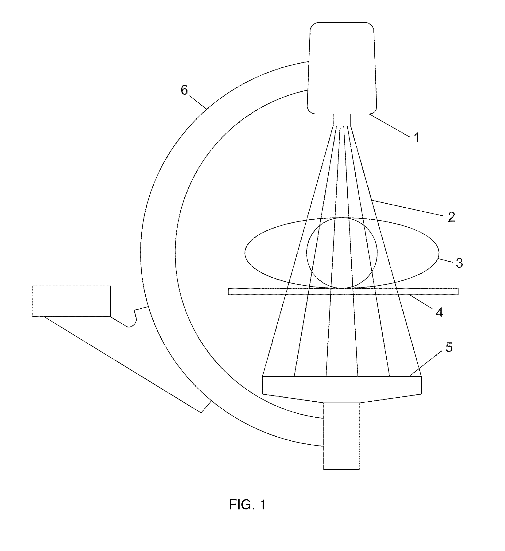
[0053]Acquisition of digital x-ray images is performed, for example, by means of the x-ray device shown in FIG. 1. It comprises x-ray tube 1 which emits a flow of x-rays 2. X-rays 2 go through the patient's body 3 placed on the table 4 and enter the receiver 5. The receiver 5 provides conversion of x-rays into a digital image. In one of the possible versions of embodiment the receiver can comprise a scintillating screen (not shown) converting x-rays into visible light and a photosensitive matrix array (not shown). The x-ray tube 1 the receiver 5 are fixed on the support 6 having 4 degrees of freedom concerning motion against the table.

[0054]According to the claimed method patient's exposure resulted in acquisition of pre-contrast series of N digital images (where N≧1). Having injected contrast agent into patient's vessel system one can obtain post-contrast series of M digital images (where M≧1). For every image of post-contrast series one can select an appropriate reference image wh...


PUM


Abstract

The invention relates to the field of digital processing of x-ray images and can be used in digital subtraction angiography to compensate for impact of involuntary patient movement and movement of internal organs on image of the vascular system. The technical result of the claimed invention is the improvement of diagnostic value of subtraction angiographic images by eliminating artifacts caused by the motility of anatomical structures. Technical result is achieved by the that at the stage of digital images registration a search for characteristic details is performed for each image. According to the shift of the said details determine expectable shift of a patient organs. Then perform segmentation of image from the pre-contrast series in the region of homogenous warping; for each region a geometrical transformation is calculated and corresponding geometrical transformations are performed for each region of a series of pre-contrast digital images.

Description

RELATED APPLICATIONS[0001]This application claims priority to Eurasian Patent Application No. EA201200924, filed Jul. 10, 2012, which is incorporated herein by reference in its entirety.FIELD OF THE INVENTION[0002]The invention relates to the field of digital X-ray image processing and can be applied to digital subtraction angiography aimed at compensating the impact of patient's involuntary movement and its internal organs normal movement on images of the vascular system.BACKGROUND OF THE INVENTION[0003]An angiography is a medical imaging technique used to visualize the inside of blood vessels based on injecting a contrast medium into the patient's blood vessels under examination. Besides vessels in the angiograms can be seen the surrounding organ and tissue images that often interfere with recognizing vessels filled with contrast medium. In order to make surrounding tissue less visible in digital subtraction angiography (DSA) they generate a pre-contrast image that is an x-ray ima...

Claims


Application Information

Patent Timeline
IPC IPC(8): G06T5/50
CPCG06T5/50G06T2207/10116G06T2207/20012G06T2207/20016G06T2207/20224G06T2207/30101G06T7/32A61B6/00G06T5/00
Inventor AFANASENKO, ARSENIY SERGEEVICH
Owner ZAKRYTOE AKCIONERNOE OBSHCHESTVO IMPULS
Why Eureka
Try Eureka