

Stereoscopic endoscope system
a stereoscopic endoscope and endoscope technology, applied in the field of stereoscopic endoscope systems, can solve the problems of white halation likely to interfere with observation, endoscope image obtaining method does not take into account the processing method of reducing white halation in a compound-eye stereoscopic endoscope, and the removal of white halation overlaps the area of white halation generated in images of at least one image sensor, etc., to achieve the effect of whi
- Summary
- Abstract
- Description
- Claims
- Application Information
AI Technical Summary
Benefits of technology
Problems solved by technology
Method used
Image

Examples
example 1
[0055]Example 1 according to the exemplary embodiment will be described hereinafter.
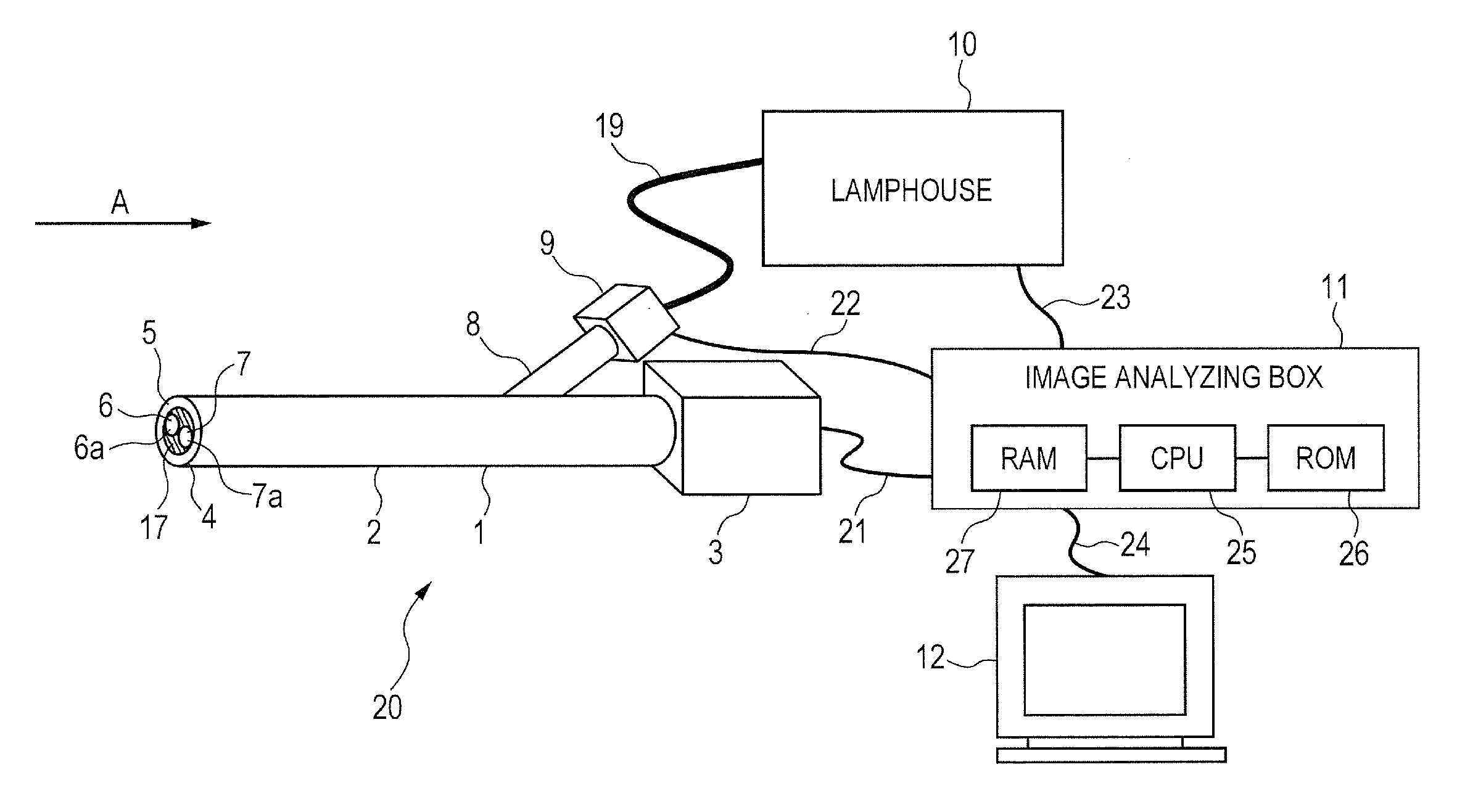
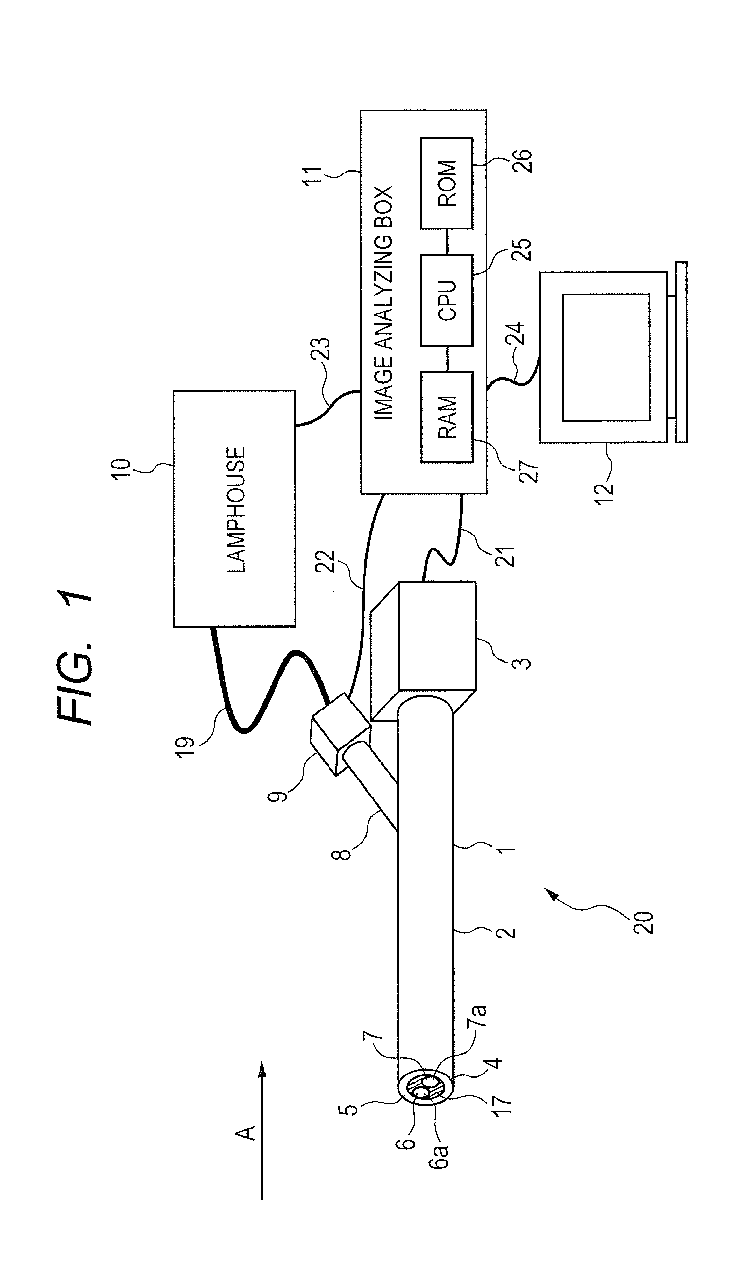
[0056]The stereoscopic endoscope system 20 used in Example 1 employs a twin-lens rigid stereoscopic endoscope having a diameter of 10 mm and a length of 250 mm for the endoscope 1. Moreover, a CCD of 960×540 (=518,400) pixels is used for each of the right-eye-side imaging system 6 and the left-eye-side imaging system 7 and a xenon lamp of 300 W is used for the lamp housed in the lamphouse 10.
[0057]In addition, a liquid crystal shutter unit is used as a shutter 9. The light distribution was changed by shielding a part of the optical fiber bundle and controlling the brightness of the lamp in the lamphouse 10 with a change in the applied voltage.
[0058]FIG. 3 is a flowchart illustrating imaging processing of an observed portion of a subject using the stereoscopic endoscope system 20 of Example 1. A program for the imaging processing is stored in the ROM (storage medium) 26. The CPU 25 reads the program f...
example 2
[0078]Subsequently, Example 2 will be described.
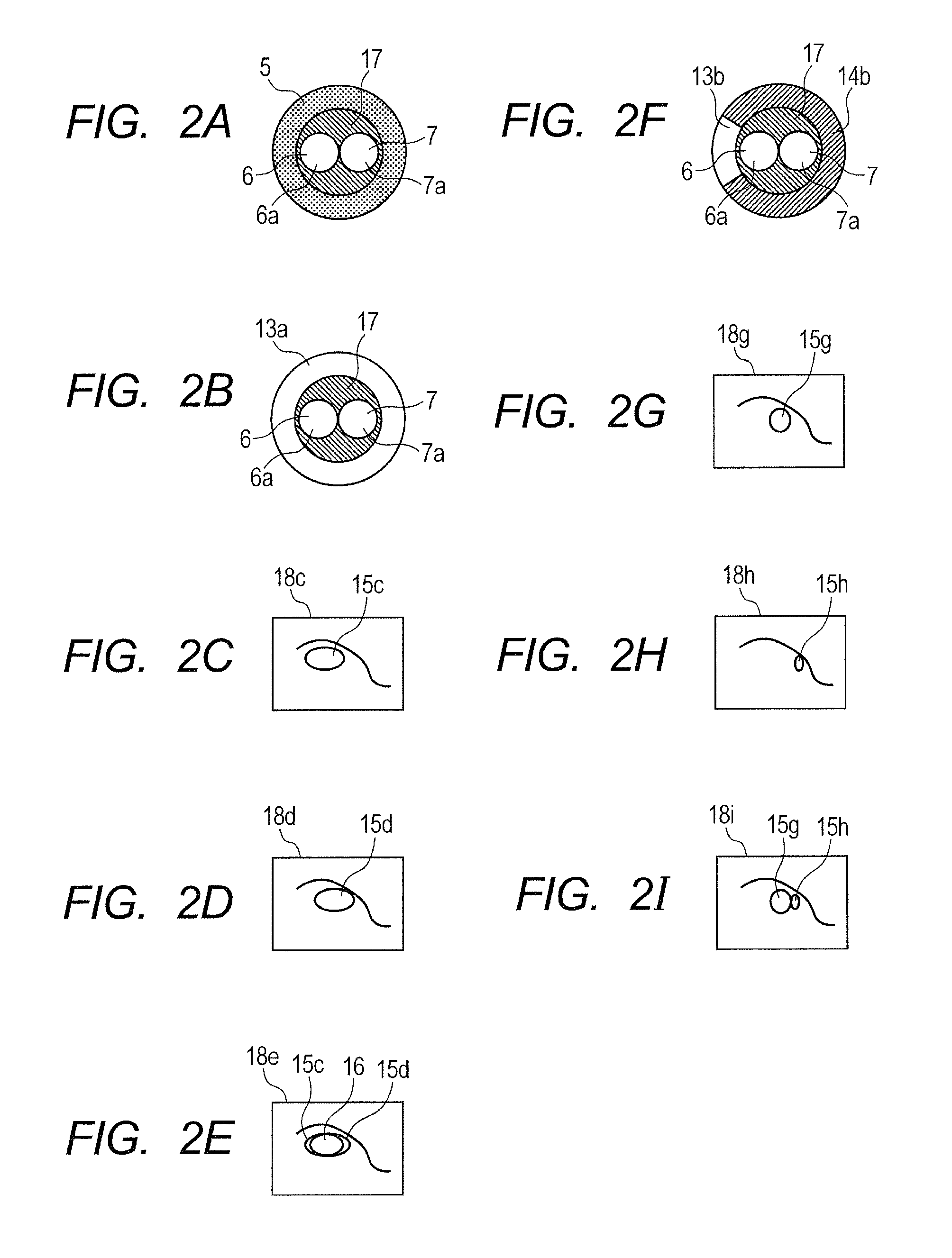
[0079]In Example 2, unlike Example 1, it is assumed that a light distribution to be selected next should be determined according to a place where the overlapped area between white halation images occurs when selecting a new light distribution. Specifically, when an image of a convex observed portion is taken in the center, an algorithm according to the location where the overlapped area between white halation images occurs was employed based on a database showing that moving the observed portion to the right is effective to reduce the overlapped area between the white halation images.
[0080]In concrete terms, five types of new light distributions were selected on the right side of the observed image if there are many overlapped areas on the right side of the observed image, and five types of new light distributions were selected on the left side of the observed image if there are many overlapped areas on the left side of the observed im...
PUM
 Abstract
Description
Claims
Application Information