Patient-Specific Segmentation, Analysis, and Modeling from 3-Dimensional Ultrasound Image Data

a patient-specific segmentation and ultrasound technology, applied in image analysis, image enhancement, instruments, etc., can solve the problems of limited tools for analyzing patient data and preoperative imagery for surgeons and cardiologists

Inactive Publication Date: 2014-03-13
THE JOHN HOPKINS UNIV SCHOOL OF MEDICINE
View PDF9 Cites 72 Cited by
  • Summary
  • Abstract
  • Description
  • Claims
  • Application Information

AI Technical Summary

Benefits of technology

[0007]Disclosed herein are methods and systems to analyze and predict patient-specific physiological behavior of a human organ or anatomical entity such as the heart complex and the heart subco

Problems solved by technology

Surgeons and cardiologists have limited tools for analyzing patient data and preoperative imagery, an

Method used


Image

  • Patient-Specific Segmentation, Analysis, and Modeling from 3-Dimensional Ultrasound Image Data
  • Patient-Specific Segmentation, Analysis, and Modeling from 3-Dimensional Ultrasound Image Data
  • Patient-Specific Segmentation, Analysis, and Modeling from 3-Dimensional Ultrasound Image Data

Examples


example implementations

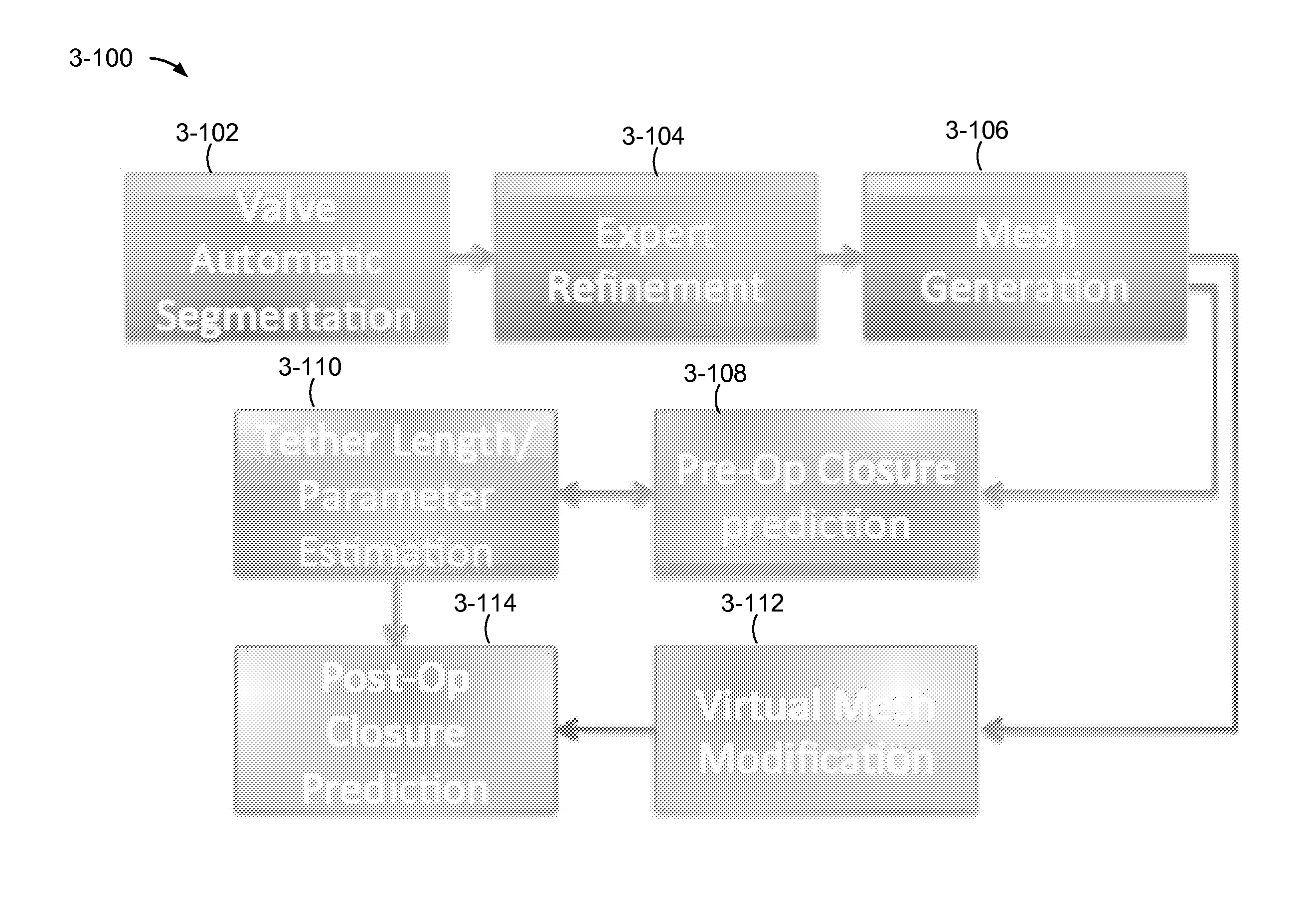
VI. Example Implementations

[0616]Methods and systems disclosed herein may be implemented in a system and / or machine, which may include hardware, firmware, a computer system, and combinations thereof, including discrete and integrated circuitry, application specific integrated circuits (ASICs), and / or microcontrollers, and may be implemented as part of a domain-specific integrated circuit package or system-on-a-chip (SOC), and / or a combination of integrated circuit packages.

[0617]FIG. 6-1 is a block diagram of a computer system 6-100, configured to process ultrasound data, such as 3DTEE.

[0618]Computer system 6-100 includes one or more computer instruction processor units and / or processor cores, illustrated here as a processor 6-102, to execute instructions of a computer program, which may also referred to herein as software, code, and / or computer program logic. Processor 6-102 may include a general purpose instruction processor, a controller, a microcontroller, or other instruction-b...


PUM


Abstract

Methods and systems to analyze and predict patient-specific physiological behavior of a human organ or anatomical entity such as the heart complex and the heart subcomponents from 3-dimensional volumetric ultrasound (3D) or time-sequential volumetric (4D) ultrasound image data, to assist physicians in performing diagnostics and cardiac preoperative planning. Also disclosed herein are methods and systems to segment patient-specific anatomical features from 3D/4D ultrasound. Also disclosed herein are methods and systems to compute patient-specific tissue motion and blood flow from 3D/4D ultrasound and contrast-enhanced 3D/4D ultrasound image data. Also disclosed herein are methods and systems to simulate the patient-specific mechanical behavior of the organ and anatomical entity using both 3D/4D ultrasound and mechanical models. Also disclosed herein are methods and systems to estimate tissue stress and strain and physiological parameters of the tissues from 3D/4D ultrasound.

Description

CROSS REFERENCE TO RELATED APPLICATIONS[0001]U.S. Provisional Patent Application No. 61 / 422,778, titled, “Estimating Blood Flow Motion in the Left Intraventricular Cavity using Contrast Enhanced Ultrasound,” filed Dec. 14, 2010, is incorporated herein by reference in its entirety.BACKGROUND[0002]1. Technical Field[0003]Patient-specific preoperative planning based on segmentation, motion analysis, simulation and modeling of anatomical entities and organs from 3-dimensional volumetric (3D) ultrasound image data and 3-dimensional volumetric time-sequenced (4D) ultrasound image data; Preoperative planning for heart and valve surgery (valvuloplasty); Patient-specific simulation, modeling, and prediction of the mechanical behavior of anatomical entities and organs from 3D and 4D ultrasound image data; Determination of tissue motion from 3D / 4D ultrasound and blood motion flow from contrast-enhanced 3D / 4D ultrasound (CEUS) image data.[0004]2. Related Art[0005]Surgeons and cardiologists have...

Claims


Application Information

Patent Timeline
IPC IPC(8): G06T17/00
CPCG06T17/00G06T2207/10076G06T2207/10136G06T2207/30048G06T7/246G06T7/00
Inventor BURLINA, PHILIPPE M.MUKHERJEE, RYAN N.SPROUSE, CHAD R.
Owner THE JOHN HOPKINS UNIV SCHOOL OF MEDICINE
Why Eureka
Try Eureka