Clear margin determination during cancer surgery

a cancer and surgical technique, applied in the field of specific moieties of biomarkers, can solve the problems of no reliable intraoperative margin assessment method, surgical procedures, additional cost, time and pain for patients,

Active Publication Date: 2014-03-20
DREXEL UNIV
View PDF11 Cites 4 Cited by
  • Summary
  • Abstract
  • Description
  • Claims
  • Application Information

AI Technical Summary

Problems solved by technology

Generally, re-excision is required if the tissue margin is found to contain cancer cells, which often results in additional cost, time, and pain to the patients.
Currently there is no reliable intraoperative margin assessment method.
Conventional surgical procedures, such as preparation cytology (hereinafter “TPC”) and frozen section analysis (hereinafter “FSA”), do not provide sufficiently reliable findings.
The reliability of FSA and TPC are questionable due to histological artifacts.

Method used


Image

  • Clear margin determination during cancer surgery
  • Clear margin determination during cancer surgery
  • Clear margin determination during cancer surgery

Examples


example i

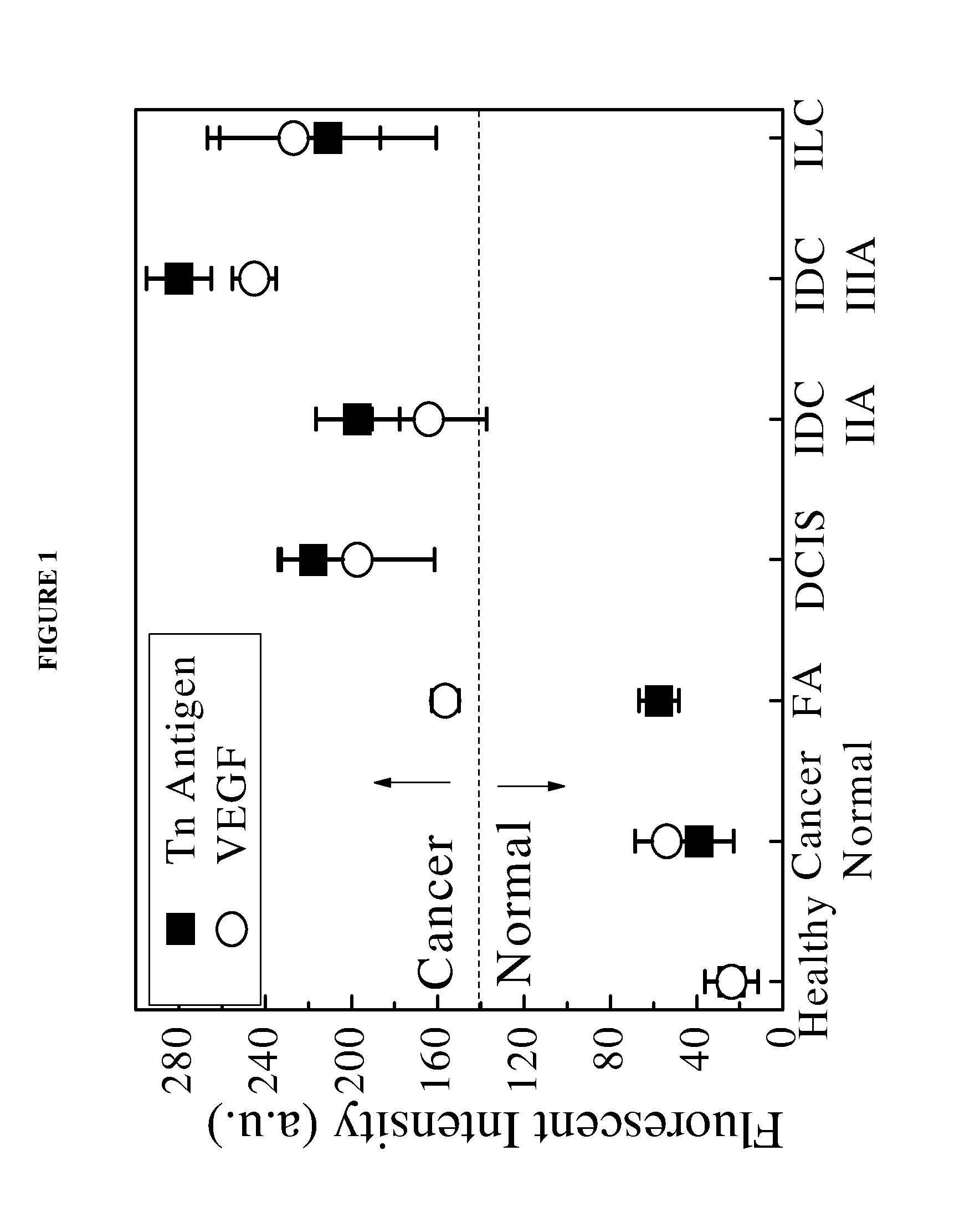
[0063]Aqueous CdSe QDs conjugated with anti-Tn and antibodies anti-VEGF antibodies were used to image HT-29 colon cancer cells in the margin of excised tissue samples. An aqueous solution of the conjugated QDs was applied to pathological excised tissue sample slides containing tumor masses. The intensity of the photoluminescence was measured to determine if cancer cells were present on the surface of or embedded within the tissue margins.

[0064]In this study, breast tissue sections were obtained from multiple patients. Each breast tissue section was divided into three regions: tumor mass, normal surrounding tissue and margin area in which the tissue contained both tumor and normal tissue. Within each region, multiple blocks were cut by pathologists. For each patient sample, two blocks from six margin areas containing tumor mass and normal tissue were examined. The sample tissue blocks were embedded in paraffin, cut to a thickness of about 5 μm and subsequently mounted on glass slides...

example ii

Antibody Conjugated MPS replaced MPA Capped Cd0.3Pb0.7S Quantum Dots

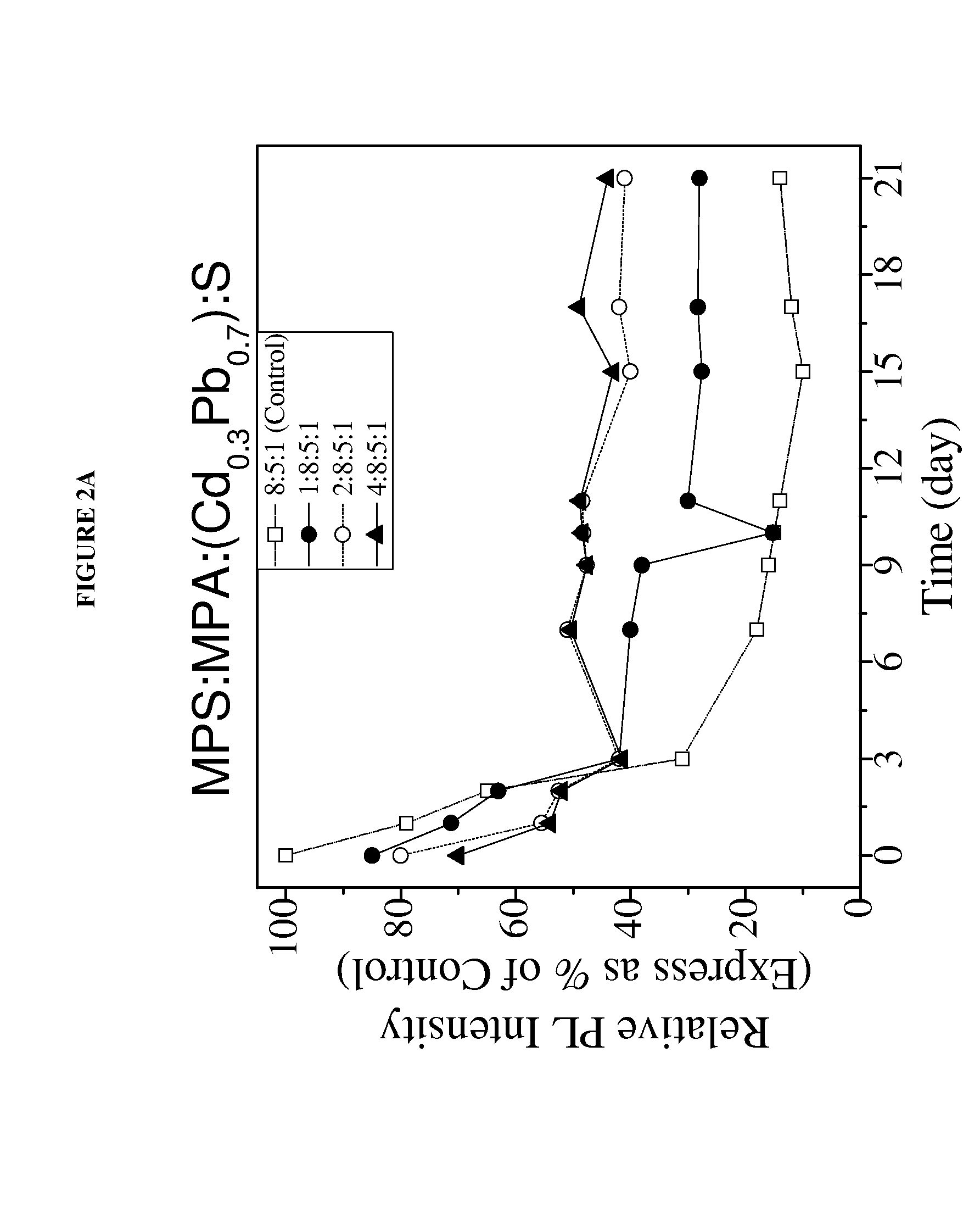
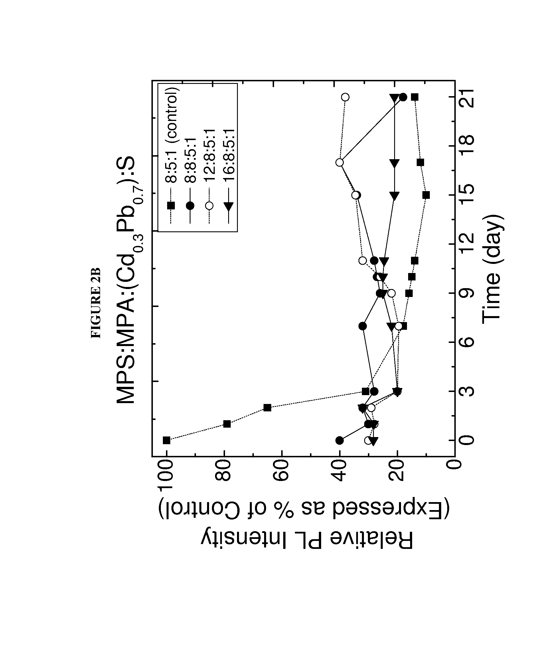
[0076]In this study MPS replaced MPA capped Cd0.3Pb0.7S QDs of the present invention were synthesized. To optimize photoluminescent intensity, the CdPbS QD was synthesized to have an MPA:cation:anion molar ratio of about 8:5:1. The synthesized Cd0.3Pb0.7S QD having a 8:5:1 MPA:[excess Cd+(Cd1-xPbx)]:S molar ratio was stable for a sufficient period of time to enable the detection of cancer cells within the sample excised tissues. To further improve the stability of the QD, some of the MPA capping molecules were replaced with MPS capping molecules. FIG. 2 shows various MPS replaced MPA capped Cd0.3Pb0.7S. These QDs were made by reacting a predetermined number of moles of MPS (as indicated by the first of the four numbers) to the 8:5:1 MPA capped Cd0.3Pb0.7S QDs. The MPS replaced QDs were found to remain stable for at least 20 days, maintaining a photoluminescent intensity of at 50% of the initial photoluminescent inte...

example iii

Cancer Detection Study Using Biotinylated Antibody Conjugated to MPS Replaced MPA Capped CdSe QDs Using SMCC and SA

[0079]Biotinylated antibodies were conjugated to MPS replaced MPA capped CdSe QDs having an 8:5:1 molar ratio of MPA:Cd:Se using the conjugation linkers, sulfosuccinimidyl 4[N-maleimidomethyl]cyclohexane-1-carboxylate (SMCC) and streptavidin (SA) to form a biotin-streptavidin conjugation scheme. The conjugated QDs were used to detect the presence of cancer cells within the margins of breast tissue excised from breast cancer patients.

[0080]SMCC and SA were used to bind biotinylated antibodies to the MPS replaced MPA capped CdSe QDs in a biotin-streptavidin conjugation scheme. SMCC was bound to MPS replaced MPA capped CdPbS QDs and was also bound to SA, which in turn was bound to a biotinylated antibody. SMCC was allowed to react with the QD at a pH of about 7 for an initial period of about 45 minutes at about room temperature. Excess SMCC was then removed from the soluti...


PUM

PropertyMeasurementUnit
excitation wavelengthaaaaaaaaaa
excitation wavelengthsaaaaaaaaaa

Abstract

One or more aqueous, near infrared emitting, high yield, highly photoluminescent, stable quantum dots conjugated to one or more biomarkers specific moieties. The conjugated quantum dots have an enhanced detection sensitivity and selectivity and may be formed using a novel and efficient method for conjugating one or more biomarker specific moieties to the quantum dots. The invention is further directed to a method for using the conjugated quantum dots for cancer detection in the margin of excised tissue.

Description

STATEMENT OF GOVERNMENT INTEREST[0001]This invention was reduced to practice with Government support under Grant No. W81XWH-09-1-0701 awarded by the Department of Defense; the Government is therefore entitled to certain rights to this invention.BACKGROUND OF THE INVENTION[0002]1. Field of the Invention[0003]The present invention relates to biomarker specific moieties conjugated quantum dots capable of highly sensitivity and selective biomarker detection.[0004]2. Brief Description of the Prior Art[0005]Breast cancer surgery relies on accurately assessing whether cancer cells are present in excised tissue to ensure complete tumor removal. As expected, the local cancer recurrence rate is higher for tissue margins testing positive for cancer cells within 3 mm of the margin surface (16%) in comparison to tissue margins that test negative (6%). Generally, re-excision is required if the tissue margin is found to contain cancer cells, which often results in additional cost, time, and pain t...

Claims


Application Information

Patent Timeline
Patent Type & Authority Applications(United States)
IPC IPC(8): G01N33/574
CPCG01N33/574G01N33/532G01N33/588B82Y15/00G01N33/57492Y10S977/774Y10S977/824Y10S977/892Y10S977/927G01N33/551B82Y5/00B82Y40/00C07K16/22C07K16/30G01N33/533G01N33/57484G01N33/57488G01N2333/475G01N2333/705G01N2400/00G01N2458/00
Inventor SHIH, WEI-HENGSHIH, WAN Y.AU, GIANGBROOKS, ARI D.SWAMI, VANLILA K.
Owner DREXEL UNIV
Why Eureka
Try Eureka