
Method and medicament for inhibiting lymphangiogenesis

Inactive Publication Date: 2014-07-10
TSINGHUA UNIV +1
View PDF0 Cites 3 Cited by
  • Summary
  • Abstract
  • Description
  • Claims
  • Application Information

AI Technical Summary

Benefits of technology

The patent describes a new treatment that can inhibit the growth of tumor lymphatic vessels and prevent the cancer from spreading to the lymph nodes. The treatment involves blocking two pathways, one controlled by a chemokine called CXCL12 and the other controlled by a growth factor called VEGF-C. This combination treatment is more effective than using just one substance alone. The patent also describes using antibodies to neutralize these substances and preventing tumor lymphangiogenesis and metastasis in mice.

Problems solved by technology

However, due to the lack of specific markers that can distinguish blood vessels from lymphatic vessels, intensive studies on lymphangiogenesis and the functions of lymphatic vessels have been carried out for no more than 20 years.
Tumor metastasis is the leading cause of cancer death.
However, it has not yet been fully and clearly investigated how lymphangiogenesis is induced in tumor tissues and what the regulation mechanisms are.

Method used


Image

  • Method and medicament for inhibiting lymphangiogenesis
  • Method and medicament for inhibiting lymphangiogenesis
  • Method and medicament for inhibiting lymphangiogenesis

Examples


example 1

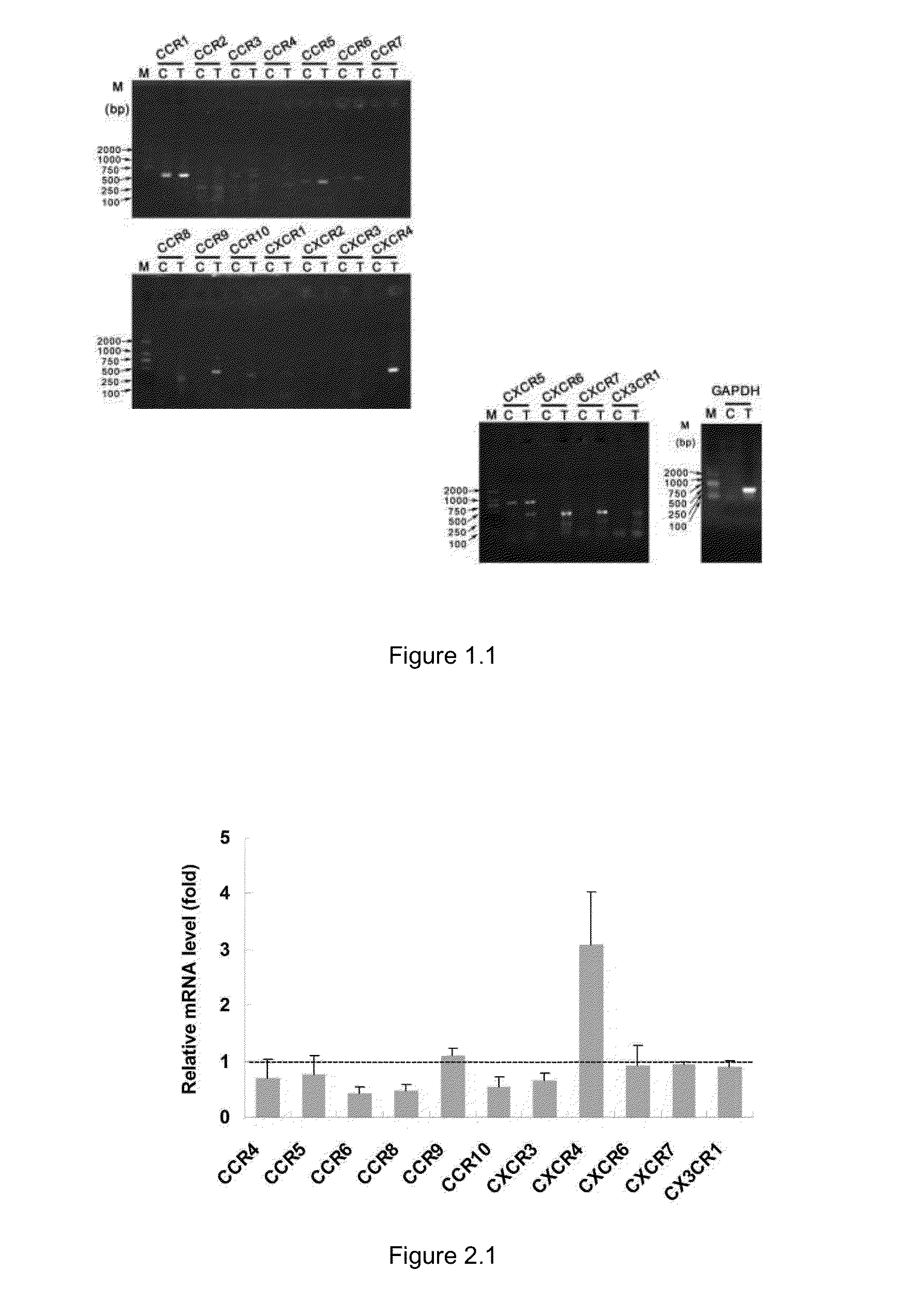
Lymphatic Endothelial Cells Express a Variety of Chemokine Receptors

Methods

[0070]1. Extraction of Total RNA from Cells and Detection of Chemokine Receptor Expression in Lymphatic Endothelial Cells by RT-PCR

[0071]The isolation and extraction of total RNA from cells was performed using TRIZOL reagent (purchased from Invitrogen) following the standard operations as described in the reagent instructions. 1 mL of TRIZOL was added to the primary lymphatic endothelial cells (about 1×10⁶) collected by centrifugation, repeatedly pipetted up and down for 30 times, and then set aside at room temperature for 5 minutes. After centrifugation at 10,000×g at 4° C. for 15 minutes, the supernatant was removed gently. 0.2 mL of chloroform was added to the supernatant, vortexed vigorously for about 15 seconds, and then set aside at room temperature for 3 minutes. After centrifugation at 10,000×g at 4° C. for 15 minutes, the sample was stratified into three layers, i.e., a yellow organic phase, an inter...

example 2

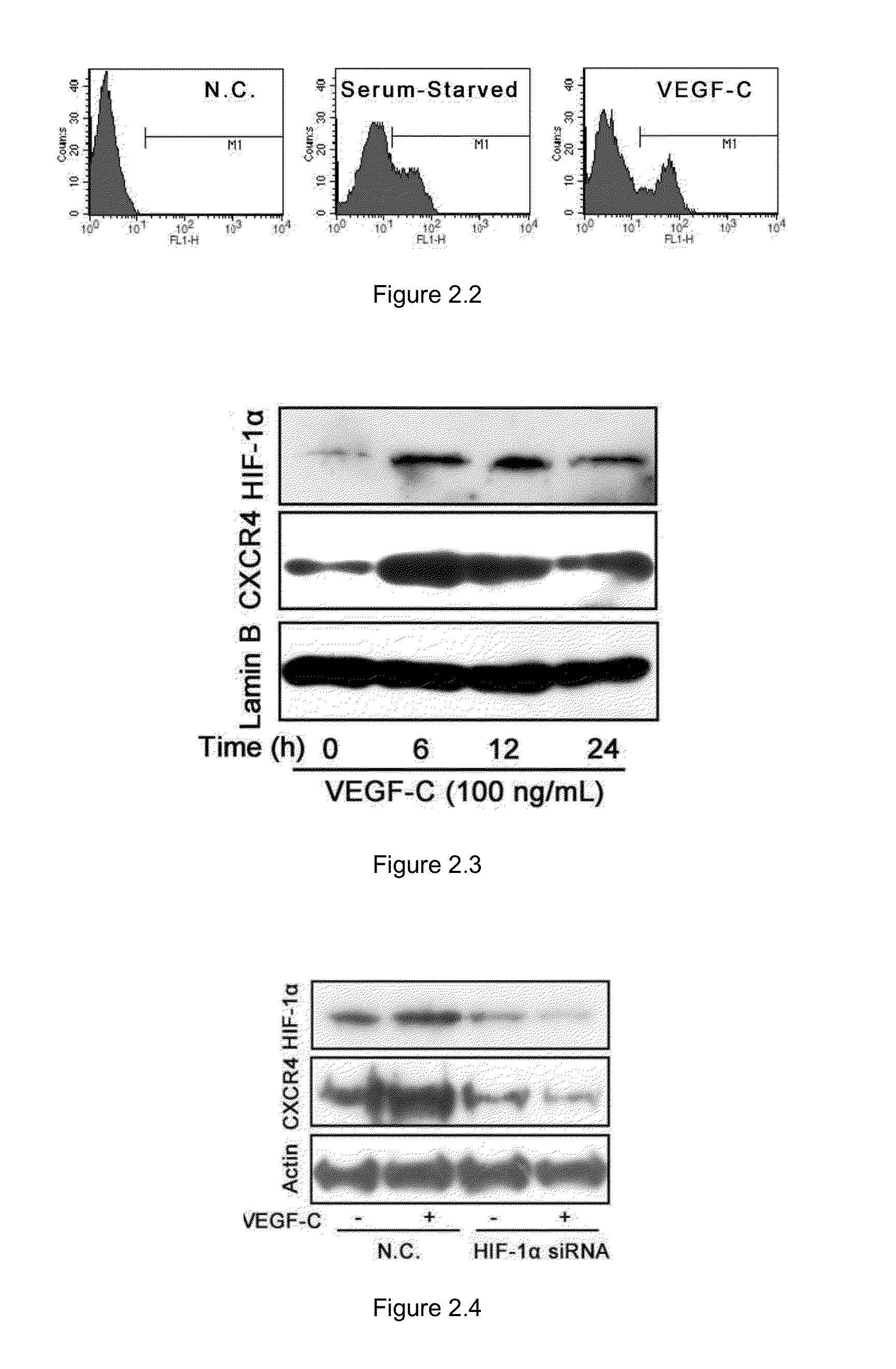
Chemokine Receptor CXCR-4 is Highly and Specifically Expressed on VEGF-C-Activated Lymphatic Endothelial Cells

Methods

1. Detection of the Expression of the Chemokine Receptors on Lymphatic Endothelial Cells by RT-PCR

[0075]Fluorescence Quantitative Real-Time PCR was performed using a kit from Stratagene (Brilliant II SYBR® Green QRT-PCR Master Mix). The fluorescence quantitative PCR instrument was MX3000P (purchased from Stratagene), the fluorescence dye was SYBR Green, the volume of the reaction system was 20 μL, and the cycle number of the reaction was 40. GAPDH was used as an internal control. The ΔCt value was obtained from the fluorogram provided by the instrument, and the relative Δ(ΔCt) value was calculated, thereby calculating the relative change in the level of the corresponding gene.

2. Verification of Vascular Endothelial Growth Factor C (VEGF-C)-Induced Expression of Chemokine Receptor CXCR4 on the Surface of Lymphatic Endothelial Cells by Flow Cytometry

[0076]Mouse primary ...

example 3

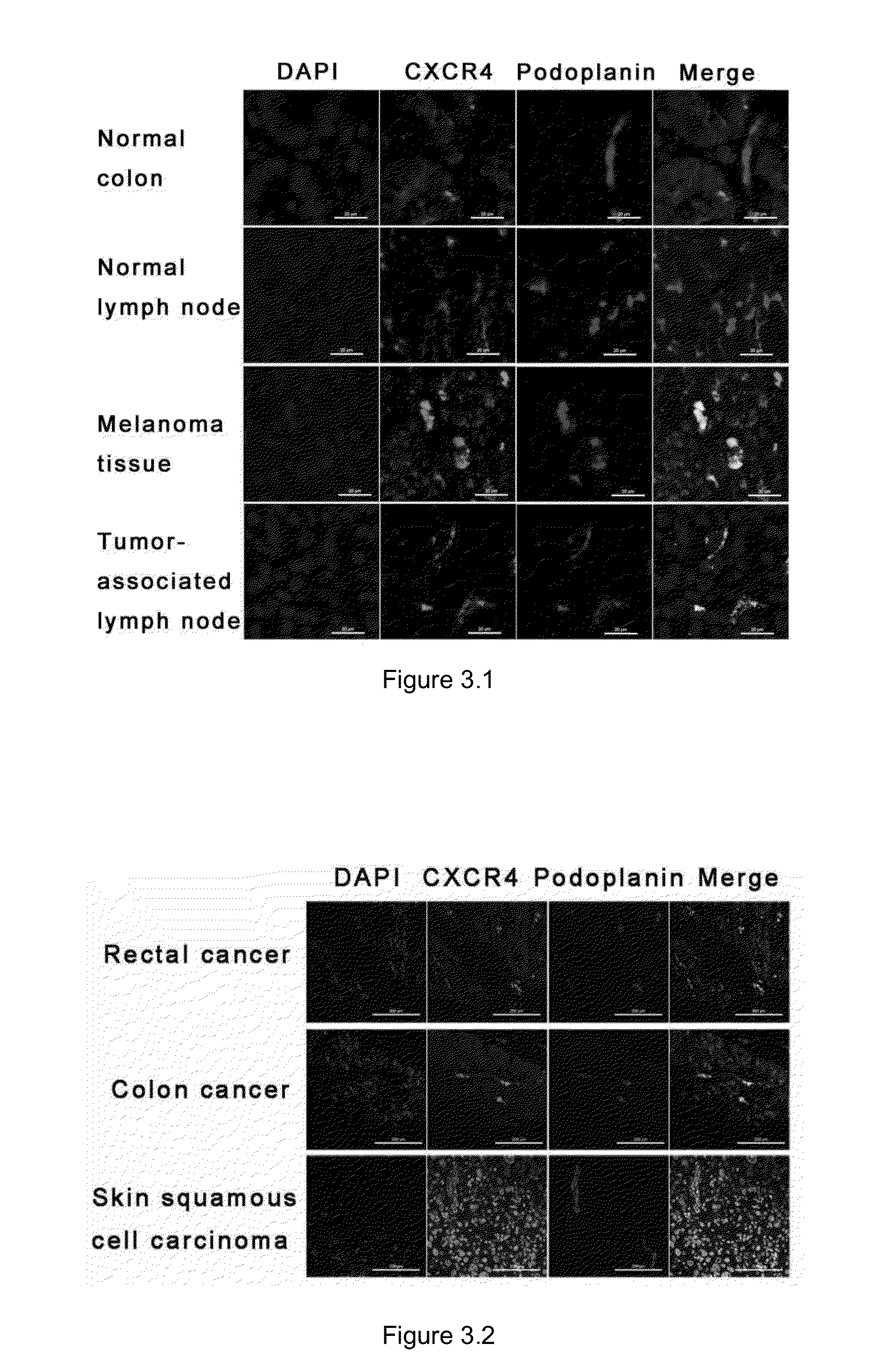
CXCR4 is Highly and Specifically Expressed on the Surface of Newly Formed Lymphatic Vessels In Vivo

Methods

1. Detection of the Distribution of Chemokine CXCR4 on Lymphatic Vessels In Vivo by Tissue Immunofluorescence

[0089]Eight healthy C57BL / 6 mice (6-8 weeks old, female, purchased from Vital River Laboratories, Beijing) were divided into two groups, one of which includes three mice normally fed, and the other includes five tumor-bearing mice intracutaneously inoculated with 5×10⁶ B16 / F10 mouse melanoma cells (American Type Culture Collection, ATCC). 14 days after inoculation, the tumor tissues and peritumoral axillary lymph node tissues were removed from the tumor-bearing mice, and the colon tissues and lymph node tissues were removed from the normal mice.

[0090]Fixing and embedding: The removed tissues were fixed with 4% formaldehyde solution overnight, and then rinsed with tap water overnight to completely wash off formaldehyde (the tissue blocks could be wrapped with gauze, placed...


PUM


Abstract

The present invention provides a method for inhibiting lymphangiogenesis in a subject, comprising administering a therapeutically effective amount of a CXCR4 inhibitor and / or a CXCL12 inhibitor to the subject. The invention further provides a method for inhibiting tumor lymphatic metastasis in a cancer patient, comprising administering to the subject (a) a therapeutically effective amount of a CXCR4 inhibitor and / or a CXCL12 inhibitor, in combination with (b) a therapeutically effective amount of a VEGF-C inhibitor and / or a VEGF-D inhibitor and / or a VEGFR-3 inhibitor.

Description

FIELD OF THE INVENTION[0001]The present invention relates to the field of biopharmaceuticals, in particular, to a method and medicament for inhibiting lymphangiogenesis.INCORPORATION BY REFERENCE OF SEQUENCE LISTING[0002]The Sequence Listing in an ASCII text file, named 30339_SEQ.txt of 7 KB, created on Mar. 7, 2014, and submitted to the United States Patent and Trademark Office via EFS-Web, is incorporated herein by reference.BACKGROUND OF THE INVENTION[0003]In the body, blood vessels are responsible for delivering oxygen, nutrients and other substances to various tissues, and exchanging substances with surrounding tissues through capillaries. The presence of blood pressure causes plasma to leak continuously from capillaries into interstitial space, which is called interstitial fluid. The main function of lymphatic vessels is collecting and returning such protein-rich fluid to the blood circulation. Water, macromolecules and cells may be absorbed by the lymphatic capillaries locate...

Claims


Application Information

Patent Timeline
IPC IPC(8): A61K39/395
CPCA61K38/19C07K16/22C07K16/2863C07K16/2866A61K2039/505A61K2039/507C07K2317/76A61K38/179A61P29/00A61P35/00A61P35/04A61P37/06A61K2300/00
Inventor LUO, YONGZHANGZHUO, WEIJIA, LINFU, YANCHANG, GUODONG
Owner TSINGHUA UNIV
Learn More