

Volume rendering of medical images
a volume rendering and medical image technology, applied in the field of volume rendering of medical images, can solve the problems of not being able to visualize thickness metrics or fuse them with structural data, and the user's perception of volume rendered images is artificial,
- Summary
- Abstract
- Description
- Claims
- Application Information
AI Technical Summary
Benefits of technology
Problems solved by technology
Method used
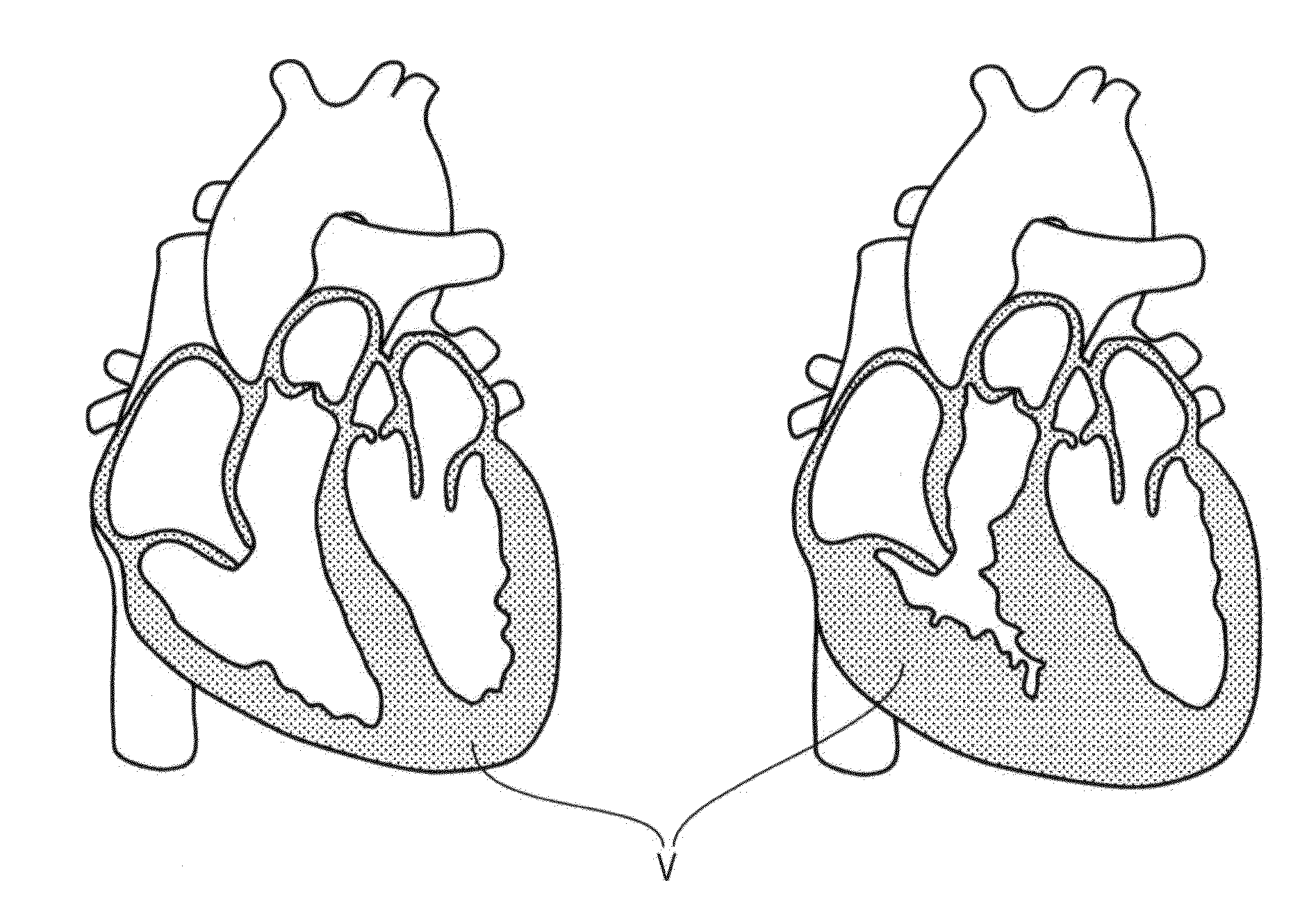
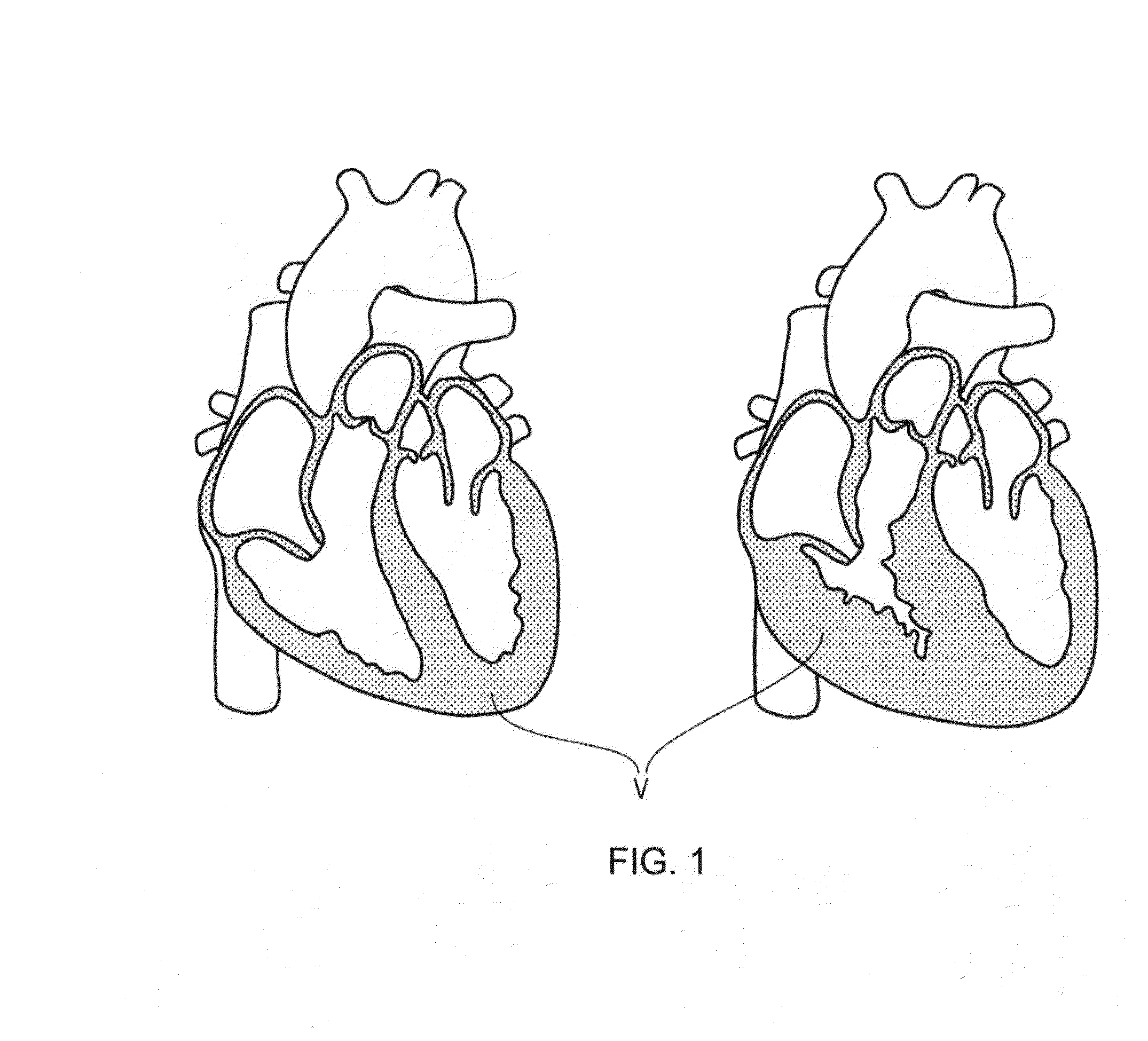
Image

Examples
Embodiment Construction
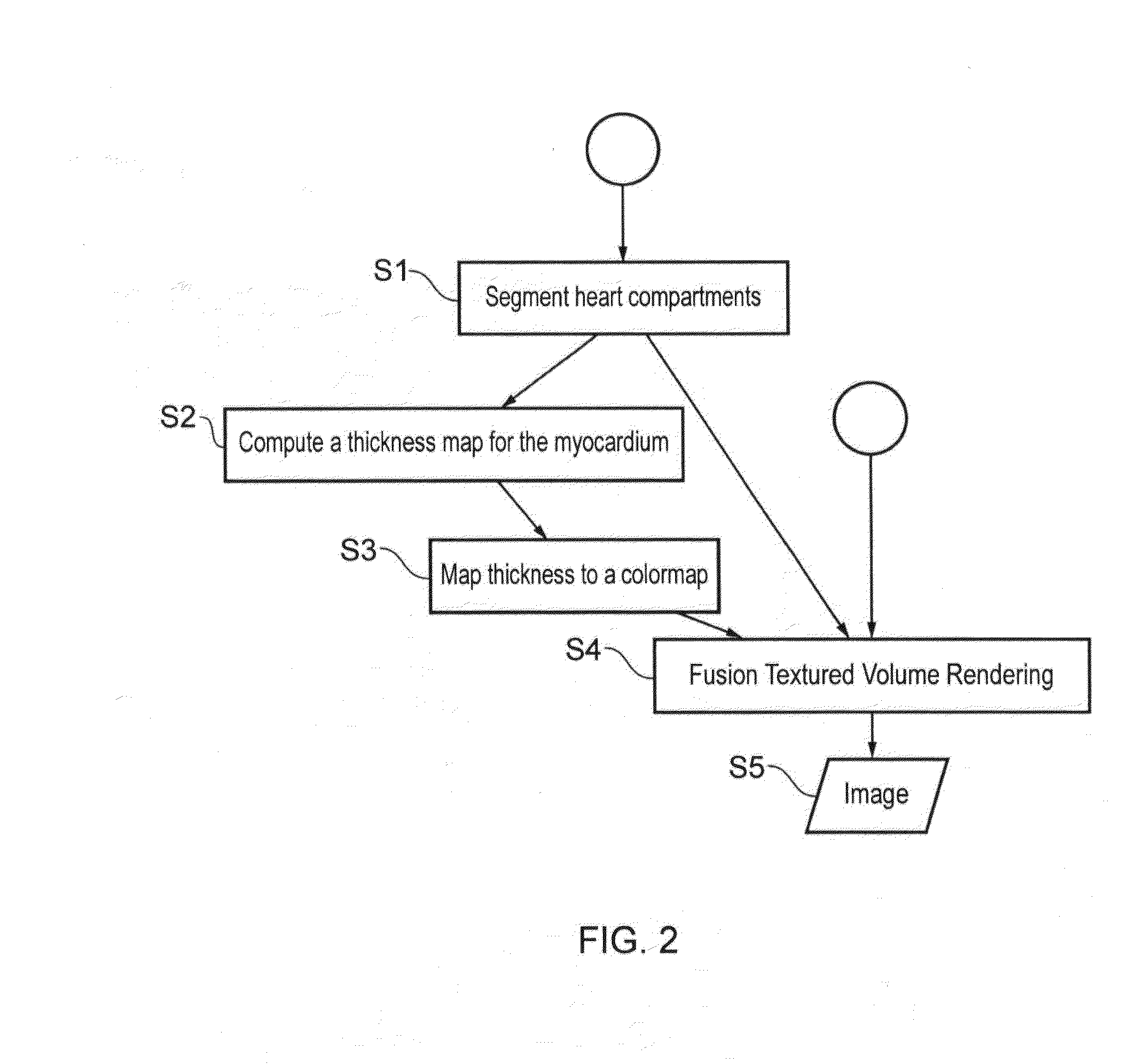
[0037]Certain embodiments of the invention provide a computer apparatus comprising: memory; a processor; a display; a network output connection and a rendering application loaded in the memory operable to cause the computer apparatus to:[0038]a) load a three-dimensional patient image data set of voxels which forms a scene for processing by the rendering application;[0039]b) assign one or more of the voxels to act as a light source, wherein said voxels are adjacent a region of tissue of interest;[0040]c) render an image of the scene from the perspective of a view point or view plane by calculating how light emitted from the light source voxel(s) travels through the tissue of interest to the view point or view plane using an optical model which includes absorption and scattering; and[0041]d) show the image on the display and / or store the image in the memory and / or output the image to the network output connection.
[0042]Certain embodiments of the invention provide a computer-automated ...
PUM
 Abstract
Description
Claims
Application Information