[0018]Reference now will be made in detail to various embodiments of the disclosed subject matter, one or more examples of which are set forth below. Each example is provided by way of explanation, not limitation. In fact, it will be apparent to those skilled in the art that various modifications and variations may be made in the present disclosure without departing from the scope or spirit of the subject matter. For instance, features illustrated or described as part of one embodiment, may be used on another embodiment to yield a still further embodiment. Thus, it is intended that the present disclosure covers such modifications and variations as come within the scope of the appended claims and their equivalents.
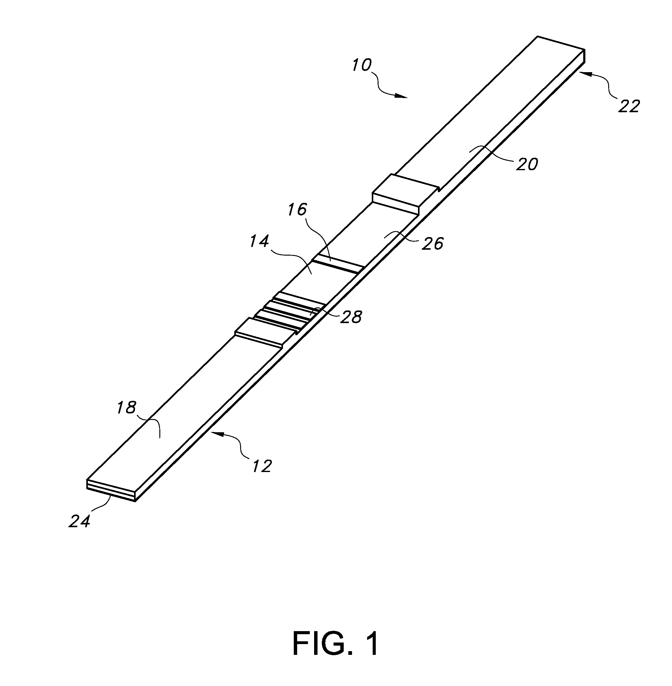
[0019]This disclosure describes a device to rapidly classify bacteria in a sample of a bodily fluid as either Gram positive (GP) or Gram negative (GN). Such fluids include respiratory fluids (e.g. from the lung, throat, nasal cavity), digestive fluids, blood, spinal fluids, etc. A sample from the lungs, for example, can originate from the upper airway (e.g. expectorated sputum), but is desirably from the lung. A bronchoalveolar lavage (BAL), both bronchoscopic and non-bronchoscopic/blind, is a common procedure used to acquire lung sputum samples from ventilated patients. One embodiment of this device interfaces directly with a sputum collection container, although it should be understood that it can be used as a stand-alone unit, either at the bedside or in the laboratory/field.
[0020]The device utilizes membrane-based lateral flow immunoassay (also known as the “assay”) technology to perform the Gram classification test. One or more multi-component test strips are used to receive the sample and perform the assay. In one embodiment of this device, two individual, discreet test strips are employed: one specifically designed for GN detection (FIG. 1) and one specifically designed for GP detection (FIG. 2). Both test strips incorporate protein detection via an antibody-antigen sandwich arrangement. It should be understood that other arrangements are possible (e.g. non-sandwich) and furthermore, that the individual GP and GN test strips are not required to utilize the same arrangement simultaneously.
[0021]Each test strip has both capture and detection antibodies, both of which may be the same. For both cases, matched antibody pairs are used. The antibodies can be either monoclonal or polyclonal and can be derived from any common host sources. One embodiment of the device includes two monoclonal antibodies targeting lipoteichoic acid (LTA) and peptidoglycan of S. aureus for GP detection and a combination of one monoclonal antibody targeting lipopolysaccharide (LPS) and one polyclonal antibody targeting multiple Klebsiella antigens for GN detection. Furthermore, the detection antibodies are desirably conjugated (attached) to colloidal gold nanoparticles and placed on the test strip to result in an assay with at least one visual signal. The nanoparticles and attached antibodies are known as “the conjugate”. Gold particles of approximately 40 nanometers (nm) in size are desirably used but other sizes between about 20 and 80 nm are feasible as well. Other possibilities with which to conjugate the detection antibodies include latex particles between about 100 and 300 nm, magnetic particles and fluorescent particles.
[0022]Each test strip 10, 40 is assembled and laminated from a series of discreet components. A backing card 12, 42 serves as the physical support for the other components, and this is commonly made from Mylar, though other materials may also be used. A porous matrix, 14, 44 e.g. nitrocellulose membrane, cellulose, porous polyethylene pads, and glass fiber filter paper is either laminated or directly cast onto a region of the backing card 12, 42. (The terms “nitrocellulose membrane” or “nitrocellulose” are used to represent suitable porous matrices.) The nitrocellulose contains the capture antibodies.
[0023]The capture antibodies are immobilized in a specific detection zone, desirably as a test line 16, 46 on the porous matrix 14, 44 via standard liquid dispensing equipment, including both contact and non-contact heads, as well as spraying. In one embodiment the capture antibodies form a stripe on and/or in the matrix to yield a test line 16, 46 of approximately 1 mm in width perpendicular to the longest dimension of the matrix. It has been demonstrated that larger pore size matrices improve sample flow through the system and allow for samples with whole cell organisms to be tested without the need for any sample processing steps. An exemplary nitrocellulose for this application has a measured flow rate between 40-75 cm/4 sec in the longest dimension of the matrix. Specific commercial examples include Prima 40 (GE Whatman) and 70CNPH-N-SS40 (MDI).
[0024]In one embodiment, a conjugate pad 18, 48 is used to receive the sample. As can be seen in FIGS. 1 and 2, the conjugate pads 18, 48 are supported on their respective backing cards 12, 42 proximal to the porous matrix 14, 44. The conjugate pad 18, 48, which may be glass fiber (e.g. G041 Glass Fiber Conjugate Pads, from Millipore), is used to initially contain the conjugate (e.g. detection antibodies and gold nanoparticles). The conjugate is immobilized on the conjugate pad 18, 48 until coming into contact with the sample. The conjugate can be placed onto the conjugate pad using a liquid carrier by several methods including spraying, striping, and soaking. In addition to the conjugate, the conjugate pad 18, 48 can have dried chemicals (e.g. surfactants) that aid in sample flow, particularly for those samples with high viscosity, elasticity, or organism counts. The addition of chemicals can be accomplished with the same methods. Lastly, there is a physical overlap and contact between the conjugate pad 18, 48 and the matrix 14, 44 that allows for fluid communication between them and allows the assay to run correctly.
[0025]Alternative embodiments may have an additional and separate sample pad positioned at the proximal end of the strip to receive the sample and be in physical contact with the conjugate pad to allow for fluid flow through the test strip. The sample pad may be made of conventionally known materials, e.g. commonly available cellulose materials.
[0026]A wicking pad 20, 50 for each strip 10, 40 is located at the distal end 22, 52 of the backing card 12, 42 and is used to draw the fluid sample through the system by capillary action. The sample is deposited near or on the proximal end 24, 54 of the test strip 10, 40. The wicking pad 20, 50 is an absorptive material and can be made via a number of different cellulosic materials and can vary in size and absorptive capacity as generally conventionally known. There is an overlap between the wicking pad 20, 50 and the porous matrix 14, 44 that allows for fluid communication. A suitable example material is A22 Absorptive Paper (Ahlstrom).
[0027]In some embodiments, a “running buffer” reagent in liquid form is used to help facilitate sample flow through the test strip 10, 40 and promote even, uniform color change in the test line. This buffer is water-based and has proteins (e.g. Bovine serum albumin, BSA) and surfactants (e.g. Tween 20). It is desirably mixed with the sample prior to addition to the test strip 10, 40 either via mechanisms that are part of the sampling device or through stand-alone actions such as pipetting. Alternatively, it is possible to add the running buffer to the test strip 10, 40 immediately after the sample has been introduced to the test strip 10, 40 either via mechanisms that are part of the device or through stand-alone actions such as pipetting.
[0028]In one format, a 2:1 ratio is used; 2 parts sample to 1 part running buffer, although other combinations are possible, including a range between 4:1 and 1:2 (sample: running buffer). In some embodiments 100 μl of sample is mixed with 50 μl of running buffer for each assay. The total volume of fluid applied to a single test strip can vary depending on the material selection of individual test strip components.
[0029]One feature unique to the GN (FIG. 1) assay is the need to prevent GP organisms cross-reacting to the GN antibodies on the GN test strip 10. There are several potential causes for this cross-reaction, including Protein A from Staphylococcus species and Protein G from Streptococcus species. Several arrangements have been found to be effective at reducing the GN test line signal from these agents; in some embodiments, targeting antibodies are immobilized on the test strip 10 in a zone prior to the test line 16 with respect to the proximal end 24 of the strip 10, and are termed “guard lines”28 as illustrated in FIG. 1. Suitable antibodies for these guard line can bind either Protein A and Protein G specifically to capture them, or can effectively neutralize the reactivity of whole cell Staphylococcus and whole cell Streptococcus. Additionally, other anti-GP antibodies (e.g. Rabbit polyclonal antibodies developed against whole cell Enterococcus faecium)) have been shown to be effective. There is no preference of antibody type (e.g. monoclonal vs. polyclonal, host animal, etc.) for this application. By using this configuration, the cross-reactive factors present in the sample are “trapped” before the test line 16 and thus do not contribute to the test line signal. One or more of these guard lines 28 can be incorporated into the test strip 10 on the nitrocellulose via striping, spraying, or other similar conventionally known methods.
[0030]Another way of preventing GP organisms from cross-reacting to the GN antibodies is to use targeting antibodies in liquid form instead of immobilizing them in a defined zone on the test strip. In this embodiment, these targeting antibodies are introduced to the sample prior to addition to the test strip and the sample must be segregated between test strips 10, 40 so as to not interfere with the GP assay. Addition to the sample of targeting antibodies in a liquid carrier does not require additional mixing or incubation time, although the inclusion of both is possible.
[0031]Another way to reduce GP organism cross-reactivity on GN test strips is to modify the physical structure of the capture antibodies in the test line. Protein A and Protein G are known to bind to the FC portion of the antibodies on the test line. Removing this portion and only including the remaining F(ab′)2 region in the antibodies of the test line 16 has been demonstrated to be effective at removing the cross-reactive signal while maintaining the true detection of GN organisms. This modified antibody (FC portion removed), once prepared, can be applied to and immobilized on the test strip 10 in the same manner as unmodified antibodies. Lastly, as it relates to this cross-reactivity, it should be noted that any combination of these embodiments can be employed together and simultaneously.
[0032]Confirmation that the appropriate amount of sample and conjugate have passed through the system is also desirable. This confirmation can involve a physical zone on the test strip 10, 40 distal to the test line 16, 46 that forms a visible signal with a specified amount of sample and/or conjugate exposure. This zone can contain either a specific antigen (e.g. Protein A) that is known to bind the conjugate antibodies or a separate antigen that requires a non-specific conjugate antibody to be added to the test strip 10, 40. In both cases, the formation of a visible signal, e.g. a line of particular intensity, signifies a proper assay. This line is referred to as the control line 26, 56 and is desirably located after the test line 16, 46. Alternatively, the confirmation can be accomplished through an associated reader (not shown) which is able to assess sample and conjugate flow via either reflectance or transmission measurements.
[0033]In order to prevent additional fluid from flooding the strip 76 (a known problem with lateral flow assays) and ameliorate flow onto the strip, the device can include strip compression features as shown in FIG. 3. The first compression feature is a well 72 above the conjugate pad 74 that prevents a large release of sample from initially flooding this pad 74 and having the sample transfer off the strip 76 and onto uncontrolled surfaces. This can be further enhanced by containing the end of the proximal end 78 of the strip 76 in a pocket. A second compression feature is a set of one or more ribs 80 against the upper surface 82 of the strip 76 over the conjugate pad 74. The ribs 80 allow some of the sample that is added to the conjugate pad 74 to accumulate on the surfaces of the pad 74 and the upstream rib(s) 80 while staying on the strip 76 and thus allow the gold conjugate to solubilize and mix effectively before wicking onto and through the nitrocellulose membrane 84. The final compression feature is at the conjugate pad 74 and nitrocellulose membrane 84 interface 86. This feature acts as a final regulator to encourage the sample to flow through the nitrocellulose 84 and not remain on the surface 82 and flood the system.
[0034]The result of the assay is able to be read and interpreted visually or with the inclusion of a reader. The inclusion of a reader system can take the form of either an integrated unit or a stand-alone unit. For a visual assay system based on colloidal gold, either reflectance or transmission-based measurements are possible. Additionally, reader measurements can be collected kinetically as the assay is running or at the end of a specified time once the assay has completed.
[0035]While the human eye or any number of commercially available lateral flow assay scanners may be capable of reading the test line after formation, a desirable embodiment would include a disposable integrated electronic reader. This electronic reader could include more than one or differing types of lateral assay sensors.
[0036]The simplest arrangement for an electronic reader would be an optical sensor arrangement. A light emitting diode (LED) 88, for example, would emit light onto the test line 100 and a photo-detector 90 could capture the optical density of the test line 100 being reflected. To eliminate background noise and imperfections of the strip 76 and sample, another photo-detector 92 could determine the optical density of the background noise of the strip and subtract this reading level from the test line reading level to produce a reading of the sample alone. All the components used for a digital reader could be mounted and or attached to a printed circuit board 94 including the LED(s), photo-detector(s), micro-controller chip, energy source (e.g. battery) 96, outputs (e.g. LED or LCD display) 98, and other components (e.g. resistors and capacitors).
[0037]Desired features to GP/GN Classification: [0038] Detection/capture antibodies targeting common, bacterial surface antigens like LTA (GP) and LPS (GN). [0039] Flow control to enable samples with a wide range of characteristics to be run: [0040] Large pore size nitrocellulose. [0041] Addition of chemicals/surfactants, both in the running buffer and the test strip pads, to facilitate flow. [0042] Housing configuration—inclusion of compression points, both on the conjugate pad and the overlap between materials. [0043] Mechanisms to reduce the cross reactivity of GP organisms to GN antibodies.
[0044]Table 1 below provides exemplary materials that may be used for the production of a test strip for the detection of Gram positive bacteria. Table 2 below provides exemplary materials that may be used for the production of a test strip for the detection of Gram negative bacteria.
TABLE 1 GP Assay Specifications Nitrocellulose MDI 70CNPH-N-SS40, 2.5 cm Test Line 1 Anti-GP antibody, e.g. Mouse monoclonal antibodies raised against whole cell S. aureus cells Mouse monoclonal antibodies raised against whole cell S. epidermidis cells 1-3 mg/ml in 1X PBS Control Line None Top Pad Ahlstrom 222, 27 mm Conjugate G041 glass fiber (27 mm) treated with 150 mM NaCl & Pad 0.5% Triton X-100 Sprayed with 40-80 gold units (GU) of gold conjugate Gold 40 nm gold colloid conjugated to anti-GP antibody, Conjugate e.g. Mouse monoclonal antibodies raised against whole cell S. aureus cells Mouse monoclonal antibodies raised against whole cell S. epidermidis cells 10% sucrose Sample Pad None Running 50 μl added to 100 μl sample Buffer Composition: 50 mM Tris, possible range 25 mM-200 mM 150 mM NaCl, possible range 50 mM-1.0M 1% BSA, possible range 0.1%-2% 1% Tween 20, possible range 0.1%-2% pH 8.0, possible range 7.0-10.0
TABLE 2 GN Assay Specifications Nitrocellulose MDI 70CNPH-N-SS40, 2.5 cm Test Line 1 Anti-GN antibody, e.g. Mouse monoclonal antibodies raised against whole cell E. coli cells Rabbit polyclonal antibodies raised against whole cell K. pneumoniae cells. 1-3 mg/ml in 1X PBS (phosphate buffered saline) Guard Lines Anti-GP antibody, e.g. Rabbit polyclonal antibodies developed against whole cell Enterococcus faecium Mouse monoclonal antibodies raised against whole cell S. aureus cells4.5 mg/ml (3 guard lines total) Control Line None Top Pad Ahlstrom 222, 27 mm Conjugate G041 glass fiber (27 mm) treated with 150 mM NaCl & Pad 0.5% Triton X-100 Sprayed with 40-80 gold units (GU) of gold conjugate Gold 40 nm gold colloid conjugated to anti-GN antibody, e.g. Conjugate Mouse monoclonal antibodies raised against whole cell E. coli cells Rabbit polyclonal antibodies raised against whole cell K. pneumoniae cells. 10% sucrose Sample Pad None Running 50 μl added to 100 μl sample Buffer Composition: 50 mM Tris, possible range 25 mM-200 mM 150 mM NaCl, possible range 50 mM-1.0M 1% BSA, possible range 0.1%-2% 1% Tween 20, possible range 0.1%-2% pH 8.0, possible range 7.0-10.0
[0045]While the subject matter has been described in detail with respect to the specific embodiments thereof, it will be appreciated that those skilled in the art, upon attaining an understanding of the foregoing, may readily conceive of alterations to, variations of, and equivalents to these embodiments. Accordingly, the scope of the present disclosure should be assessed as that of the appended claims and any equivalents thereto.