Systems and methods for evaluating a brain scan

Active Publication Date: 2014-09-18
VESTEVICH JACQUELINE K
View PDF12 Cites 11 Cited by
  • Summary
  • Abstract
  • Description
  • Claims
  • Application Information

AI Technical Summary

Benefits of technology

[0026]An advantage of the invention is that it provides a solution to subjective or faulty interpretation of scans during evaluation and diagnosis.
[0027]Another advantage of the invention is that it provides a superior and objective evaluation of brain scan data.
[0028]Yet another advantage is that the system is capable of detecting and quantifying plaque and tau proteins, biomarkers for

Problems solved by technology

Brain injury, such as a concussion, can cause physiological and psychological problems, and even disability or death.
Repeated brain injury, an unfortunately common occurrence in professional athletics, such as football or boxing, may cause serious conditions later in life, such as chronic traumatic encephalopathy.
However, the lack of the highly noticeable external signs and symptoms cause clinicians to fail to identify brain injuries caused by head trauma.
But, in the case of mild injury, such as that associated with concussions, the physiological or anatomical changes may not be present in scan images or may be faint.
Further, detection of injury by physicians from visual inspection of scan images may be unreliable due to the skill level of the physician or subjective interpretation of the faint injury in the scan images.
Moreover, these tests and devices require dedicated facilities and are not readily available at the point of injury.
Currently there is no practical objective method of diagnosing concussions.
The visual output provided by conventional models of diagnostic medical imaging, such as computed tomography (CT) and magnetic resonance imaging (MRI), are not sensitive enough to detect the subtle brain injuries incurred in concussive events.
And while newer neuroimaging technologies and histological testing techniques have proven successful in detectin

Method used


Image

  • Systems and methods for evaluating a brain scan
  • Systems and methods for evaluating a brain scan
  • Systems and methods for evaluating a brain scan

Examples


Embodiment Construction

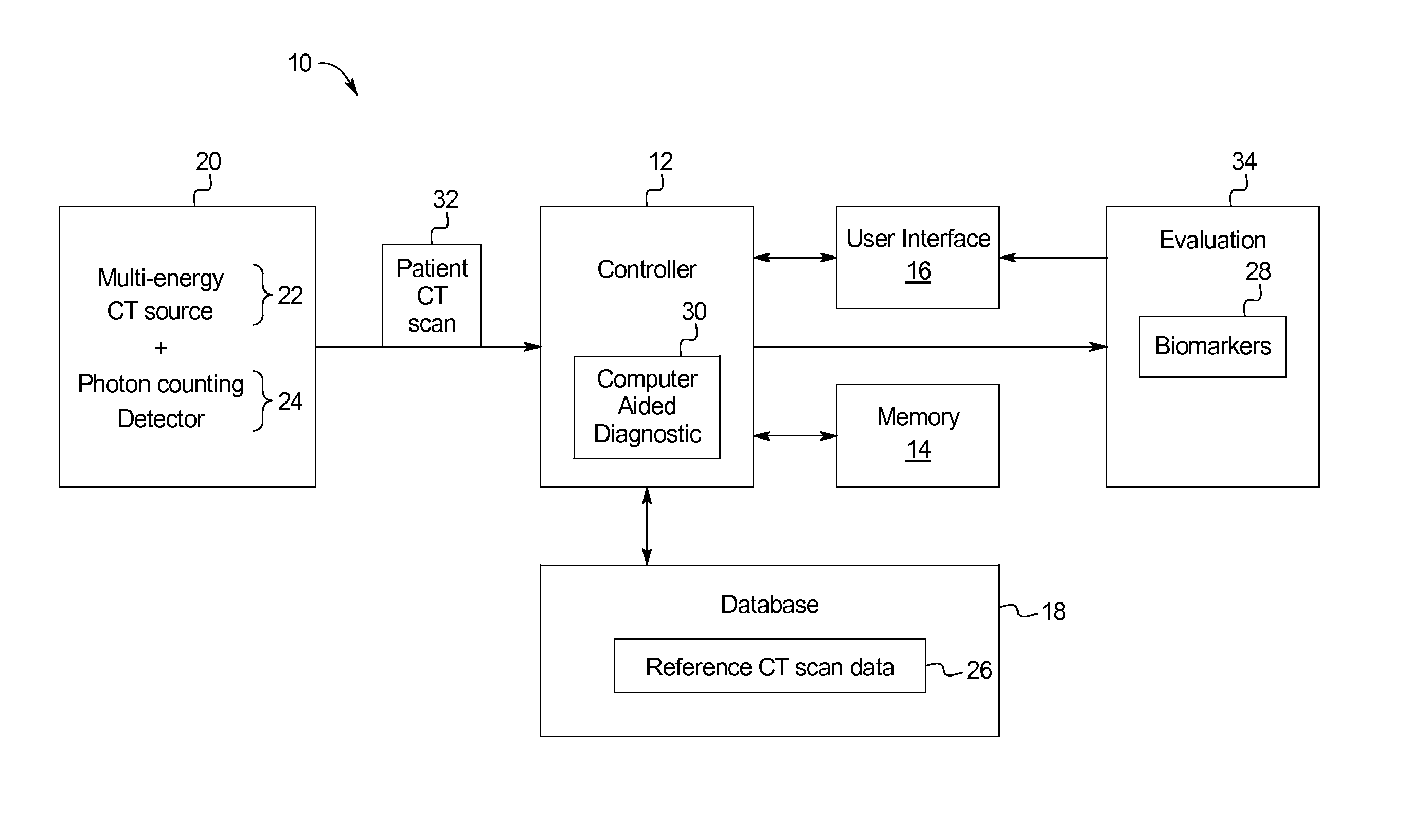
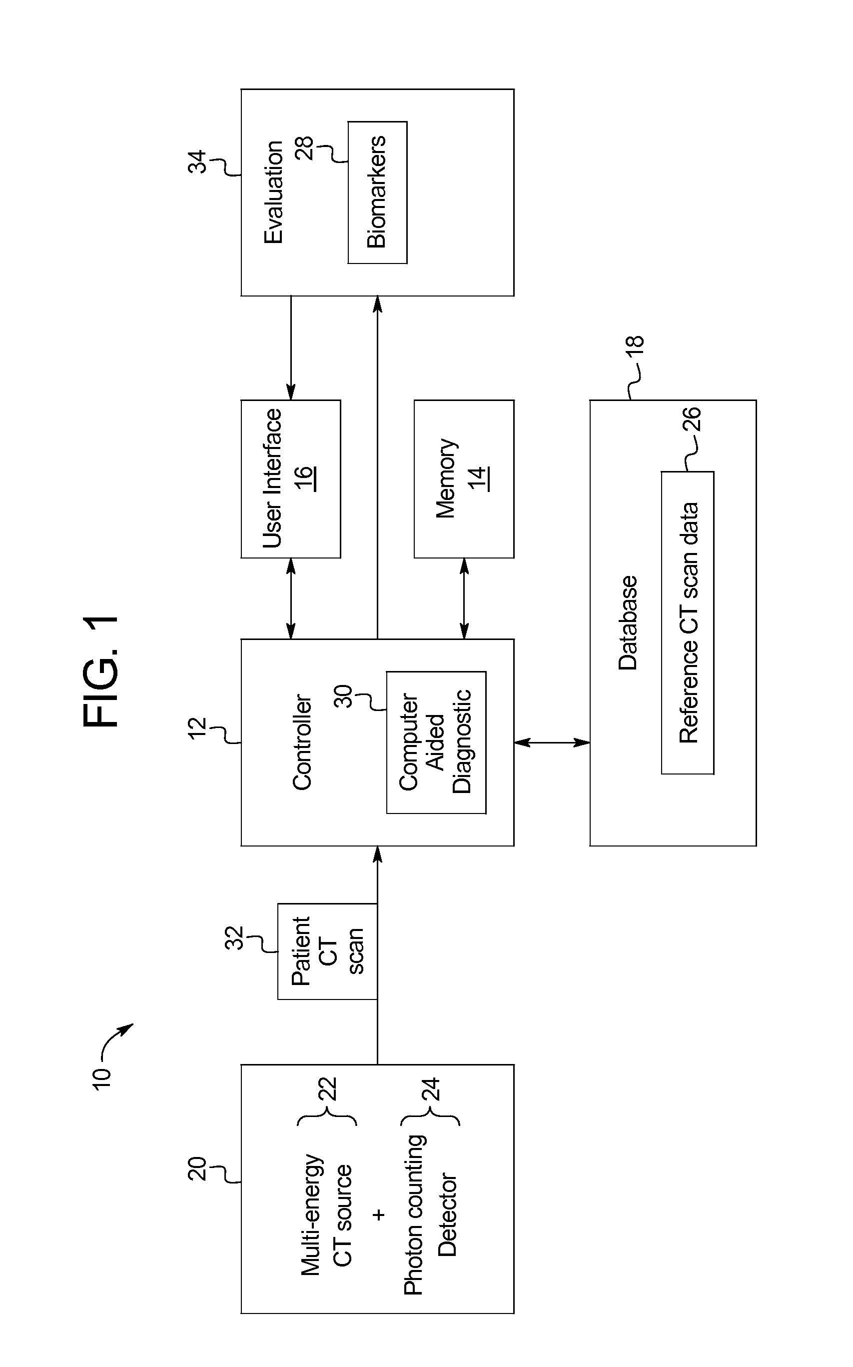
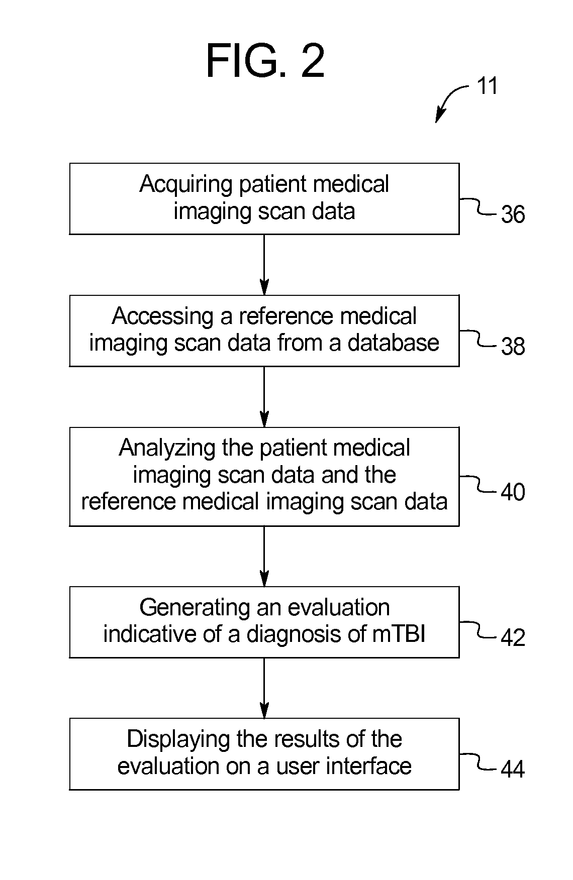
[0033]The following description details embodiments of inventive systems and methods for evaluating a brain scan using reference data. The systems and methods use a dataset provided from a medical imagining device to derive an objective diagnosis of concussions or mTBI. The examples below rely primarily on the example of using a CT scanner as the medical imagining device, but it is understood that the advantages of the systems and methods provided herein can be attained using alternative, or additional, medical imaging devices.

[0034]FIG. 1 illustrates an example of the present subject matter using a computed tomography system 10. As shown in FIG. 1, the computed tomography system 10 includes a CT scanner 20 including a multi-energy x-ray source 22 in connection with a photon counting detector 24 configured to collect a patient scan data 32 of a patient.

[0035]The CT scanner 20 uses a rotating anode (x-ray source 22) to generate x-rays that pass through an object and are collected on ...


PUM


Abstract

The present disclosure provides systems and methods for evaluating a brain scan using reference data. Specifically, the systems and methods include a computed tomography (CT) scanner for the purpose of diagnosing concussions or mild traumatic brain injuries (mTBI). The system includes a multi-energy x-ray source (i.e., spectral CT), a photon counting x-ray detector, and a content-aware computer aided diagnostic (CAD) algorithm designed to detect imperceptible changes indicative of structural and physiological damage caused by a concussive event by comparing raw volumetric datasets of baseline (healthy) reference scan data to patient scan data taken after a concussive event.

Description

CROSS-REFERENCE TO RELATED APPLICATIONS[0001]This application incorporates by reference and claims priority to U.S. Provisional Application 61 / 798,445 filed on Mar. 15, 2013.BACKGROUND OF THE INVENTION[0002]The present subject matter relates generally to systems and methods for evaluating a brain scan. More specifically, the present invention relates to objective systems and methods for evaluating a brain scan using reference data to detect acute injury, such as a concussion, or detecting chronic injury such as chronic traumatic encephalopathy.[0003]Brain injury, such as a concussion, can cause physiological and psychological problems, and even disability or death. Repeated brain injury, an unfortunately common occurrence in professional athletics, such as football or boxing, may cause serious conditions later in life, such as chronic traumatic encephalopathy. However, the lack of the highly noticeable external signs and symptoms cause clinicians to fail to identify brain injuries c...

Claims


Application Information

Patent Timeline
IPC IPC(8): A61B6/00A61B6/03
CPCA61B6/501A61B6/482A61B6/032A61B6/4241A61B6/5217G16H50/30
Inventor VESTEVICH, JACQUELINE K.
Owner VESTEVICH JACQUELINE K
Why Eureka
Try Eureka