
Method and Device for Fetal Heart Rate Monitoring with Maternal Contribution Detection

a heart rate monitor and fetal heart rate technology, applied in the field of fetal monitoring, can solve the problems of limited ultrasound penetration, difficult application of doppler ultrasound to detect fetal heart rate (fhr) in obese patients, and the risk of competing physiological signals of mothers, and achieve the effect of maximizing signal quality

Active Publication Date: 2014-09-18
GENERAL ELECTRIC CO
View PDF3 Cites 20 Cited by
  • Summary
  • Abstract
  • Description
  • Claims
  • Application Information

AI Technical Summary

Benefits of technology

The patent describes a device that can monitor the fetal heart rate using ultrasound. The device transmits ultrasound waves over a specific area and receives signals that reflect those waves. The device analyzes those signals to create a fetal heart rate waveform and calculates the average heart rate over that waveform. The device also compares this average heart rate to a reference heart rate to determine if there is a close match and if the heart rate waveform is stable. The device also determines the quality of the ultrasound signal and selects specific parts of the signal for analysis. The device can produce a maternal artifact alarm if there is no coincidence between the selected parts of the signal and can provide information on when the ultrasound transducer should be used. The technical effect of this patent is to provide a reliable and accurate way to monitor fetal heart rate during pregnancy.

Problems solved by technology

Current methods to detect fetal heart rate (FHR) using Doppler ultrasound have difficulty when applied to obese patients as the ultrasound scanning depth of the solutions has a limited ultrasound penetration range that, when thickness of abdominal fat is taken into account, does not reach the heart of the fetus.
Therefore, ultrasound fetal heart rate monitors with extended penetration depth create a risk to pick up competing physiological signals of the mother, rather than the heart activity of the fetus.
However, the quality of the fetal heart rate determined using fetal ECG through abdominal ECG is typically lower than the quality of fetal heart rate detection experienced in the clinical setting for non-obese maternal patients using Doppler ultrasound.
The abdominal ECG requires that a plurality of electrodes be applied to the patient, such electrodes are at risk of producing poor quality signals if careful skin preparation or maintenance of the electrodes is not considered.
This can occur in a case wherein the fetal heart signal is weak or lost due to worsening fetal health or due to movement of the fetal heart beyond the ultrasound beam as a result of maternal or fetal movements.
Also, if the MHR is elevated, then the monitoring device may lock on the MHR and erroneously produce a result indicating fetal well-being, when in fact the fetal health may be deteriorating.
The addition of a still further technology and transducer adds complexity and cost to systems and such combinations present further challenges due to the operation of competing transducers in the same space.
Specifically, SpO2 is known to be sensitive to motion artifact and the tocodynamometer membrane can be a source of such motion artifacts, limiting the quality of the MHR determined by the SpO2 transducer.

Method used


Image

  • Method and Device for Fetal Heart Rate Monitoring with Maternal Contribution Detection
  • Method and Device for Fetal Heart Rate Monitoring with Maternal Contribution Detection
  • Method and Device for Fetal Heart Rate Monitoring with Maternal Contribution Detection

Examples


Embodiment Construction

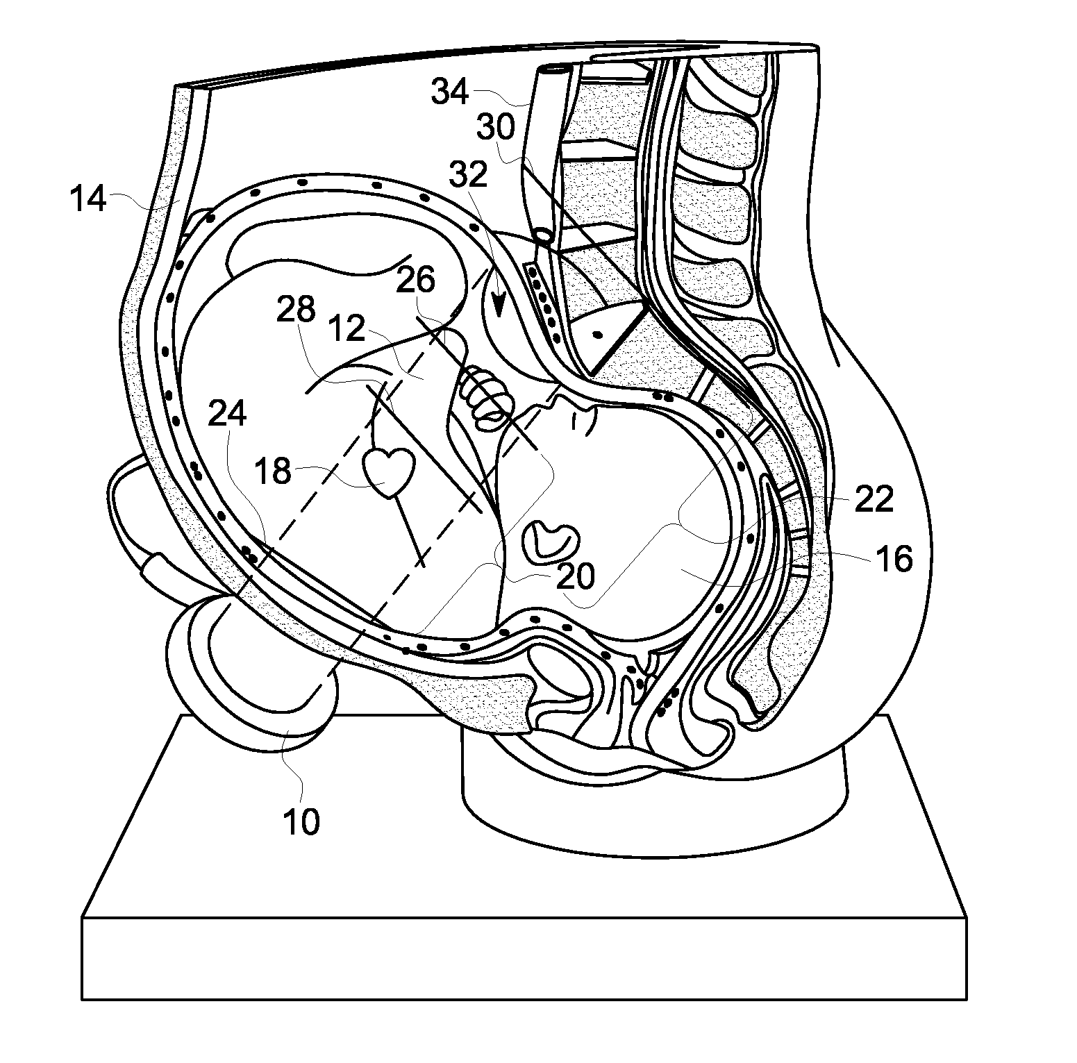
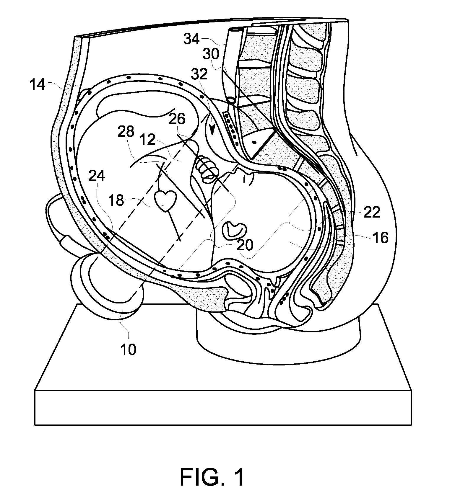
[0016]FIG. 1 diagrammatically depicts the operation of an embodiment of an ultrasound transducer 10 to project an ultrasound beam 12 into the body of a maternal patient 14 and a fetus 16. The ultrasound beam 12 is particularly directed at the heart 18 of the fetus 16. FIG. 1 exemplarily depicts a first ultrasound sensitivity zone 20 and a second ultrasound sensitivity zone 22. In the exemplary embodiment depicted in FIG. 1, the first ultrasound sensitivity zone 20 is a near zone and the second ultrasound sensitivity zone 22 is a far zone. It is to be understood that these ultrasound sensitivity zones are merely exemplary and such zones may exemplarily be defined in other manners than that described herein, including a single ultrasound sensitivity zone.

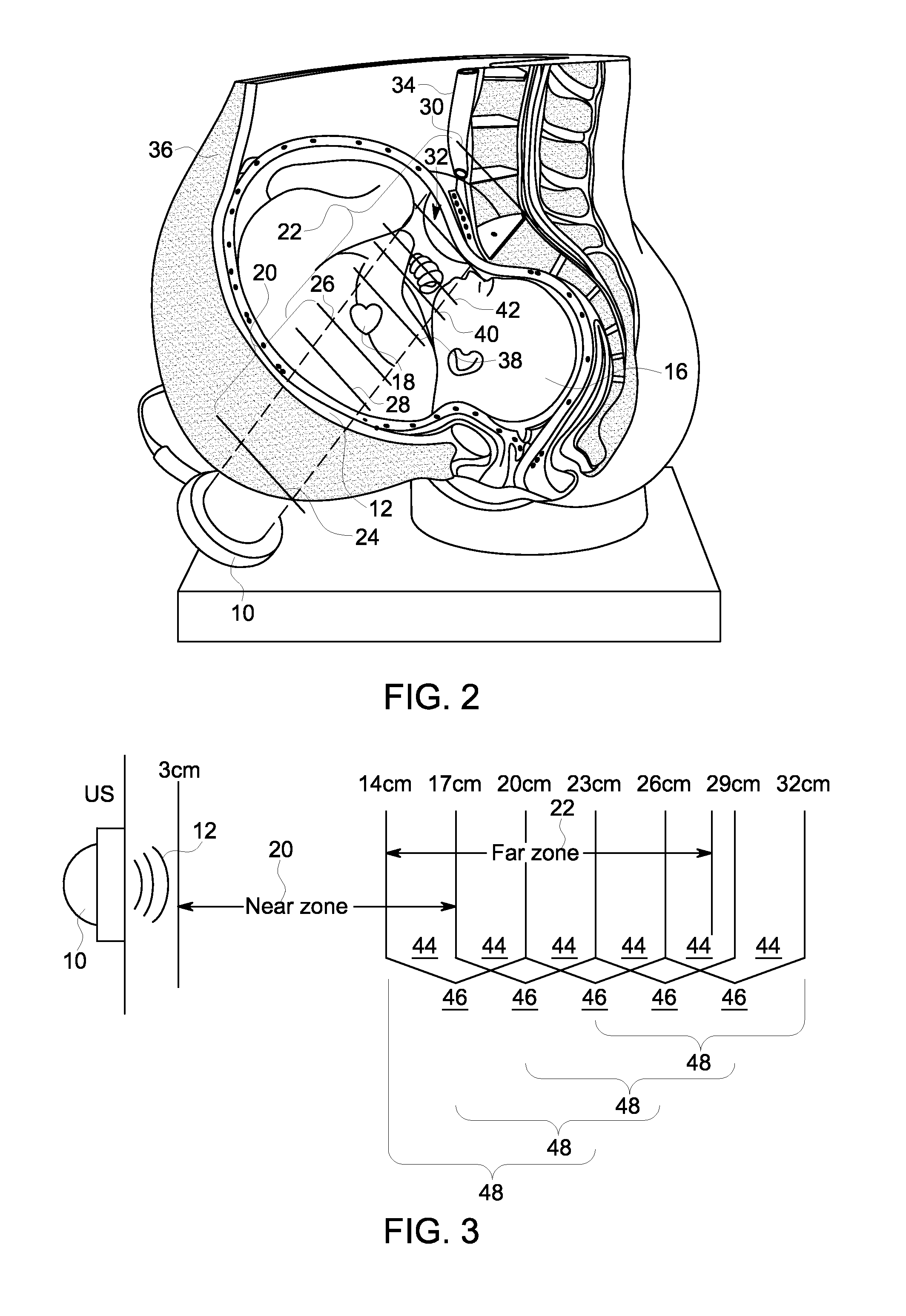
[0017]In the exemplary embodiment depicted in FIG. 1, the near zone 20 extends across a depth range between 3 centimeters indicated at 24 to 17 centimeters indicated at 26. In an exemplary embodiment the far zone may extend across a d...


PUM


Abstract

A method and device for monitoring a fetal heart rate includes a reference fetal heart rate detected across an ultrasound depth zone of sensitivity. The ultrasound depth zone of sensitivity is scanned in overlapping increments of a first depth. An average fetal heart rate detected for each overlapping increment is tested for a coincidence with the reference fetal heart rate. Overlapping increments with the coincidence and a maximized signal quality rate are identified. An ultrasound depth increment of a second depth is selected representing the selected adjacent increments. Fetal heart rate is determined from an ultrasound signal returned from a scan depth of the second depth.

Description

BACKGROUND[0001]The present disclosure is related to the field of fetal monitoring. More specifically, the present disclosure is related to fetal heart rate monitoring with the detection of maternal artifacts.[0002]Current methods to detect fetal heart rate (FHR) using Doppler ultrasound have difficulty when applied to obese patients as the ultrasound scanning depth of the solutions has a limited ultrasound penetration range that, when thickness of abdominal fat is taken into account, does not reach the heart of the fetus. Thus, the heart of a fetus with an obese mother is beyond the typical range of ultrasound sensitivity.[0003]While ultrasound penetration depth can be increased either by increasing a duration of an ultrasound transmit pulse and a duration of a receive window or by increasing the duration of the receive window with a fixed transmit pulse, the increased penetration depth causes a decrease in the signal to noise ratio in the received reflected ultrasound signals. The...

Claims


Application Information

Patent Timeline
IPC IPC(8): A61B8/02A61B8/08
CPCA61B8/02A61B8/5269A61B8/488A61B5/7221A61B5/7235A61B8/0866A61B8/0883A61B8/5276A61B8/54A61B8/543A61B5/02411A61B5/0255
Inventor KABAKOV, SERGUEIFALK, STEVEN M.
Owner GENERAL ELECTRIC CO
Learn More