Real-time feedback for preventing high dose c-arch geometry positions

a real-time feedback and geometry technology, applied in the direction of radiation beam directing means, medical science, diagnostics, etc., can solve the problems of patient exposure to some x-ray radiation which poses a health risk, turns into a burden, etc., to reduce the dose, reduce the dose, and reduce the effect of dos

Active Publication Date: 2014-10-16
KONINKLJIJKE PHILIPS NV
View PDF2 Cites 77 Cited by
  • Summary
  • Abstract
  • Description
  • Claims
  • Application Information

AI Technical Summary

Benefits of technology

[0016]In interventional C-Arch X-ray imaging, some imaging geometry positions or angles are far more patient “X-ray dosage friendly” than others so less dosage is required. This dosage required to maintain a desired image quality is directly correlated with the distance travel of the X-ray in the tissue. For instance, in a cardiac application, a “high angle” spider view requires up to 6 times the X-ray dose than for a normal frontal (AP—anterior-posterior) image. The apparatus harnesses the fact that relatively minor variations of the geometry angle have littl...

Problems solved by technology

When using the X-ray equipment, the patient is inevitably exposed to some X-ray radiation which poses a health risk.
This freedom however may turn into a burden on medical personal desiro...

Method used


Image

  • Real-time feedback for preventing high dose c-arch geometry positions
  • Real-time feedback for preventing high dose c-arch geometry positions
  • Real-time feedback for preventing high dose c-arch geometry positions

Examples


Embodiment Construction

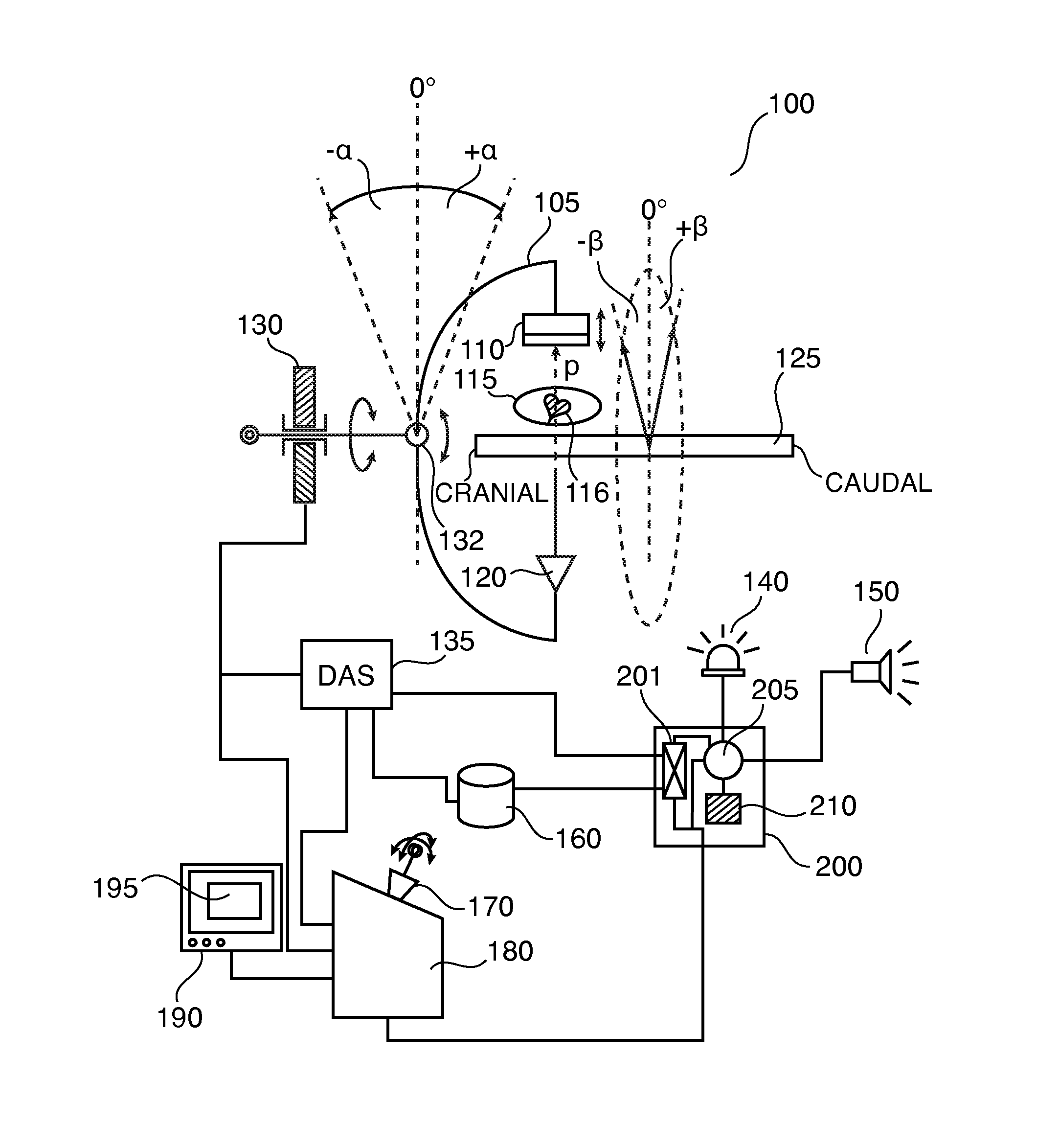
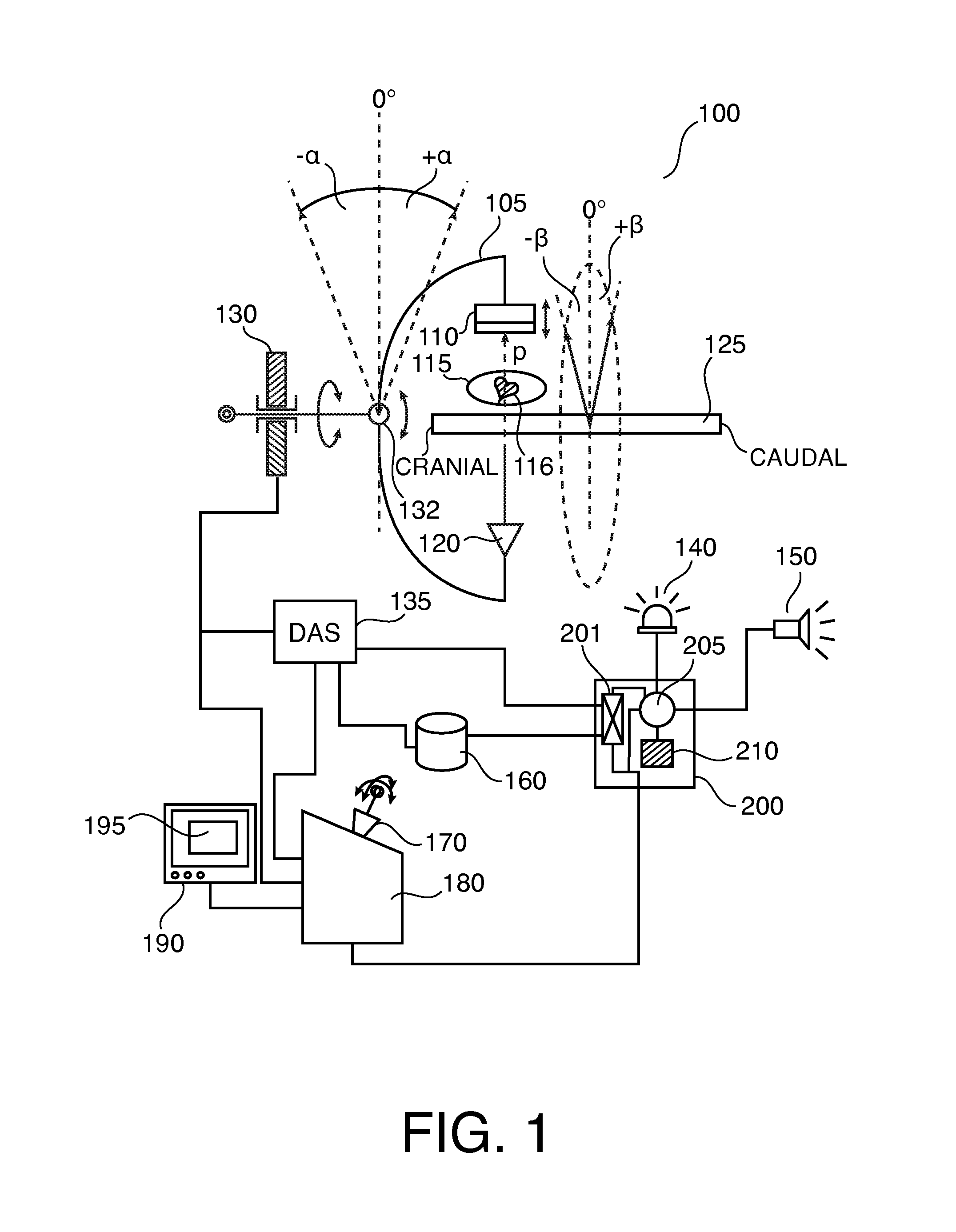
[0051]With reference to FIG. 1, an interventional x-ray imaging equipment 100 (X-ray imager) of the C-arm type is shown. A “C” shaped arm 105 or fame has attached to one of its end an x-ray source 120 and in opposed relationship on the other end a detector 110 configured to detect x-rays emitted by said x-ray source 120.

[0052]Rigid C arm 105 is connected to a shaft by way of a joint. Shaft is journaled on a bearing 130 to allow rotation of arm 105 carrying with it the ensemble of X-ray source 120 and detector 105 (“source-detector ensemble”). Bearing 130 includes a slip ring arrangement so that electronic signals between the source-detector ensemble and an operator console 180 can be exchanged via a suitable communication network.

[0053]A table or patient stretcher 125 is arranged centrally between a circle swept out by x-ray source 120 and detector 105 whilst arm 105 is rotating. A patient 115 is disposed on table 125 such that an anatomical object of interest is arranged substantia...


PUM


Abstract

The present invention relates to an apparatus for aiding operation of an interventional x-ray imager during image acquisition, to a method of aiding operation of an x-ray imager, to an interventional x-ray imager, to a computer program element and to a computer readable medium. The X-ray imager is capable of varying X-ray dosages depending on differences in X-ray attenuation levels across an object of interest to be imaged and is capable of assuming any one of a plurality of imaging geometry positions when acquiring an image. An indication, visual, acoustic or haptic, to the operator of an X-ray imager is provided on the incurred change in X-ray dosage when changing from a current projection view to an updated projection view, provided that a given constant image quality is to be maintained throughout the different views.

Description

FIELD OF THE INVENTION[0001]The present invention relates to an apparatus for aiding operation of an interventional x-ray imager during image acquisition, to a method of aiding operation of an x-ray imager, to an interventional x-ray imager, to a computer program element and to a computer readable medium.BACKGROUND OF THE INVENTION[0002]X-ray imaging equipment is widely used by medical personnel to gain clues about a patient's condition or during medical interventions. WO 2011 / 042834 discloses X-ray imaging equipment.[0003]When using the X-ray equipment, the patient is inevitably exposed to some X-ray radiation which poses a health risk. Modern X-ray equipment allows the operator to vary the X-ray dosage according to the medical need at hand. This freedom however may turn into a burden on medical personal desirous to strike the right balance between the level of patient's X-ray exposure used in a given image acquisition and the medical relevance of the image material obtained during...

Claims


Application Information

Patent Timeline
IPC IPC(8): A61B6/10A61B6/00
CPCG06F19/3406A61B6/4441A61B6/461A61B6/5294A61B6/503A61B6/542A61B6/10A61B6/467G16H40/63
Inventor WITHAGEN, PETRUS JOHANNESDEN HARTOG, MARKUS JOHANNES HARMENHOORNAERT, BART PIERRE ANTOINE JOZEF
Owner KONINKLJIJKE PHILIPS NV
Why Eureka
Try Eureka