
Method and device for measuring haemoglobin in the eye

Inactive Publication Date: 2014-11-20
INSTRACION Y OFTALMOLOGIA INSOFT
View PDF1 Cites 1 Cited by
  • Summary
  • Abstract
  • Description
  • Claims
  • Application Information

AI Technical Summary

Benefits of technology

The present invention provides a method and device for measuring the amount of haemoglobin in the optic nerve tissue, which can identify and differentiate different regions of the optic nerve. This is useful for monitoring and diagnosing glaucomatous disease or glaucoma. The method compensates for variations in light intensity and spectral composition, so it is not affected by changes in the lens or detecting equipment. The invention is based on a colorimetric analysis, which does not require prior knowledge of the spectral composition of the incident light or capture equipment.

Problems solved by technology

Currently, in vivo oximetry measurements have been limited to isolated experiments using imaging spectroscopy and are based on monochromatic systems or well-known spectral composition methods in illumination or in detection.
All these previously cited spectral analysis methods have the drawback that they disregard the specific value of the volume of blood (amount of haemoglobin) in which oxygen saturation was evaluated and therefore the results obtained do not directly reflect the volume of blood on which the measurement is made.
That is why the images obtained with these methods are not able to clearly distinguish the poorly irrigated regions (regions of atrophy or regions with little vascularised tissue) from well irrigated regions such as the neuroretinal ring.
Therefore, these existing methods do not allow complete and adequate information of optic nerve perfusion, so it is necessary to evaluate not only the degree of oxygen saturation of haemoglobin, but also the amount of haemoglobin present in the optic nerve.

Method used


Image

  • Method and device for measuring haemoglobin in the eye
  • Method and device for measuring haemoglobin in the eye
  • Method and device for measuring haemoglobin in the eye

Examples


Embodiment Construction

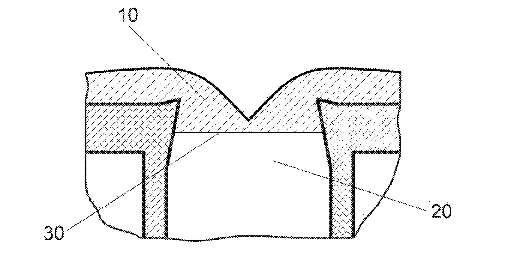
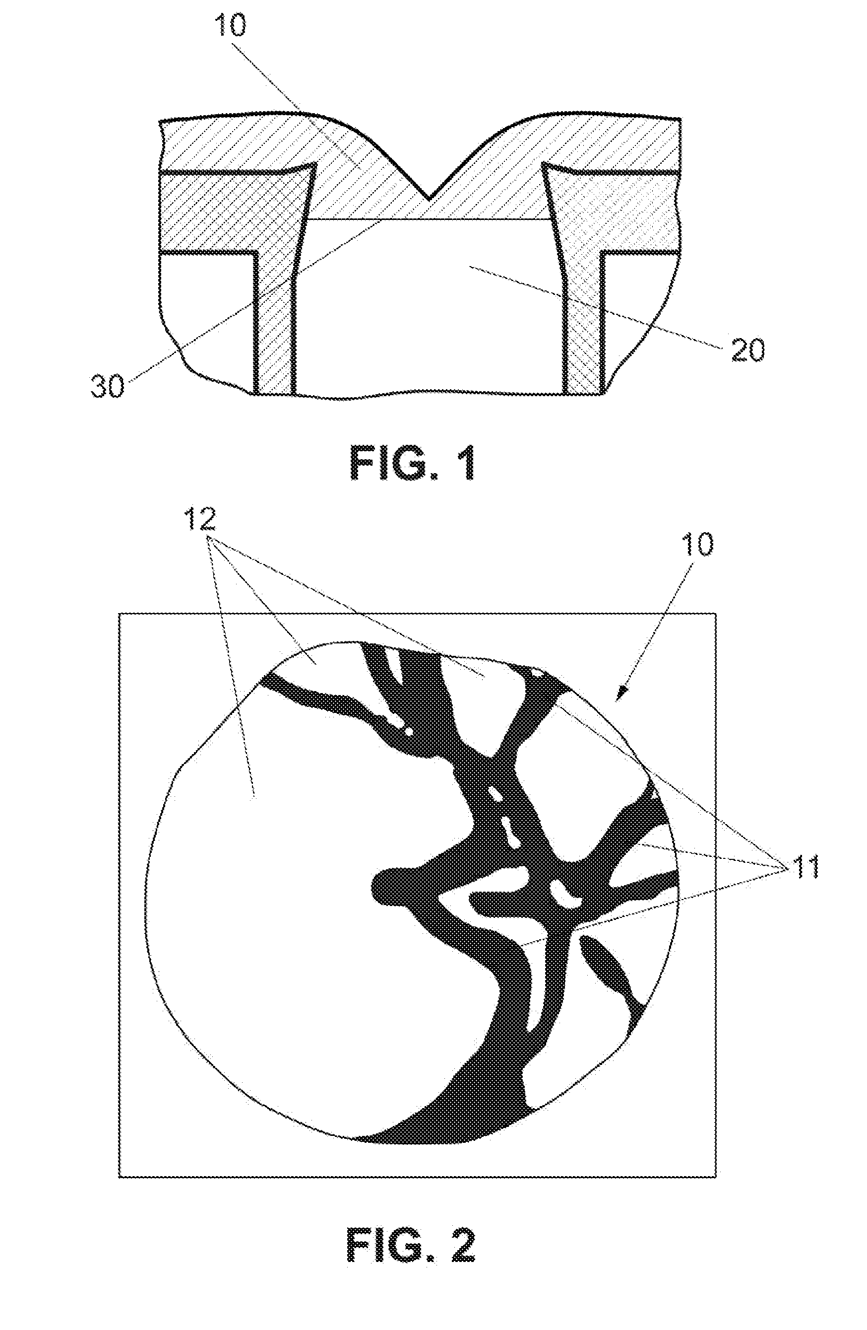
[0010]The present invention resolves the previously cited drawbacks, providing a method and device for measuring the amount of haemoglobin in the optic nerve tissue and more specifically in the optic nerve head, through which it is possible to identify and clearly differentiate the irrigated regions of the optic nerve from the non-irrigated regions, this thereby constituting an extremely useful and interesting tool in monitoring and diagnosing glaucomatous disease or glaucoma. In addition, this method enables taking into account the effect of loss of lens transparency in patients with lens ageing or “cataracts”, so that the effects of spectral absorption and light diffusion are compensated without affecting the final results of the estimation and amount of haemoglobin.

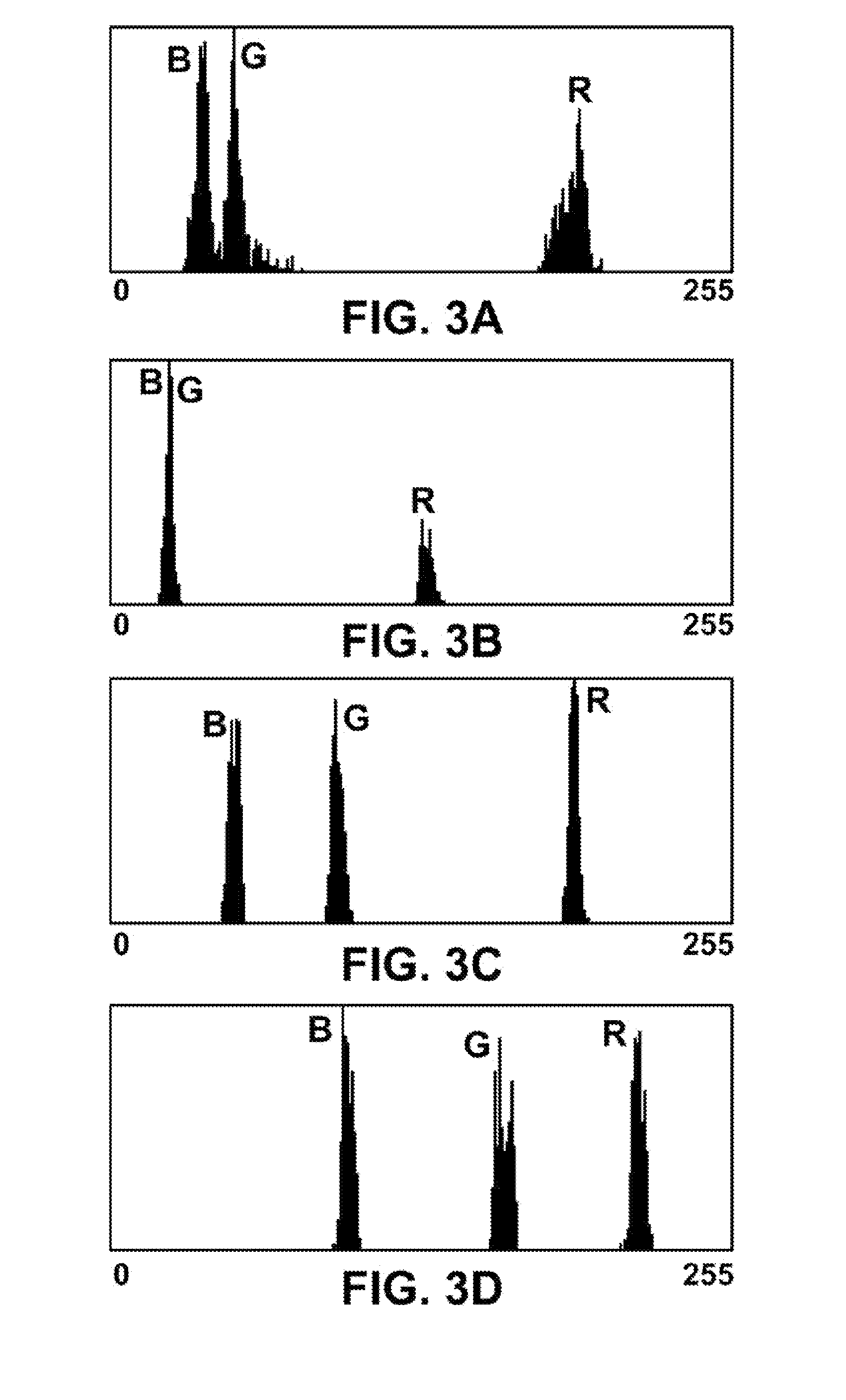
[0011]The method for measuring haemoglobin (Hb) “ex vivo” in an individual, the object of the invention, is mainly based on the identification of primary colours red, green and blue (R, G, B) contained in digital image...


PUM


Abstract

Based on the identification by colorimetry of a colour image of the fundus of the eye of an individual, obtained by specialised cameras (fundus camera or retinograph). The method and the device of the present invention enable establishing a direct relation between the chromatic properties of the various tissues of the optic nerve (10) and the amount of haemoglobin (Hb) in the optic nerve head (10), allowing identification and clearly distinguishing the irrigated regions of the optic nerve (10) from the non-irrigated regions or those with atrophy, constituting a tool of great interest in monitoring and diagnosis of glaucomatous disease and other optic diseases affecting vascularisation. This method also allows taking into account the effect of loss of transparency of the lens in patients with cataracts, as this effect is compensated without affecting the final results of the determination of the amount of haemoglobin (Hb).

Description

[0001]The present invention pertains to the sectors of Ophthalmology and Optometry, more specifically to the diagnosis of diseases of the retina and optic nerve.[0002]The main object of the present invention is a method and device for measuring the amount of haemoglobin in the optic nerve head, of particular application to monitoring and diagnosis of glaucomatous disease.BACKGROUND OF THE INVENTION[0003]It is currently known that glaucoma mainly acts through the influence of intraocular pressure on the perfusion of the optic nerve. Currently, in vivo oximetry measurements have been limited to isolated experiments using imaging spectroscopy and are based on monochromatic systems or well-known spectral composition methods in illumination or in detection. That is, they are in the field of spectrophotometry.[0004]Spectral analysis of the image of the optic disc or optic nerve head has been mainly used to measure “oxygenation” of the haemoglobin contained. These techniques for measuring ...

Claims


Application Information

Patent Timeline
IPC IPC(8): A61B3/12A61B3/14A61B3/00A61B5/1455
CPCA61B3/1241A61B3/0025A61B3/14A61B5/1455A61B3/1176A61B5/489
Inventor GONZALEZ DE LA ROSA, MANUEL
Owner INSTRACION Y OFTALMOLOGIA INSOFT
Learn More