System and method for displaying anatomy and devices on a movable display

a technology of anatomy and movable display, applied in the field of diagnosis and treatment of disorders, can solve problems such as difficult viewing of the screen, and achieve the effects of easy navigation, quick selection of access points, and light angular bias

Active Publication Date: 2014-12-04
AURIS HEALTH INC
View PDF16 Cites 235 Cited by
  • Summary
  • Abstract
  • Description
  • Claims
  • Application Information

AI Technical Summary

Benefits of technology

[0024]In one embodiment, a live image of the patient anatomy is shown on a repositionable display screen located over the patient. The physician can move the display over the patient while sensors track the motion of the display so that the image shown on the display screen may be periodically or constantly updated to show the medical device, and the patient anatomy substantially aligned with the patient from the perspective of the user with a slight angular bias toward the user. The position of the user relative to the patient may be entered by the user at the start of the procedure by touching a button on the display labeled “patient left,”“patient right,”“patient head,” or “patient feet.” In this manner, the image shown on the display provides a view of the medical device and patient anatomy that is intuitive, ergonomic, and allows for easy navigation of the medical device within the patient anatomy shown on the display screen. While the image of the anatomy is frequently based on a pre-operative image, a live image may be supplemented with other anatomical images from live or static sources which are sensored, registered, and displayed in the same substantially co-aligned manner on the display screen.
[0025]In additional embodiments, a sensor on the medical device provides position and orientation data of the device to a data processor. A sensor on the patient provides position and orientation data of the patient to the processor, and sensors on the display screen provide the viewing position and orientation of the display screen to the processor. With data from the medical device, the patient, and the display, the processor unifies the three coordinate systems so that the image shown on the display screen

Problems solved by technology

For instance, moving the display with a unity ratio may cause the display to interfere with o

Method used


Image

  • System and method for displaying anatomy and devices on a movable display
  • System and method for displaying anatomy and devices on a movable display
  • System and method for displaying anatomy and devices on a movable display

Examples


Embodiment Construction

[0033]While preferred embodiments of the present invention have been shown and described herein, it will be obvious to those skilled in the art that such embodiments are provided by way of example only. Numerous variations, changes, and substitutions will now occur to those skilled in the art without departing from the invention. It should be understood that various alternatives to the embodiments of the invention described herein may be employed in practicing the invention. It is intended that the following claims define the scope of the invention and that methods and structures within the scope of these claims and their equivalents be covered thereby.

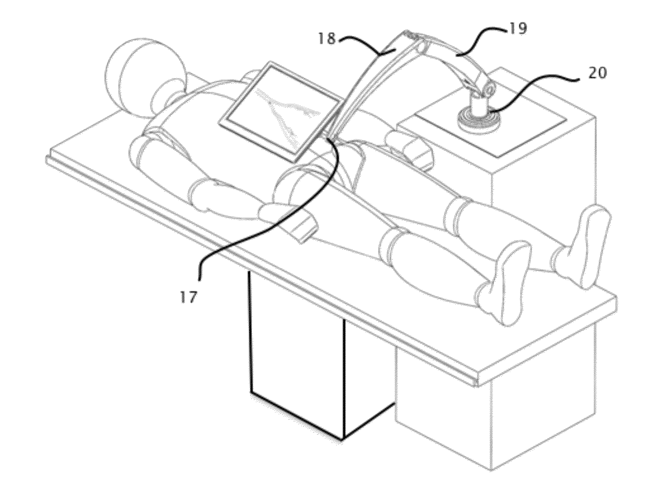
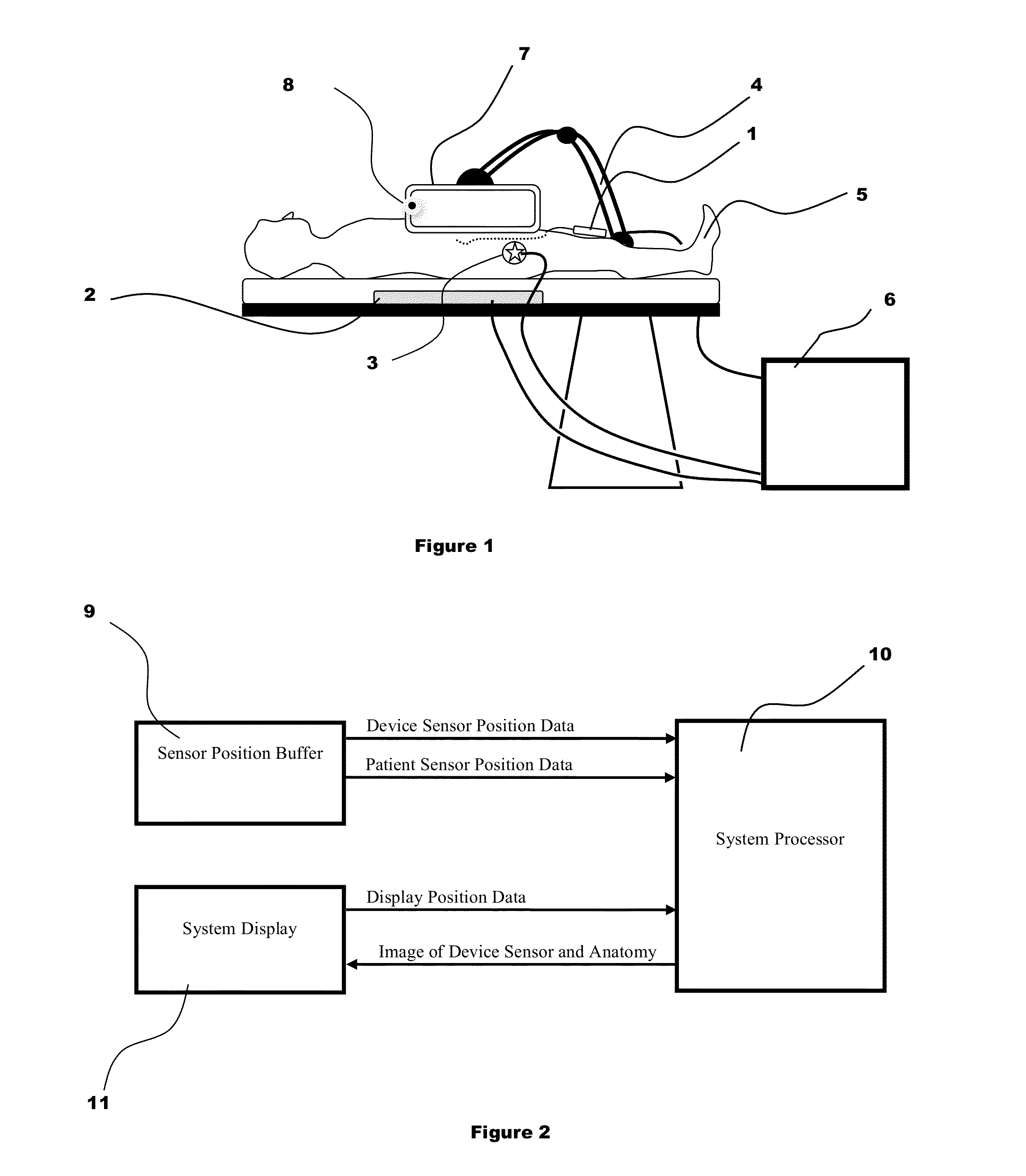
[0034]FIGS. 1-2 describe an embodiment for navigating a minimally invasive medical device within the patient using an acquired three-dimensional anatomical image shown in a display 7 that is substantially aligned to the patient anatomy. A sterile cover may be used to separate the display from the sterile operating field and the steril...


PUM


Abstract

An image display system is provided comprised of a virtual window system that creates a visual coherency between the patient's anatomical images and the actual patient by aligning the image on the display to the patient and then presenting the image to the user in a way that feels as if the user is looking directly into the patient through the display. The image shown within the image display system is dependent upon the position of the image display apparatus and the position of the user so that the display orientation of the image may be biased slightly toward the user to improve ergonomics and usability.

Description

CROSS-REFERENCE[0001]1. This application claims the benefit of U.S. Provisional Application No. 61 / 829,078 filed May 30, 2013.BACKGROUND OF THE INVENTION[0002]1. Field of the Invention[0003]The invention relates generally to the diagnosis and treatment of disorders using minimally invasive techniques. In many minimally invasive procedures very small devices are manipulated within the patient's body under visualization from a live imaging source like ultrasound, fluoroscopy, or endoscopy. Live imaging in a minimally invasive procedure may be supplemented or replaced by displaying the position of a sensored medical device within a stored image of the patient anatomy.[0004]Many minimally invasive procedures are conducted in expensive settings by specialized physicians. Often small, percutaneous medical devices are visualized during the procedure by using live fluoroscopic or ultrasonic imaging. While the live imaging provides a real-time image of anatomy, it has many drawbacks:[0005]Ti...

Claims


Application Information

Patent Timeline
IPC IPC(8): A61B5/06A61B6/00A61B8/08A61B5/00
CPCA61B5/062A61B8/0833A61B6/487A61B5/7425A61B5/7445A61B5/055A61B90/37A61B2034/2051G16H40/63G16H20/40
Inventor WALLACE, DANIELSTAHLER, GREGORYGROGAN, AARON
Owner AURIS HEALTH INC
Why Eureka
Try Eureka