Device and method for determining border of target region of medical images

a target region and image technology, applied in the field of physiological parameters determination, can solve the problems of reducing ejection function, valvular dysfunction and other symptoms, reducing ejection function, and reducing so as to achieve timely correct diagnosis, improve and enhance the accuracy of physiological parameters, and effective and accurate

Inactive Publication Date: 2015-01-22
HONGEN HANGZHOU CHINA MEDICAL TECH +1
View PDF8 Cites 18 Cited by
  • Summary
  • Abstract
  • Description
  • Claims
  • Application Information

AI Technical Summary

Benefits of technology

[0023]In view of the defects in the prior art, the invention aims at seeking a more effective and accurate image processing and calculating device and a method based on the current medical imaging technology, to improve and enhance the ...

Problems solved by technology

However, as an echocardiogram contains a lot of noise and the endocardium of the cardiac chamber and the edge of a myocardium are irregular (in particular in the cases of a cardiac chamber and a myocardium with pathological changes), the relevant quantitative calculation thereof becomes difficult.
Particularly, how to accurately obtain the endocardial border and how to accurately make calculation regarding a heart with irregular changes are difficult.
At present, it is relatively common in clinical use to apply a method for determining a heart ejection fraction (EF value) which includes defining some control points in an interactive way, and modeling the cardiac chamber using a series of simulated geometric shapes, so that the result is very inaccurate.
The shape changes of the cardiac chamber result in reduction of ejection function, valvular dysfunction and other symptoms.
However, since such prior model is obtained by calculation according to the normal heart, in the actual clinical application, it is difficult to obtain accurate results when images of a h...

Method used


Image

  • Device and method for determining border of target region of medical images
  • Device and method for determining border of target region of medical images
  • Device and method for determining border of target region of medical images

Examples


embodiment 1

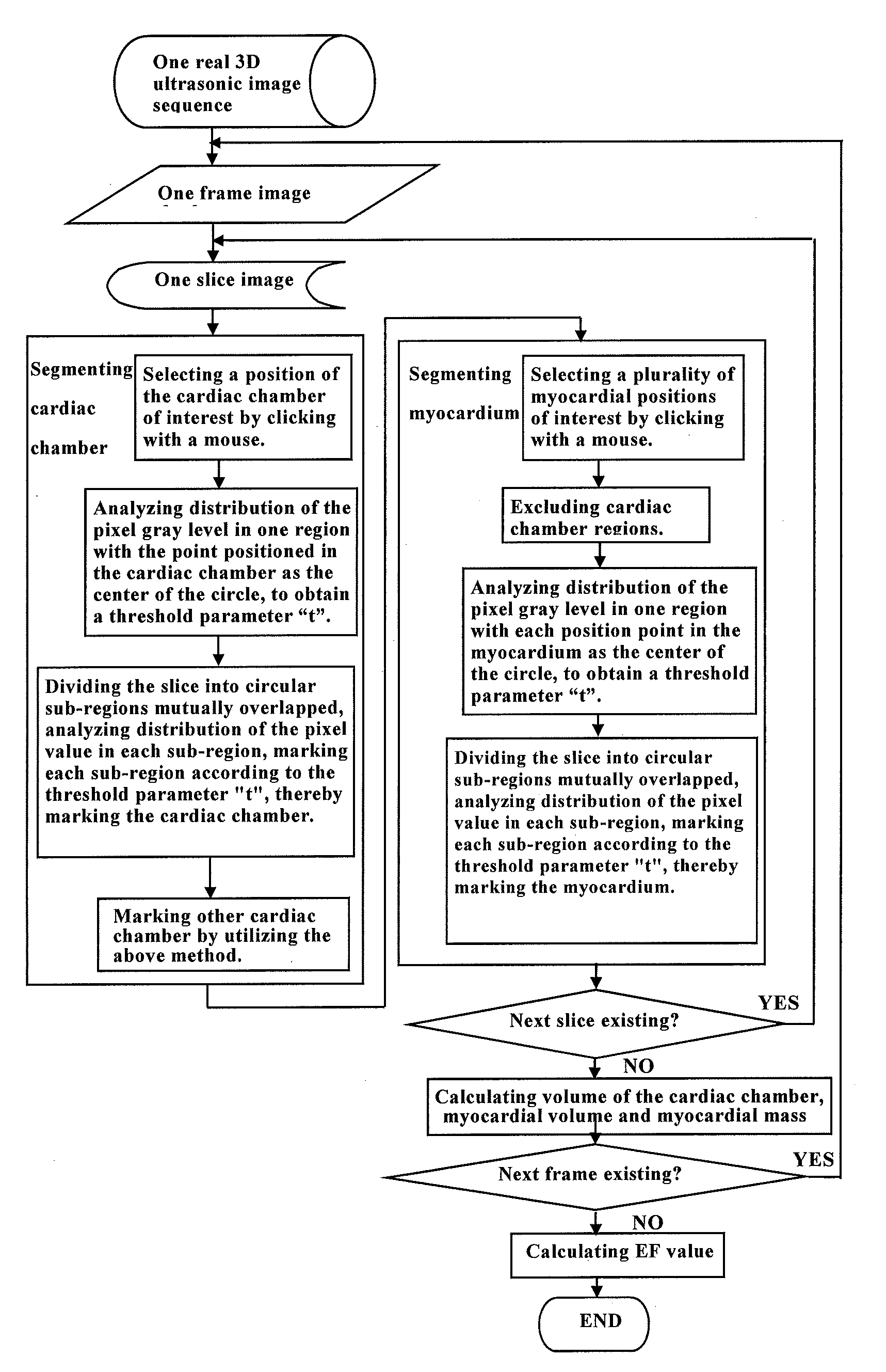
[0073]The invention is applied to real three-dimensional (3D) ultrasonic image data processing with respect to the heart of a patient, and in the embodiment, the invention is used for obtaining the volume of a cardiac chamber and an ejection fraction.

[0074]In Step 1, medical image data of the patient are obtained by utilizing an ultrasonic imaging device. In the embodiment, a real 3D ultrasonic probe is used to scan the region of the heart, then multiple time sequences of a 3D is ultrasonic image are obtained, each time sequence contains a series of frames recording one or more complete cardiac cycles, and each frame contains 3D voxel data consisting of multiple slices. The imaging device, such as Siemens SC2000 echocardiographic instrument and Philips IE33, is used.

[0075]In Step 2, the contour of the cardiac chamber is extracted from all slice images of all the frames in the real 3D ultrasonic image time sequence. In the specific embodiment, generally, 5-8 time sequences are scanne...

embodiment 2

Calculation of Myocardial Volume and Mass

[0097]The step 1 and step 2 of Embodiment 4 are the same as those of Embodiment 1, so that detailed explanation thereof are omitted.

[0098]After the step 1 and the step 2 are completed, the step a), the step b) and the step c) in step 2 are repeated to mark out other cardiac chamber regions on the slice, for the step of excluding the cardiac chambers in subsequent myocardial segmentation. Said other cardiac chamber regions refer to the cardiac chambers not completely exposed and unclear, on which similar segmentation operation is performed for the purpose of marking out all the cardiac chambers to avoid affecting the myocardial segmentation. This step is an additional pretreatment step performed before the myocardial segmentation, for the purpose of excluding all the cardiac chambers.

[0099]In Step 3, the myocardial contour is extracted from all the slice images of all the frames in the real 3D ultrasonic image time sequence.

[0100]a) Selecting ...


PUM


Abstract

The invention discloses a device for determining a border of a target region of a medical image, which is used for differentiating the border of the target region according to corresponding physical quantitative properties reflected by tissue distribution in the medical image. The device comprises: an interactive unit, by which an operator can select the target region on the medical image; a threshold setting unit, which determines threshold values of the physical quantitative properties in the target region selected; and a thresholding segmentation unit, which divides the region to be analyzed or the region of interest into sub-regions, or fills image cells (pixels) in the region to be analyzed, wherein the region to be analyzed contains the target region, and the sub-regions are the sub-regions which are segmented in the image. The thresholding segmentation unit compares average parameter values of the physical quantitative properties of each sub-region with the threshold values and marks all the cells according to comparison results. The present invention provides a calculating and processing method and a device with clear physical significance and a simple and effective algorithm, and the method and the device are particularly suitable for processing special situations of various hearts with pathological changes in clinical art.

Description

FIELD OF THE INVENTION[0001]The invention relates to a method and a device for determining a border of a target region in medical images, as well as determination of physiological parameters by utilizing the determined border of the target region. More specifically, the invention relates to determination of heart physiological parameters based on real ultrasonic image data.BACKGROUND OF THE INVENTION[0002]Medical imaging has become an indispensable part of modern medical treatment, and the application of the medical imaging runs through the whole clinical work. The medical imaging is widely used in disease diagnosis, and further plays an important role in the aspects of planning, implementing, and curative effect evaluating of surgeries, radiotherapies and the like. At present, medical images can be grouped into two kinds, namely anatomical images and functional images. The anatomical images mainly describe human body's morphological information including X-ray transmission imaging,...

Claims


Application Information

Patent Timeline
IPC IPC(8): G06T7/00
CPCG06T7/0012G06T2207/10136G06T2207/30048G06T7/0085G06T7/602G06T7/0081G06T2200/04G06T2207/10016G06T2207/20101G06T2207/20148G06T7/11G06T7/136G06T7/62
Inventor LI, PENGYUAN, XINCHEN, GONG
Owner HONGEN HANGZHOU CHINA MEDICAL TECH
Why Eureka
Try Eureka