Interpolated three-dimensional thermal dose estimates using magnetic resonance imaging

a three-dimensional thermal dose and magnetic resonance imaging technology, applied in the field of magnetic resonance imaging, can solve the problems of limited volume or spatial resolution of thermal data or images acquired, particularly by magnetic resonance imaging, and achieve the effect of useful clinical endpoints

Active Publication Date: 2015-02-05
KONINKLIJKE PHILIPS ELECTRONICS NV
View PDF1 Cites 2 Cited by
  • Summary
  • Abstract
  • Description
  • Claims
  • Application Information

AI Technical Summary

Benefits of technology

[0027]Magnetic resonance data or magnetic resonance thermometry data is typically acquired from volumes called voxels or from thick slab-like volumes called slices. The magnetic resonance thermometry data from a particular slice is typically represented as a two-dimensional graph or field. Execution of the machine executable instructions further causes the processor to interpolate a three-dimensional thermal dose estimate in accordance with the magnetic resonance thermometry data. This is to say the magnetic resonance thermometry data from the multiple slices is used to interpolate a three-dimensional dose estimate. This embodiment may be beneficial because this allows the estimation of the thermal dosage in regions for which magnetic resonance data was not acquired.
[0033]The three-dimensional thermal dose estimate of individual sonications in the case of high-intensity focused ultrasound can be added for the entire session. This may provide a three-dimensional cumulative thermal dose estimate for the particular sonication session. This may be useful for clinicians or doctors to assess the effect of a particular sonication or heating or cooling treatment. A benefit of a cumulative three-dimensional thermal dose estimate may be that it provides a three-dimensional view of the region estimated to be thermally damaged during the course of the treatment session, thus providing a very useful clinical endpoint.
[0035]In another embodiment execution of the instructions further causes the processor to modify the treatment plan in accordance with the three-dimensional thermal dose estimate. This may be beneficial because the temperature control system may heat or cool the target zone in alternating temperature control time periods. Having the three-dimensional thermal dose estimate available may allow the medical apparatus to avoid temperature treating regions which are not desired to be treated or to adjust the control of the temperature control system to more accurately heat or cool the target zone.
[0036]In another embodiment execution of the instructions further cause the processor to detect an endpoint condition using the three-dimensional thermal dose estimate and the treatment plan. Execution of the instructions further cause the processor to halt at least a portion of the temperature control of the target zone by sending a halt command to the temperature control system if the endpoint condition is detected. This embodiment may be beneficial because the three-dimensional thermal dose estimate may be used to predict the thermal dosage in regions which are not being measured by thermal magnetic resonance data by the magnetic resonance imaging system. This may prevent the medical apparatus from heating or cooling the target zone longer than necessary. In some embodiments the three dimensional does estimate may also be a cumulative three dimensional dose estimate constructed by summing multiple three dimensional dose estimates.
[0037]In another embodiment the target zone has a border. Execution of the instructions further causes the processor to calculate a border thermal dose within a predetermined distance from at least a portion of the border. This embodiment may be beneficial because it may help to avoid the difficulty of cooling regions which are not intended to be heated or cooled in the treatment plan.
[0039]The predetermined interpolation volume can be used as a mask to look at heated regions or areas adjacent to the heated regions. This may also speed up the calculation of the three-dimensional thermal dose estimate.

Problems solved by technology

A difficulty that may arise is that thermal data or images acquired, particularly by magnetic resonance imaging, may be acquired from limited volumes or have limited spatial resolution due to restrictions placed on the acquisition time.
This may be problematic if the images are repositioned for additional sonications as no measure of the cumulative thermal dose is then possible.

Method used


Image

  • Interpolated three-dimensional thermal dose estimates using magnetic resonance imaging
  • Interpolated three-dimensional thermal dose estimates using magnetic resonance imaging
  • Interpolated three-dimensional thermal dose estimates using magnetic resonance imaging

Examples


Embodiment Construction

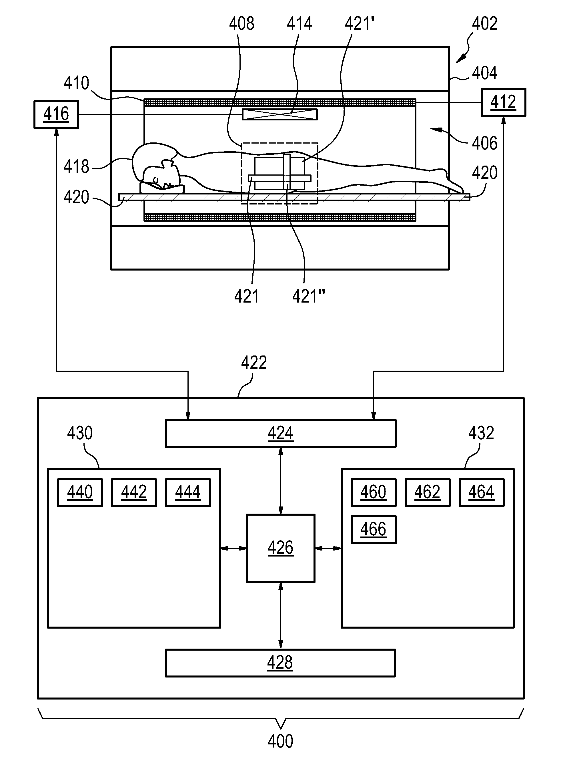
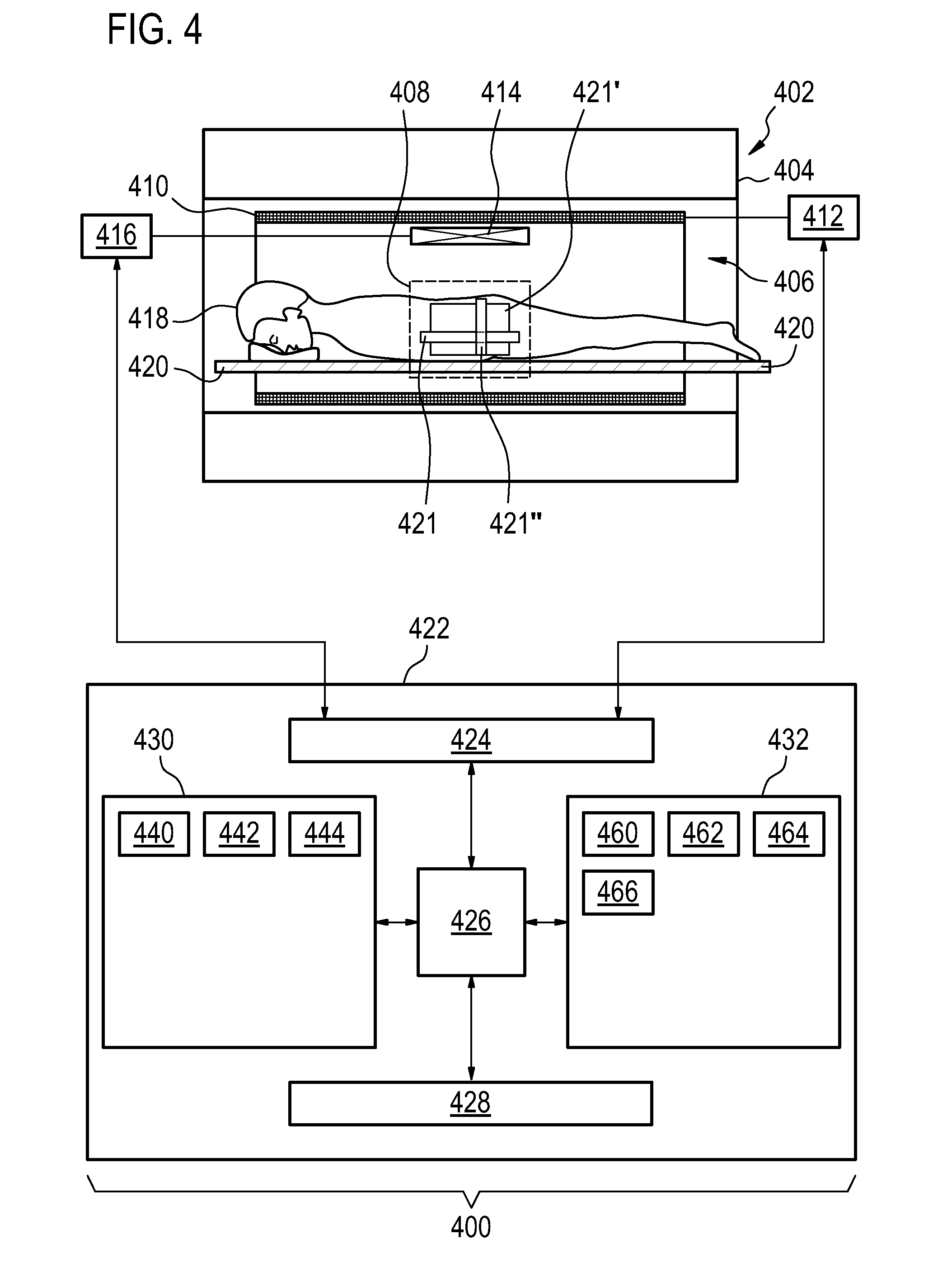
[0067]Like numbered elements in these figures are either equivalent elements or perform the same function. Elements which have been discussed previously will not necessarily be discussed in later figures if the function is equivalent.

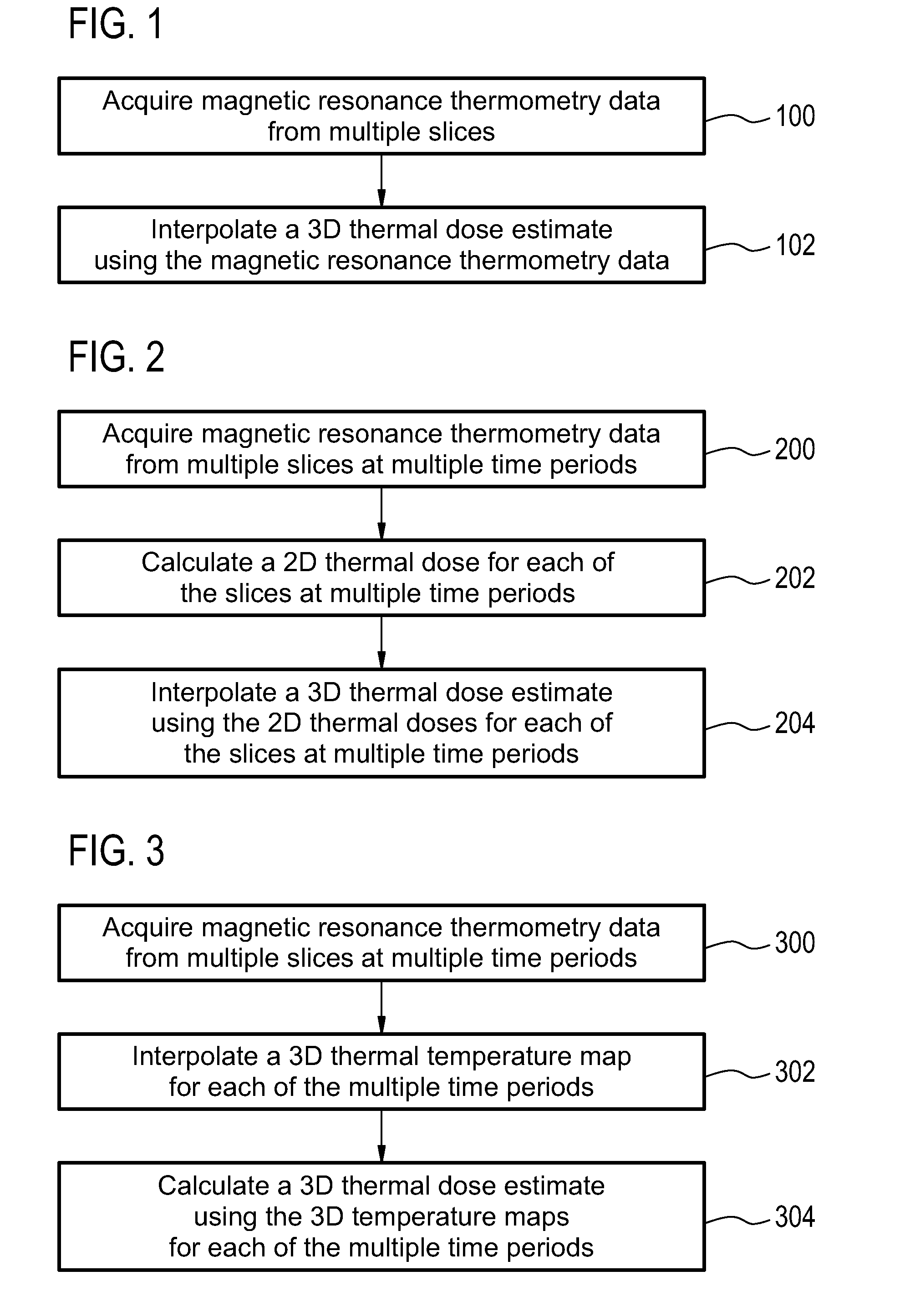
[0068]FIG. 1 shows a flow diagram which illustrates a method according to an embodiment of the invention. In step 100 magnetic resonance thermometry data is acquired from multiple slices. Next in step 102 a three-dimensional thermal dose estimate is interpolated using the magnetic resonance thermometry data.

[0069]FIG. 2 shows a flow diagram which illustrates a method according to a further embodiment of the invention. In step 200 magnetic resonance thermometry data is acquired from multiple slices. Next in step 202 a thermal dose estimate is calculated for each of the slices at the multiple time periods. Next in step 204 a three-dimensional thermal dose estimate is interpolated using the two-dimensional thermal doses for each of the slices at multiple t...


PUM


Abstract

The invention provides for a medical apparatus (400, 500, 600, 700, 800) comprising a magnetic resonance imaging system (402) for acquiring magnetic resonance thermometry data (442) from a subject (418). The magnetic resonance imaging system comprises a magnet (404) with an imaging zone (408). The medical apparatus further comprises a memory (432) for storing machine executable instructions (460, 462, 464, 466, 10, 660). The medical apparatus further comprises a processor (426) for controlling the medical apparatus, wherein execution of the machine executable instructions causes the processor to: acquire (100, 200, 300) the magnetic resonance thermometry data from multiple slices (421, 421′, 421″) within the imaging zone by controlling the magnetic resonance imaging system; and interpolate (102, 202, 204, 302, 304) a three dimensional thermal dose estimate (444) in accordance with the magnetic resonance thermometry data.

Description

TECHNICAL FIELD[0001]The invention relates to magnetic resonance imaging, in particular to the use of magnetic resonance imaging for the estimation of three dimensional thermal doses.BACKGROUND OF THE INVENTION[0002]The thermal dose is a measure of thermal tissue damage that is based on the Arrhenius equation and was originally introduced by Sapareto and Dewey in 1984 and is given byTD(t)=∫0τR43-T(τ)dτwhereR={0.25,T≤430.5,T>43and T is the temperature. The unit is typically given in equivalent minutes at 43 degrees Celsius. An increase of one degree (when above 43 degrees) doubles the thermal dose. This measure is currently the most prominently used measure for estimating when adequate thermal damage has been obtained in thermal therapies. A conventionally used limit for thermal necrosis in muscle tissue and uterine fibroids is 240 equivalent minutes at 43 degrees, although this limit has been found to be tissue dependent as some tissues are more sensitive to increases in temperat...

Claims


Application Information

Patent Timeline
Patent Type & Authority Applications(United States)
IPC IPC(8): G01R33/48G01R33/31G01R33/30A61B5/055A61B5/01
CPCG01R33/4804A61B5/055G01R33/31G01R33/30A61B5/015G01R33/4814G01R33/48
Inventor KOHLER, MAX OSKAR
Owner KONINKLIJKE PHILIPS ELECTRONICS NV
Why Eureka
Try Eureka