
Universal multiple aperture medical ultrasound probe

a multi-aperture, medical ultrasound technology, applied in the field of imaging techniques used in medicine, can solve the problems of poor lateral resolution, large lateral resolution, and small apertures

Inactive Publication Date: 2015-02-12
MAUI IMAGING
View PDF3 Cites 24 Cited by
  • Summary
  • Abstract
  • Description
  • Claims
  • Application Information

AI Technical Summary

Benefits of technology

The patent describes a multi-aperture ultrasound probe that has multiple transducer arrays. These arrays can be angled towards each other, and can be adjusted to focus on specific areas of a patient's body. The probe can be inserted into different patient cavities, such as the esophagus, rectum, vagina, or vessel. The transducer elements can be grouped and phased to produce focused or semicircular pulses, and can be used to insonify an entire slice or volume of the patient's body. The probe can also have a sensor to detect its position and movement. Overall, the multi-aperture ultrasound probe provides improved imaging capabilities and can be used in a variety of medical applications.

Problems solved by technology

This design configuration lies at the heart of one of the most significant limitations in the use of ultrasonic imaging for medical purposes; namely, poor lateral resolution.
Theoretically the lateral resolution could be improved by increasing the aperture of the ultrasonic probe, but the practical problems involved with aperture size increase have kept apertures small and lateral resolution large.
For scanners intended for abdominal and other use (e.g. intracavity or intravenous), the limitation on aperture size is a serious limitation as well.
The problem is that it is difficult to keep the elements of a large aperture array in phase because the speed of ultrasound transmission varies with the type of tissue between the probe and the area of interest.
When the size of the aperture is increased to improve the lateral resolution, the additional elements of a phased array may be out of phase and may actually degrade the image rather than improving it.
In the case of cardiology, it has long been thought that extending the phased array into a second or third intercostal space would improve the lateral resolution, but this idea has met with two problems.
The use of this assumption, however, severely limits the size of the aperture that can be used.

Method used


Image

  • Universal multiple aperture medical ultrasound probe
  • Universal multiple aperture medical ultrasound probe
  • Universal multiple aperture medical ultrasound probe

Examples


Embodiment Construction

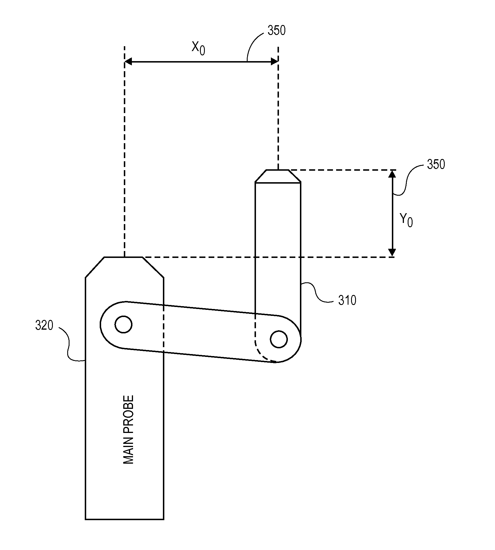
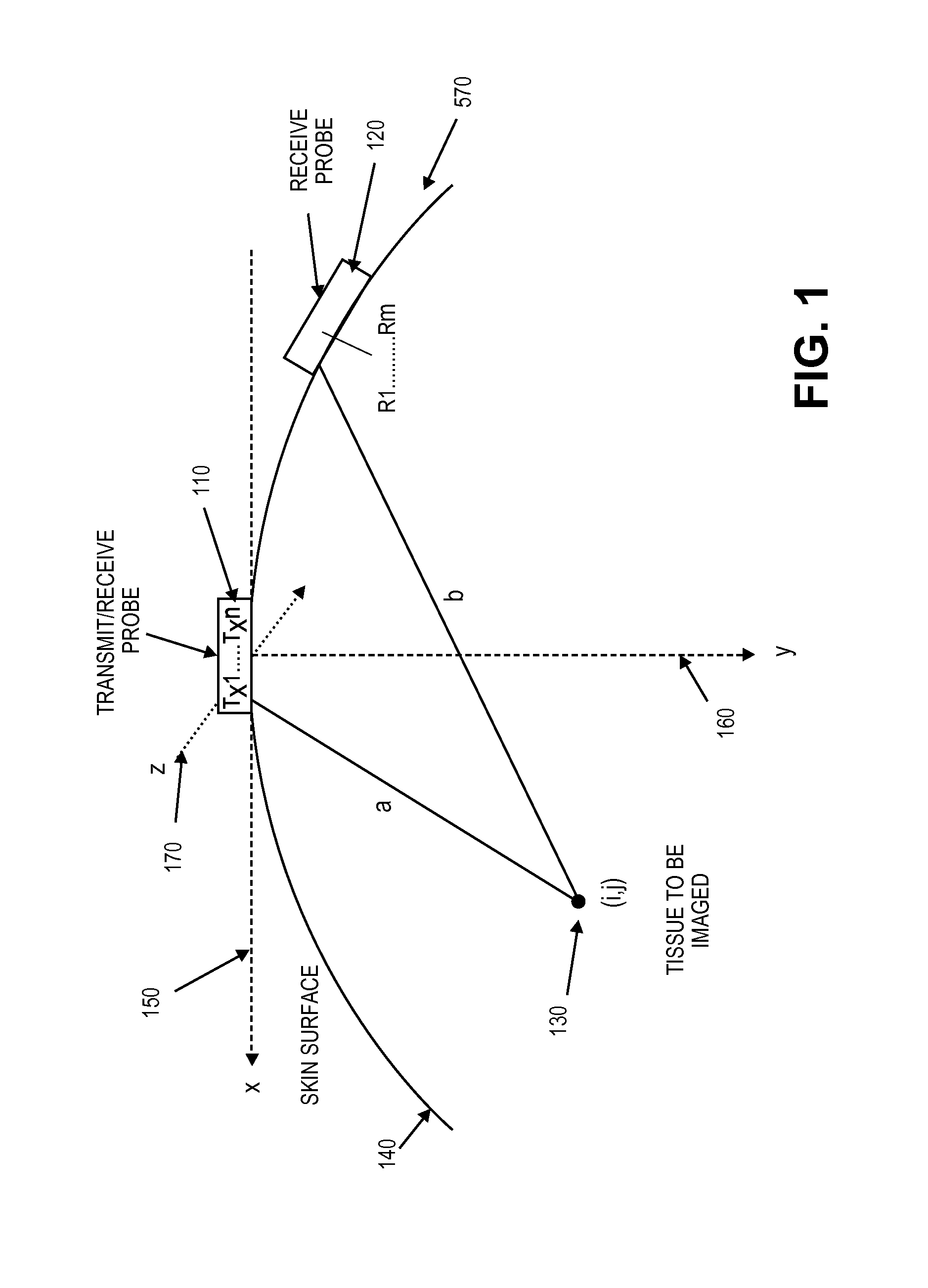
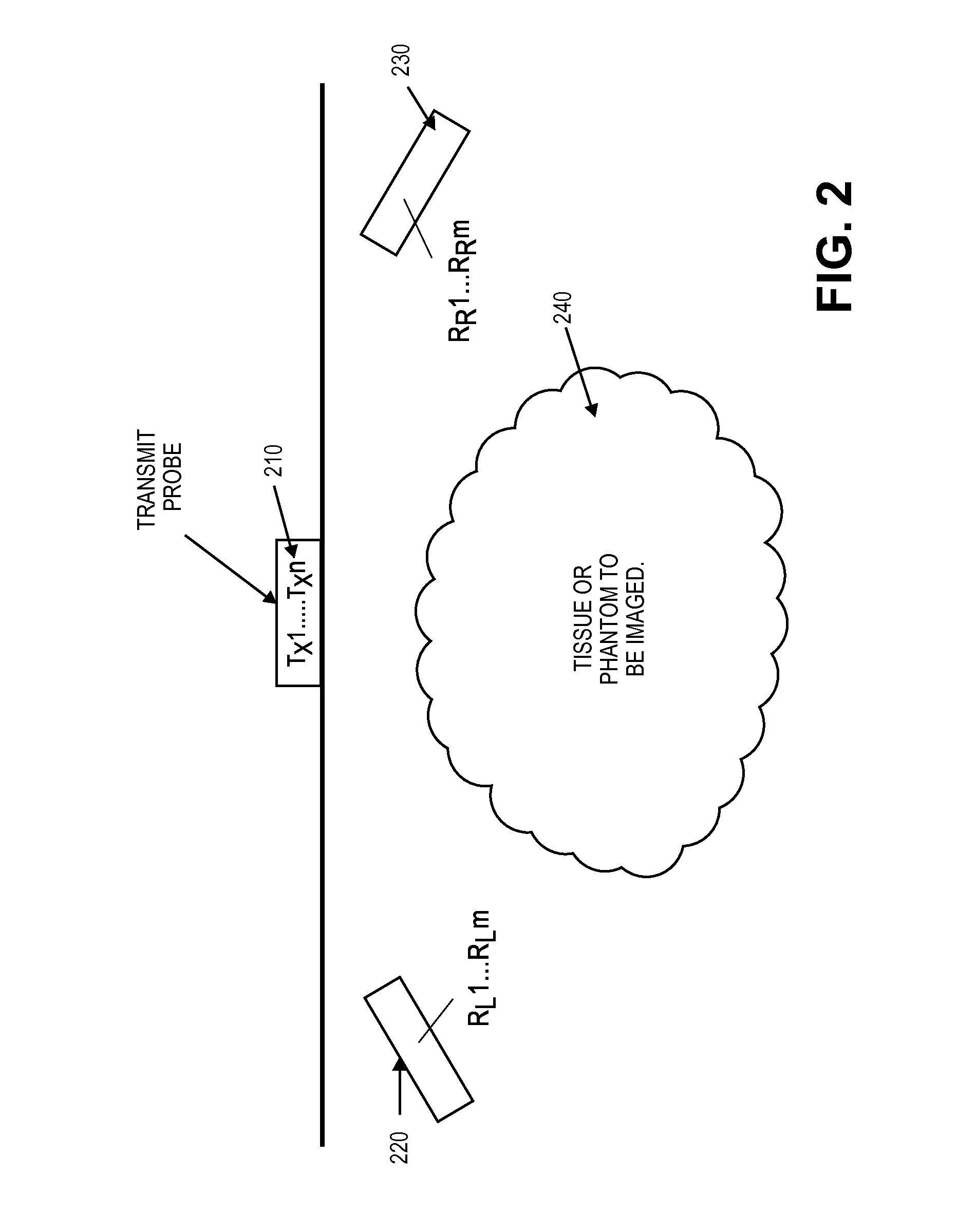
[0068]A Multiple Aperture Ultrasound Imaging (MAUI) Probe or Transducer can vary by medical application. That is, a general radiology probe can contain multiple transducers that maintain separate physical points of contact with the patient's skin, allowing multiple physical apertures. A cardiac probe may contain as few as two transmitters and receivers where the probe fits simultaneously between two or more intercostal spaces. An intracavity version of the probe, will space transmit and receive transducers along the length of the wand, while an intravenous version will allow transducers to be located on the distal length the catheter and separated by mere millimeters. In all cases, operation of multiple aperture ultrasound transducers can be greatly enhanced if they are constructed so that the elements of the arrays are aligned within a particular scan plane.

[0069]One aspect of the invention solves the problem of constructing a multiple aperture probe that functionally houses multip...


PUM


Abstract

A Multiple Aperture Ultrasound Imaging (MAUI) probe or transducer is uniquely capable of simultaneous imaging of a region of interest from separate physical apertures. Construction of probes can vary by medical application. That is, a general radiology probe can contain multiple transducers that maintain separate physical points of contact with the patient's skin, allowing multiple physical apertures. A cardiac probe may contain only two transmitters and receivers where the probe fits simultaneously between two or more intracostal spaces. An intracavity version of the probe can space transmit and receive transducers along the length of the wand, while an intravenous version can allow transducers to be located on the distal length the catheter and separated by mere millimeters. Algorithms can solve for variations in tissue speed of sound, thus allowing the probe apparatus to be used virtually anywhere in or on the body.

Description

CROSS REFERENCE TO RELATED APPLICATIONS[0001]This application is a continuation of U.S. application Ser. No. 12 / 760,375 filed Apr. 14, 2010, which application claims the benefit under 35 U.S.C. 119 of U.S. Provisional Patent Application No. 61 / 169,251, filed Apr. 14, 2009, titled “Universal Multiple Aperture Medical Ultrasound Transducer”, and U.S. Provisional Patent Application No. 61 / 169,221, filed Apr. 14, 2009, titled “Multi Aperture Cable Assembly for Multiple Aperture Probe for Use in Medical Ultrasound.”[0002]This application is related to U.S. patent application Ser. No. 11 / 865,501, filed Oct. 1, 2007, titled “Method and Apparatus to Produce Ultrasonic Images Using Multiple Apertures”, U.S. patent application Ser. No. 11 / 532,013, filed Sep. 14, 2006, titled “Method and Apparatus to Visualize the Coronary Arteries Using Ultrasound”, U.S. Provisional Patent Application No. 61 / 305,784, filed Feb. 18, 2010, titled “Alternative Method for Medical Multi-Aperture Ultrasound Imaging...

Claims


Application Information

Patent Timeline
Patent Type & Authority Applications(United States)
IPC IPC(8): A61B8/00A61B8/14
CPCA61B8/4218A61B8/145A61B8/54A61B8/4444A61B8/4494A61B8/4477A61B8/00A61B8/12A61B8/4254A61B8/4455A61B8/4488G01S15/003G01S15/8929A61B8/445A61B8/08G01N29/24
Inventor SMITH, DAVID M.ADAM, SHARON L.SPECHT, DONALD F.LUNSFORD, JOHN P.BREWER, KENNETH D.
Owner MAUI IMAGING
Learn More