Diagnosis and Treatment of Cancer Expressing ILT3 or ILT3 Ligand

a technology of ilt3 or ilt3 and cancer, applied in the direction of assay labels, drug compositions, peptides, etc., can solve the problems of difficult diagnosis and often inaccessible data

Active Publication Date: 2015-04-23
THE TRUSTEES OF COLUMBIA UNIV IN THE CITY OF NEW YORK
View PDF4 Cites 11 Cited by
  • Summary
  • Abstract
  • Description
  • Claims
  • Application Information

AI Technical Summary

Benefits of technology

[0008]In certain embodiments, cancers expressing ILT3 like CLL and AMLm4 / m5 with monocytic differentiation are diagnosed and distinguished from noncancerous cells or other forms of cancer by binding to an anti-ILT3 antibody that is detectably labeled, or that can be detected by binding to a secondary reagent that is detectably labeled. Detectable labels for use in embodiments of the invention are described below. In an embodiment, diagnosing ILT3-expressing cancers includes (a) obtaining a biological sample from a subject that has or may have cancer that expresses ILT3 on the cancer cell surface, and a control sample from a normal subject; (b) contacting the subject and control samples with an anti-ILT3 antibody or a portion thereof that selectively binds to ILT3 under conditions that permit the probe to bind to the ILT3 expressed on the cancer cell surface; (c) determining the number or percentage of cancer cells that are bound to the antibody in the subject and control samples; and (d) determining that the subject has the ILT3-expressing cancer, if the number or percentage of cells bound to the antibody in the subject sample is significantly higher than the corresponding number or percentage of cells bound to the antibody in the control sample.
[0009]Certain embodiments are directed to treating these ILT3-expressing cancers by administering therapeutically effective amounts of an antibody that selectively binds to the ILT3 on the surface of the cancer cells (including newly discovered anti-ILT3 mAbs A, B, C and D described herein), which antibody is linked either to an isotope that emits radiation at a level that kills the cancer cell, or to a cytotoxic agent, thereby treating the subject for the cancer. An exemplary embodiment is a) identifying a subject in need of treatment for a cancer that expresses ILT3 on its surface, and b) administering therapeutically effective amounts of an antibody that selectively binds to the ILT3 on the surface of the cancer cells, which antibody is linked either to an isotope that emits radiation at a level that kills the cancer cell, or to a cytotoxic agent, thereby treating the subject for the cancer.
[0010]Other embodiments are directed to methods for diagnosing and distinguishing cancers that express ILT3 ligand on their surface such as T-ALL, from noncancerous cells or other forms of cancer by (a) obtaining a biological sample from a subject that has or may have cancer that expresses ILT3 ligand on the cancer cell surface, and a control sample from a normal subject; (b) contacting the subject and control samples with a probe that selectively binds to ILT3 ligand under conditions that permit the probe to bind to the ILT3 ligand on the cancer cell surface; (c) determining the number or percentage of cancer cells that are bound to the probe in the subject and control samples; and (d) determining that the subject has the ILT3 ligand-expressing cancer, if the number or percentage of cells bound to the probe in the subject sample is significantly higher than the corresponding number or percentage bound in the control sample. In certain embodiments the probe that selectively binds to ILT3 ligand is ILT3, ILT3Fc, the extracellular domain of ILT3 or an ILT3 ligand-binding fragment of one of these molecules.
[0011]In other embodiments cancers expressing ILT3 ligand on their surface, like T-ALL are treated by a) identifying a subject in need of treatment for a cancer that expresses ILT3 ligand on its surface, and b) administering therapeutically effective amounts of ILT3, ILT3Fc, the extracellular domain of ILT3 or a fragment thereof comprising the ILT3 ligand binding site, which ILT3, ILT3Fc, extracellular domain and fragment is linked either to a radioactive isotope that emits radiation at a level that kills the cancer cell or to a cytotoxic agent, thereby treating the subject for the ILT3 ligand-expressing cancer.

Problems solved by technology

Currently no cellular markers specific for acute T-ALL are known, making diagnosis of the disease difficult.
Although the WHO classification system relies heavily on cytogenetic findings, these data are often not available at the time of diagnosis and initiation of treatment.

Method used


Image

  • Diagnosis and Treatment of Cancer Expressing ILT3 or ILT3 Ligand
  • Diagnosis and Treatment of Cancer Expressing ILT3 or ILT3 Ligand
  • Diagnosis and Treatment of Cancer Expressing ILT3 or ILT3 Ligand

Examples


example 1

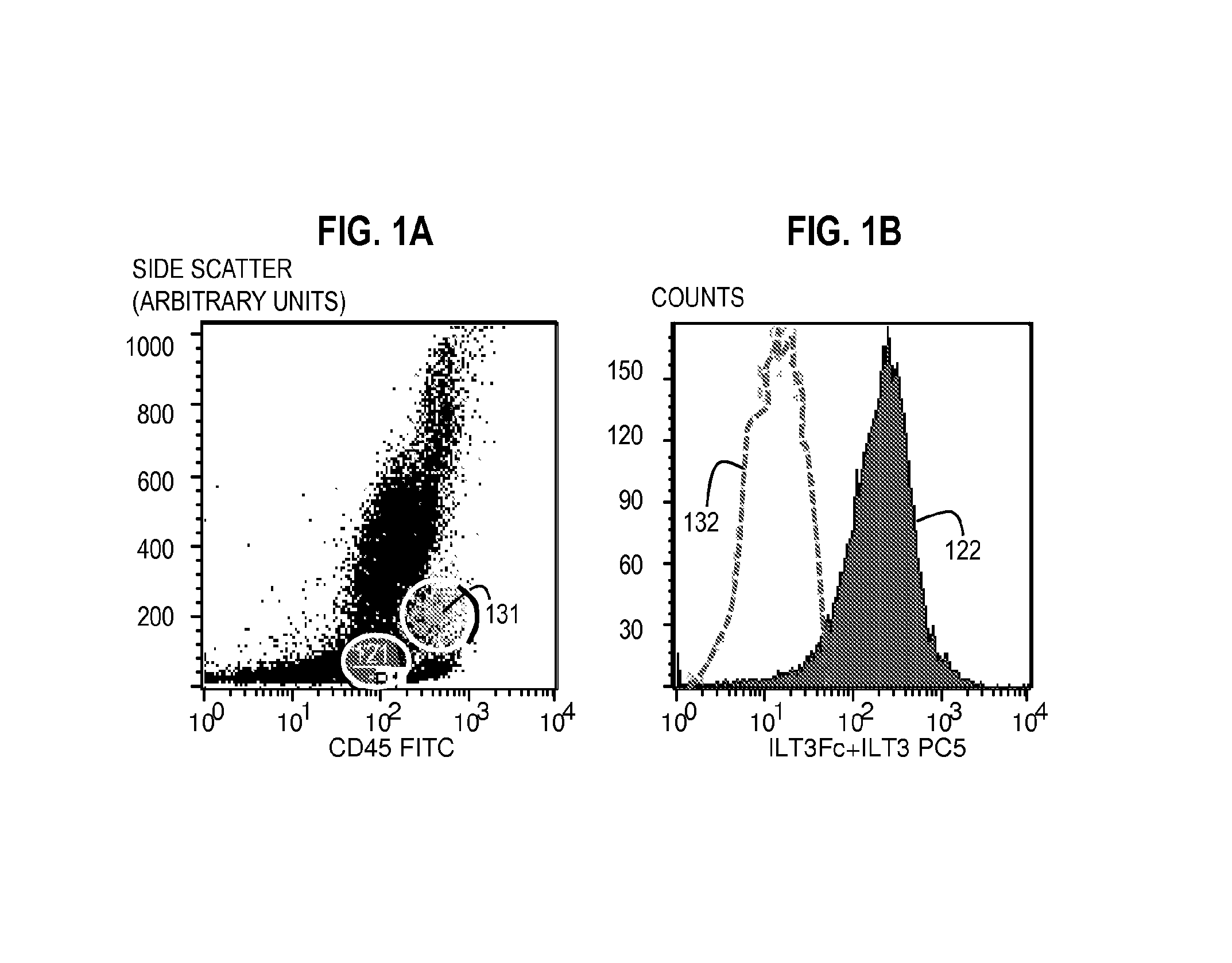
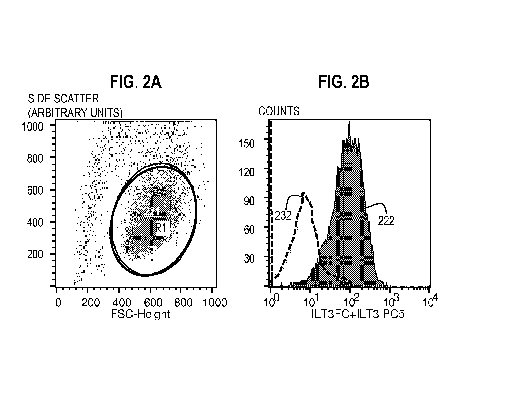
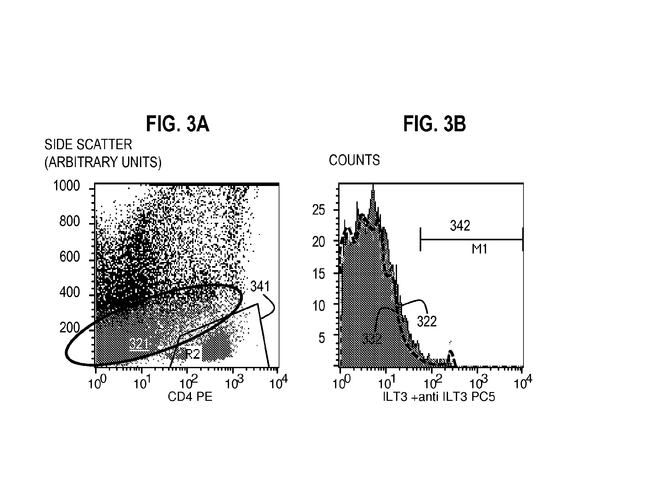
[0100]Flow Cytometry Method for Assaying the Expression of ILT3L Using Unlabeled ILT3Fc and a Labeled Secondary Reagent with Specific Affinity for ILT3Fc.

[0101]Bone marrow aspirate including peripheral blood mononuclear cells from patients with T-ALL or cultured T-ALL cell lines were incubated with 10 μg of unlabeled ILT3Fc for 30 minutes at 4° C. in 100 μl of staining buffer. The staining buffer consisted of Tris buffered saline (TBS), 1-3 mM Mn2, and 1% BSA or a similar formulation. Of note, ILT3Fc is preferably dissolved in a buffer compatible with the staining buffer used. Cells were washed three times in staining buffer. An appropriate negative control consisted of the same type of cells incubated with buffer alone (no ILT3Fc) or 10 μg human IgG during the first step.

[0102]The cells were then incubated with a secondary reagent (such as anti-Human ILT3 antibody) that binds to the ILT3 in the probe, wherein the secondary reagent is conjugated to a fluorophore such as PC5) for (30...

example 2

[0107]Targeted Lysis of T-ALL Cells that Express ILT3L with ILT3 Ligand-Binding Probes

[0108]T-ALL cells were suspended in RPMI 1640 medium at a concentration of 2×10⁶ cells / ml. First, cells were labeled with carbofluorescin diacetate (CFDA, 5 μM) for 10 minutes at 25° C., followed by thorough washing. The cells were then incubated with ILT3Fc (an ILT3 ligand-binding probe) (various concentrations, 0-250 μg / ml) for one hour at room temperature, then washed three times and incubated with rabbit complement for an additional hour.

[0109]Negative controls: the incubations were conducted without ILT3Fc, complement or both. Positive controls consisted of cells incubated with pooled human serum from allosensitized patients (containing anti HLA cytotoxic antibodies) as a first step and rabbit complement as a second step.

[0110]Finally, a viability dye (Ethidium Bromide, EtBr) was added and the cells were analyzed under a 488 nm light source. (Under 488 nm light, live cells appear green [CFDA+E...

example 3

[0113]Methods for making Anti-Human ILT3Fc Monoclonal Antibodies (mAb)

[0114]The procedures for production of monoclonal antibody were adapted from Current Protocols in Immunology (1995) 2.5 contributed by Wayne M. Yokoyama. A person of skill in the art would know how to make mAb from the antigen.

Immunization to Produce Monoclonal Antibodies.

[0115]1. Mix equal volume of human ILT3Fc protein (1 mg / ml in PBS) and CFA (Complete Freunds adjuvant, Sigma) or IFA (Incomplete Freund's adjuvant, Sigma) in a 3-way stopcock until the two parts are completely emulsified. The emulsion with CFA is used for the first time immunization. IFA emulsion is used for the rest of the immunization and boosting.

[0116]2. Inject 0.2 ml of emulsion (100 μg of ILT3Fc protein) intraperitoneally into female BALB / cj mice of 6-7 week old (JAX MICE) on day 1, 8, 15. The immunized mice are rested for 21 days, and are then boosted with 0.2 ml emulsion with IFA. Three days after boosting, mice are sacrificed, and their ...


PUM

PropertyMeasurementUnit
concentrationsaaaaaaaaaa
fluorescentaaaaaaaaaa
time courseaaaaaaaaaa

Abstract

The present invention relates to methods of using the expression of ILTL3 ligand or ILT3 on certain types of cancer cells as a diagnostic tool. Methods are provided for treating ILT3-ligand expressing cancers, such as T-cell acute lymphoblastic leukemia (T-cell acute lymphoblastic leukemia), for example by administering ILT3, the extracellular domain of ILT3 or ILT3Fc conjugated to a cytotoxic agent to kill the targeted cancer cell. Other methods are provided for treating cancers that express ILT3 on their surface, such as monocytic forms of AML, for example by administering anti-ILT3 antibodies conjugated to a cytotoxic agent.

Description

CROSS-REFERENCE TO RELATED APPLICATIONS[0001]This application claims benefit of the following U.S. Provisional Applications: 61 / 590,510, filed Jan. 25, 2012, entitled, “Diagnosis of Acute Lymphoblastic Leukemia” and, 61 / 530,936, filed Sep. 2, 2011, entitled, “Diagnosis and Treatment of Human Malignancies Using mAb Anti-ILT3 or ILT3Fc”, the entire contents of which are hereby incorporated by reference as if fully set forth herein, under 35 U.S.C. §119 (e).BACKGROUND OF THE INVENTION[0002]Acute lymphoblastic leukemia (ALL) is a form of leukemia that is characterized by the presence of excess lymphoblasts due to the continuous multiplication of malignant immature white blood cells. ALL is a disease that rapidly progresses and is characterized by crowding of normal cells in the bone marrow due to the continuous multiplication of immature white blood cells.[0003]Precursor T-cell acute lymphoblastic leukemia (T-ALL) causes 15% of acute leukemias in childhood, and approximately 40% of lymp...

Claims


Application Information

Patent Timeline
Patent Type & Authority Applications(United States)
IPC IPC(8): G01N33/574C07K14/705C07K16/28
CPCG01N33/57492C07K16/28G01N2458/00G01N2333/70596C07K14/705G01N33/57426C07K16/2803C07K14/70503C07K2319/30G01N2800/52A61K47/6867A61K47/6801G01N2333/70503A61K47/6803A61K47/6849G01N33/57484A61K2039/505C07K14/7051C07K16/3061C07K2319/33C07K2319/40C07K2319/55C07K2319/74
Inventor SUCIU-FOCA, NICOLEVLAD, GEORGECHANG, CHIH-CHAOLIU, ZHUORUCOLOVAI, ADRIANA IOANA
Owner THE TRUSTEES OF COLUMBIA UNIV IN THE CITY OF NEW YORK
Why Eureka
Try Eureka