Method of classification of organs from a tomographic image

a tomographic image and classification method technology, applied in image data processing, diagnostics, sensors, etc., can solve the problem that the identification and analysis of a previous image cannot be reused on a new image, and achieve the effect of extra robustness in classification

Active Publication Date: 2015-05-07
AMRA MEDICAL AB
View PDF6 Cites 2 Cited by
  • Summary
  • Abstract
  • Description
  • Claims
  • Application Information

AI Technical Summary

Benefits of technology

[0010]The method according to the present invention may enable a mapping of Hounsfield attenuation values defined in the prototype image to an MR (Magnetic Resonance) image. MR images may then be used for attenuation correction in PET (Positron Emission Tomography) imaging.
[0011]Extra robustness in the classification may be achieved by using the multi-field data in the water and fat image data sets. For example, the labeled volume elements for an organ in the target image may be labeled as adipose tissue. If the labeled volume elements may be multiplied with the fat image data set, and labeled volume element wrongly transferred into a water area in the target image may automatically be removed.

Problems solved by technology

The identification and analysis of a previous image is not possible to reuse on a new image.

Method used


Image

  • Method of classification of organs from a tomographic image
  • Method of classification of organs from a tomographic image
  • Method of classification of organs from a tomographic image

Examples


Embodiment Construction

[0067]The present invention will be described more fully hereinafter with reference to the accompanying drawings, in which preferred embodiments of the invention are shown. This invention may, however, be embodied in many different forms and should not be construed as limited to the embodiments set forth herein; rather, these embodiments are provided so that this disclosure will be thorough and complete, and will fully convey the scope of the invention to those skilled in the art. In the drawings, like numbers refer to like elements.

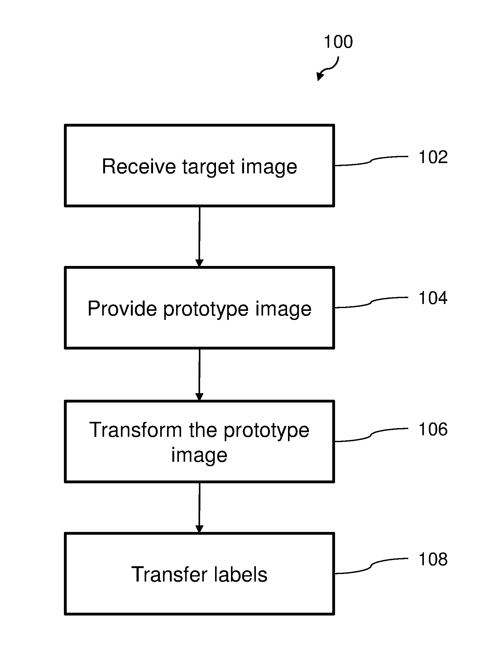
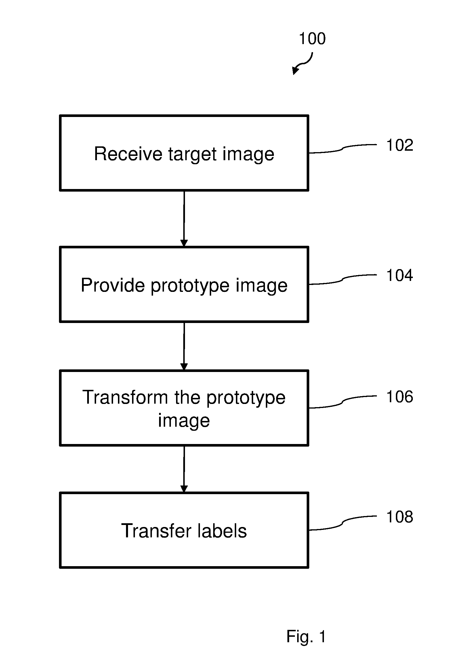
[0068]FIG. 1 illustrates a flow chart of a method 100 according to an embodiment, for registration and classification of organs in a body. The method 100 comprises a first step 102 wherein a 3-dimensional tomographic image is received. The image is provided from a medical scanning apparatus, such as a magnetic resonance (MR) scanner or a computed tomography (CT) scanner. The received tomographic image is a target image, i.e. the image in which organs is ...


PUM


Abstract

The present invention relates to a method for classification of an organ in a tomographic image. The method comprises the steps of receiving (102) a 3-dimensional anatomical tomographic target image comprising a water image data set and a fat image data set, each with a plurality of volume elements, providing (104) a prototype image comprising a 3-dimensional image data set with a plurality of volume elements, wherein a sub-set of the volume elements are given an organ label, transforming (106) the prototype image by applying a deformation field onto the volume elements of the prototype image such that each labeled volume element for a current organ is determined to be equivalent to a location for a volume element in a corresponding organ in the target image, and transferring (108) the labels of the labeled volume elements of the prototype image to corresponding volume elements of the target image.

Description

TECHNICAL FIELD[0001]The present disclosure relates to classification of organs in an image, and especially to the classification of organs in a 3-dimensional anatomical tomographic image comprising a water image data set and a fat image data set.BACKGROUND[0002]In 3-dimensional tomographic medical imaging, identification of different body tissues and organs, such as muscles, is an important part of the medical interpretation of the images. The size, shape and composition, and their variations over time, of different organs can be an important piece of information in many clinical questions. The identification of such features is in general a manual process, which includes manually identifying a specific organ and possibly labeling it in the image. The present image may, after a complete identification and analysis process, comprise a plurality of labels for different organs. Points, or voxels, in the 3-dimensional image may thereby represent a location of an organ.[0003]However, wh...

Claims


Application Information

Patent Timeline
Patent Type & Authority Applications(United States)
IPC IPC(8): G06T7/00
CPCG06T7/0014G06T7/0085G06T2207/30056G06T2207/10088G06T2207/10104G06T2207/10081A61B5/055A61B6/032A61B6/037A61B6/5217A61B6/5247G06T7/11G06T7/174G06T7/337
Inventor DAHLQVIST LEINHARD, OLOFBORGA, MAGNUS
Owner AMRA MEDICAL AB
Why Eureka
Try Eureka