
Virtual endoscopic image generation device, method, and medium containing program

Active Publication Date: 2015-07-09
FUJIFILM CORP
View PDF3 Cites 7 Cited by
  • Summary
  • Abstract
  • Description
  • Claims
  • Application Information

AI Technical Summary

Benefits of technology

The virtual endoscopic image generation device and method described in this patent allow for the creation of images that show a subject's body cavity in a way that can be observed from the view point position of a virtual endoscope. The device can extract structures within the body cavity and change their opacity based on their distance from the view point position. This allows for appropriately visibility and observation of each structure based on the virtual endoscope's position and direction of line of sight. Overall, the technology provides a better way to visualize the subject's body cavity and aid in medical diagnosis and treatment.

Problems solved by technology

This may make it difficult to see the structure of interest.

Method used


Image

  • Virtual endoscopic image generation device, method, and medium containing program
  • Virtual endoscopic image generation device, method, and medium containing program
  • Virtual endoscopic image generation device, method, and medium containing program

Examples


Embodiment Construction

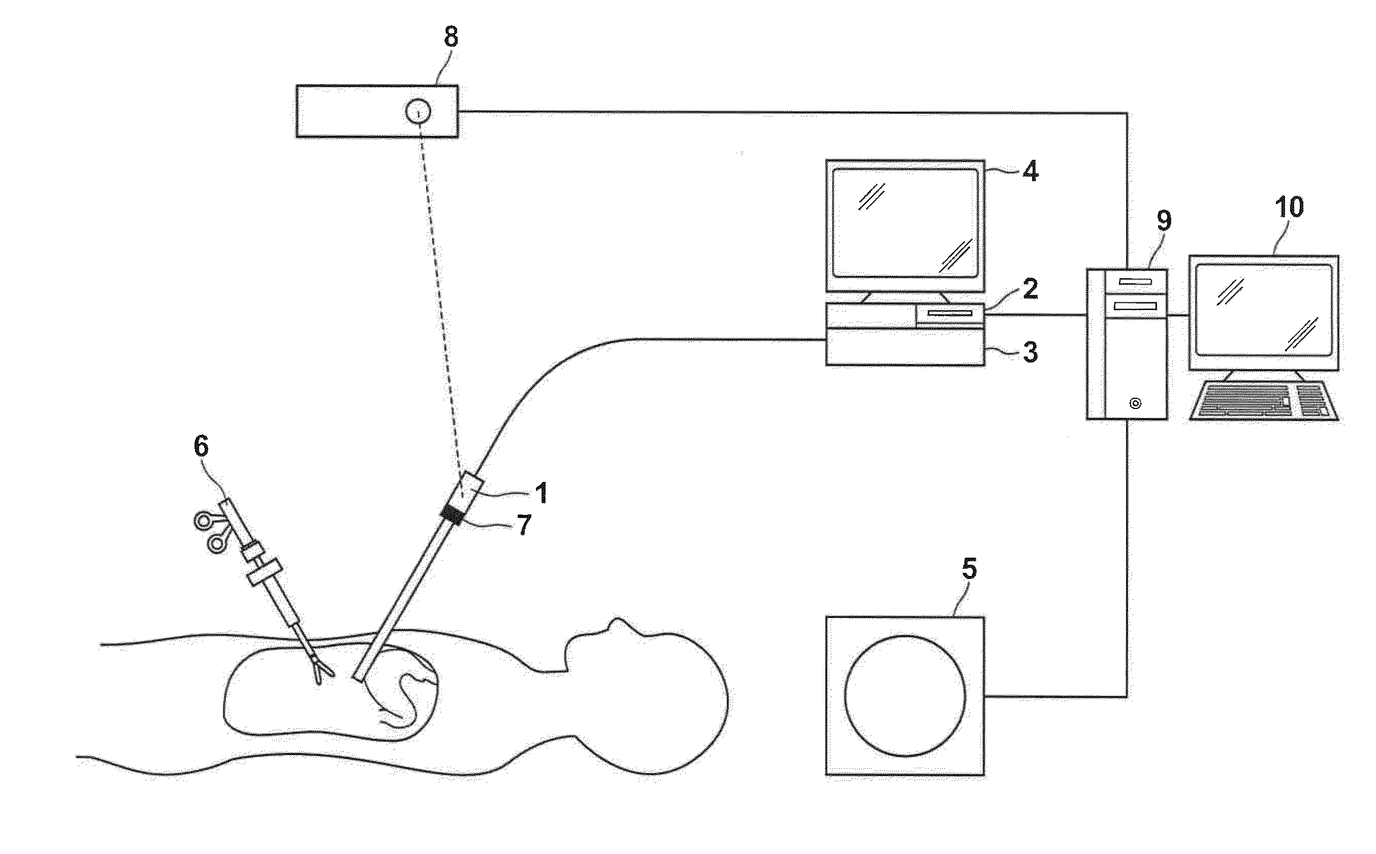
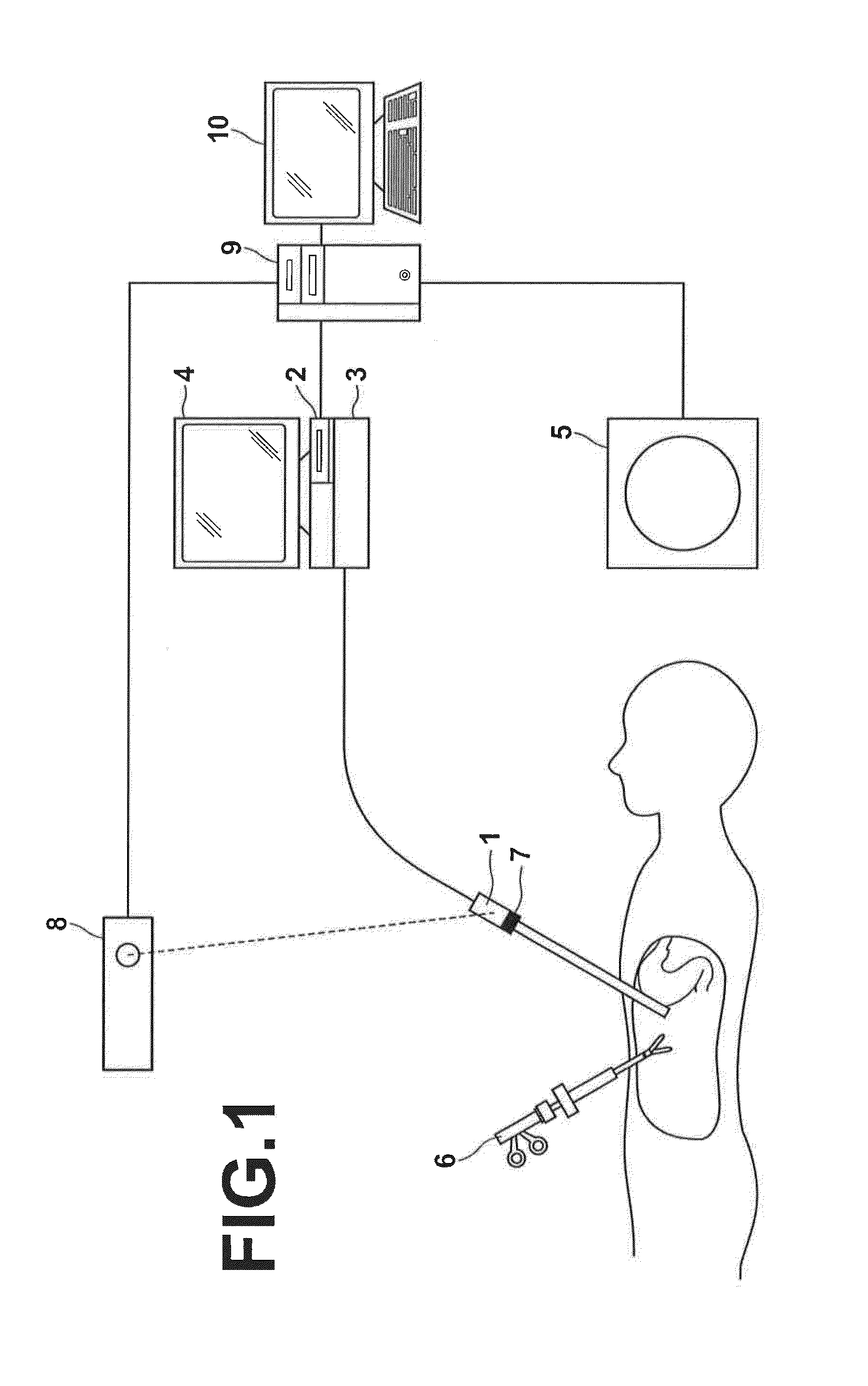
[0035]Hereinafter, an embodiment of the present invention will be described with reference to the drawings. FIG. 1 is a diagram illustrating the hardware configuration of an endoscopic observation assisting system to which a virtual endoscopic image generation device according to an embodiment of the invention is applied. As shown in FIG. 1, this system includes an endoscope 1, a digital processor 2, a light source unit 3, a real endoscopic image display 4, a modality 5, a surgical tool 6, an endoscope marker 7, a position sensor 8, an image processing workstation 9, and an image processing workstation display (which will hereinafter be referred to as “WS display”) 10.

[0036]The endoscope 1 of this embodiment is a rigid endoscope for abdominal cavity, and is inserted in the abdominal cavity of a subject. Light from the light source unit 3 is guided through optical fibers and is outputted from the distal end of the endoscope 1 to illuminate the abdominal cavity, and an image of the in...


PUM


Abstract

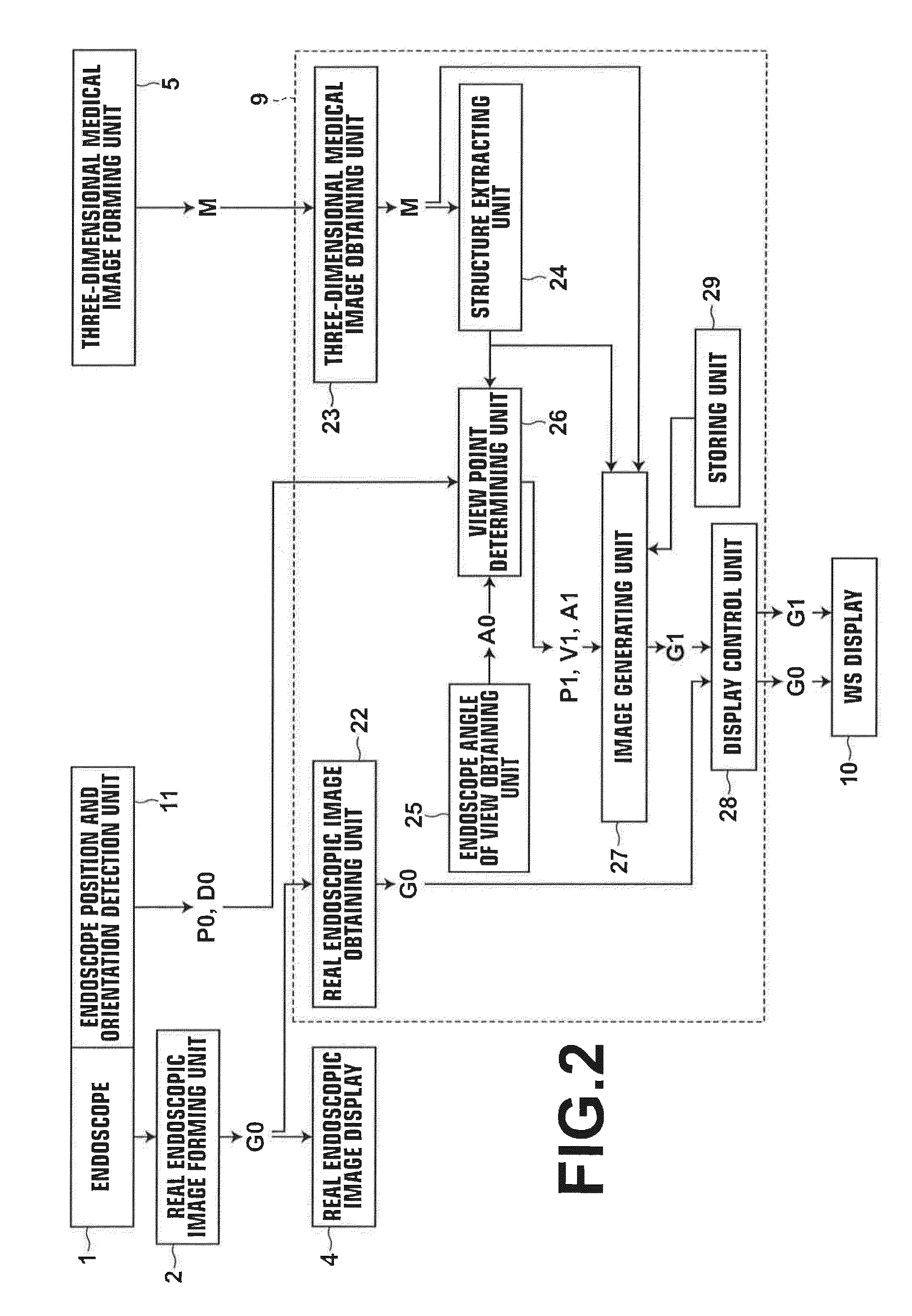
A structure extracting unit extracts a structure from a three-dimensional medical image, and a view point determining unit determines a view point position and a direction of line of sight of a virtual endoscopic image. An image generating unit calculates a distance between the view point position and the extracted structure, changes an opacity defined in a color template depending on the distance, and generates, from the three-dimensional medical image, a virtual endoscopic image containing the structure shown according to the color template with the changed opacity viewed from the view point position in the direction of line of sight. A display control unit displays the thus generated virtual endoscopic image on a WS display.

Description

CROSS-REFERENCE TO RELATED APPLICATIONS[0001]This application is a Continuation of PCT International Application No. PCT / JP2013 / 005495 filed on Sep. 18, 2013, which claims priority under 35 U.S.C. §119(a) to Japanese Patent Application No. 2012-211643 filed on Sep. 26, 2012. Each of the above applications is hereby expressly incorporated by reference, in its entirety, into the present application.BACKGROUND OF THE INVENTION[0002]1. Technical Field[0003]The present invention relates to a device, a method, and a program for generating a virtual endoscopic image that shows the interior of a body cavity of a subject for assisting endoscopic observation during surgery, examination etc., using an endoscope inserted into the body cavity of the subject.[0004]2. Background Art[0005]Endoscopic surgeries, such as laparoscopic surgery and thoracoscopic surgery, are drawing attention in recent years. The endoscopic surgery has an advantage in that the burden imposed on the patient is quite small...

Claims


Application Information

Patent Timeline
IPC IPC(8): A61B1/00G06K9/46G06T7/00G06T19/00H04N13/02
CPCA61B1/00193G06T19/006G06T2207/10068G06T7/004G06K9/468H04N13/0203A61B1/0005G06T19/20G06T2210/41G06T2219/2012A61B6/12A61B8/0841A61B1/000094A61B1/00194H04N13/204
Inventor SAKURAGI, FUTOSHI
Owner FUJIFILM CORP
Learn More