
Methods for detecting lymphedema

Inactive Publication Date: 2015-08-06
SAYEGH SAMIR I +1
View PDF0 Cites 23 Cited by
  • Summary
  • Abstract
  • Description
  • Claims
  • Application Information

AI Technical Summary

Benefits of technology

The invention provides methods for detecting and managing lymphedema, which involves using a light sensing device to capture three-dimensional images of the body and analyze them to detect abnormal swelling. The methods can detect lymphedema by measuring the size of specific regions of the body and comparing them to a reference size. The invention also includes methods for monitoring and managing lymphedema by measuring the amount of extracellular fluids and skin thickness in specific regions. The invention can help improve early detection and treatment of lymphedema by providing a non-invasive and non-intrusive method for monitoring and managing the condition.

Problems solved by technology

Lymphedema can have a profound impact on patient health as it can lead to restricted range of motion, aching or discomfort, recurring infections and fibrosis of the skin.

Method used


Image

  • Methods for detecting lymphedema

Examples


example 1

Light Sensing Devices

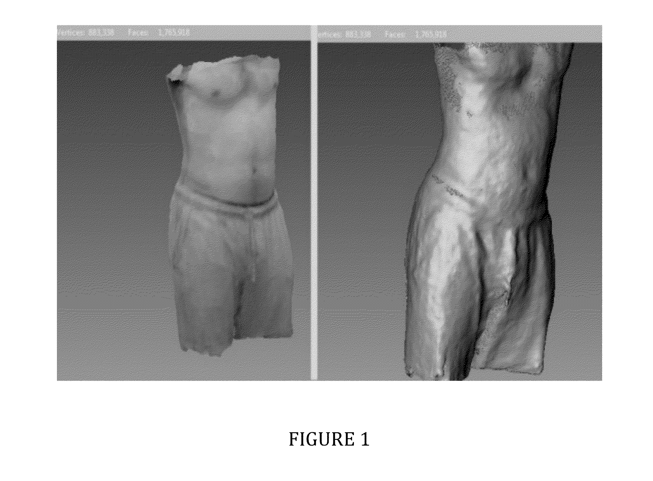
[0049]KINECT (Microsoft Corporation, Redmond, Wash.) was used to perform a three-dimensional scan of at least a portion of the body of an individual, for example, an extremity or the neck, trunk or entire body to obtain a three-dimensional digital model of an extremity or the neck, trunk or entire body as shown in FIG. 1. Methods similar to that described in Jing et al., Scanning 3D Full Human Bodies Using Kinects, IEEE T. Vis. Comput. Gr. 18:643-650 (2012) were used. In addition, one or more KINECT devices can be used in a scanning system in which the devices rotate around a stationary individual or the individual is rotated, for example using a turntable, before one or more devices as described in Jing et al. The individual is positioned about 1 meter from one or more devices, the devices or individual rotating about 360° in about 30 seconds. 1280×1024 color images and 640×480 depth images are acquired at 15 frames per second, and three-dimensional coordinates...

example 2

Bioimpedance Spectroscopy (BIS)

[0051]To obtain BIS measurements using Imp SFB7, electrodes are positioned at select positions of an individual's body, for example, at select positions on the upper or lower extremities. Identical electrode positions are used on the left and right extremities to allow comparison of bioimpedance at the left and right extremities. Impedance is calculated using the ImpediMed's software. Statistical analysis is performed using Student's 2-tailed t test where needed. The difference between the left and right extremities can be expressed as a ratio.

[0052]BIS can also be performed using the Imp XCA, which employs a single frequency below 30 kHz to measure impedance and resistance of the ECF. Methods known to those of skilled in the art can be used. For example, an individual is placed in a fully supine position with the legs not touching and the arms extended 30° from the body by their sides. Two dual-tab electrodes are placed on the dorsum of the right and ...

example 3

Ultrasound Imaging

[0054]Skin thickness can be determined by ultrasound imaging using systems known to those of skilled in the art including, for example, a Sonoline Antares ultrasound system (Siemeans, Erlanger, Germany), an Aloka SSD-1700 Diagnostic Ultrasound System (Aloka Co., Ltd., Wallingford, Conn.), DermaScan C ultrasound scanner (Cortex Technology, Smedevaenget, Denmark), and Dermcup 2020 (Atys Medical, Soucieu en Jarrest, France). Images can be obtained and analysed using methods known to those of skilled in the art, for example, see Tassenoy et al., Postmastectomy Lymphoedema: Different Patterns of Fluid Distribution Visualized by Ultrasound Imaging Compared with Magnetic Resonance Imaging, Physiotherapy 97: 234-243 (2011); Van Der Veen et al., A Key to Understanding Postoperative Lymphoedema: A Study on the Evolution and Consistency of Oedema of the Arm Using Ultrasound Imaging, The Breast 10:225-230 (2001); Mellor et al., Dual-Frequency Ultrasound Examination of Skin and...


PUM


Abstract

The invention provides methods for detecting lymphedema and methods for managing lymphedema in an individual. The methods of the invention involves utilizing a light sensing device having a video camera, depth sensor and software effective to perform a 3-dimensional scan and generate a three-dimensional digital model of at least a portion of the body from data obtained by the camera and depth sensor, thereby obtaining a three-dimensional digital model of at least a portion of the body from which the size of a select region of the body can be assessed to detect, monitor and / or manage lymphedema. The invention also provides methods to detect and manage lymphedema that involve assessing any combination of size, amount of extracellular fluids, and thickness of the skin at a select region of the body of an individual.

Description

CROSS-REFERENCE TO RELATED APPLICATIONS[0001]This application claims priority to U.S. Provisional Patent Ser. No. 61 / 936,861 filed Feb. 6, 2014, which is incorporated herein by reference in its entirety.BACKGROUND[0002]Lymphedema is swelling in regions of the body that include the upper and lower extremities and can occur in individuals who have undergone cancer treatment. Lymphedema is caused by abnormal buildup of protein-rich fluids in the interstitial space resulting from inadequate lymphatic drainage. Upper extremity lymphedema, for example, has been documented in patients who have undergone treatment for breast cancer. Lymphedema can have a profound impact on patient health as it can lead to restricted range of motion, aching or discomfort, recurring infections and fibrosis of the skin. Although there is no cure for lymphedema, early diagnosis can have a significant impact on management of the progression of lymphedema and the associated discomfort and loss of function.SUMMARY...

Claims


Application Information

Patent Timeline
IPC IPC(8): A61B5/00A61B8/08A61B5/107A61B5/053
CPCA61B5/4878A61B5/0077A61B5/1073A61B8/0858A61B5/1079A61B5/0537A61B5/418G06T7/0016G06T7/0014G06T7/62G06T2207/10028
Inventor SAYEGH, SAMIR I.TAGHIAN, ALPHONSE
Owner SAYEGH SAMIR I
Learn More