Endoscopic ultrasound-guided notched biopsy needle

a biopsy needle and ultrasound technology, applied in the field of endoscopic surgical devices, can solve the problems of limiting the diagnostic value of the procedure, the risk of false negatives,

Active Publication Date: 2015-08-20
COOK MEDICAL TECH LLC
View PDF5 Cites 4 Cited by
  • Summary
  • Abstract
  • Description
  • Claims
  • Application Information

AI Technical Summary

Problems solved by technology

As a result, this technique includes a risk of false negatives where the few cells obtained in a sample do not accurately represent the presence of a tumor or other disease condition.
The small sample size may also limit the diagnostic value of the procedure if the cells obtained are sufficiently few in number or sufficiently damaged during collection that they do not enable a definitive diagnosis.

Method used


Image

  • Endoscopic ultrasound-guided notched biopsy needle
  • Endoscopic ultrasound-guided notched biopsy needle
  • Endoscopic ultrasound-guided notched biopsy needle

Examples


Embodiment Construction

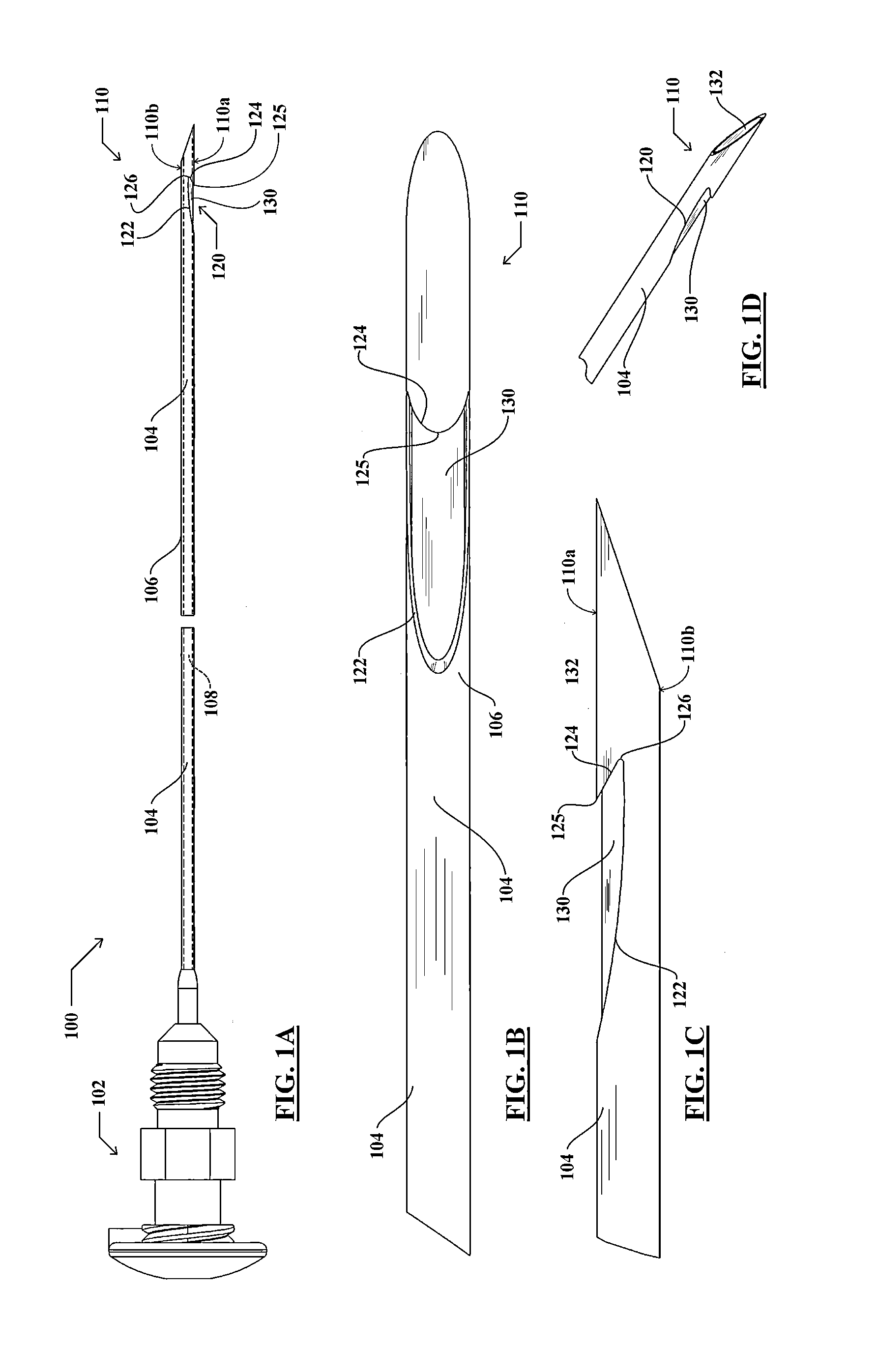
[0013]As used herein, the term “proximal” refers to the handle-end of a device held by a user, and the term “distal” refers to the opposite end. The term “surgical visualization device” refers to endoscopes including CCD, ultrasound, fiber optic, and CMOS devices, as well as other devices used for visualizing an internal portion of a patient body such as, for example, a laparoscope or bronchoscope.

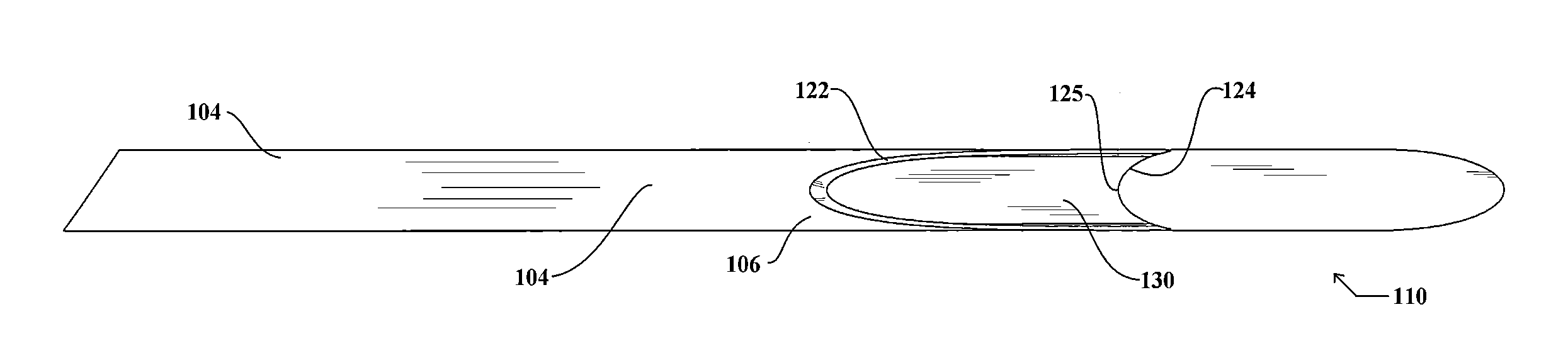
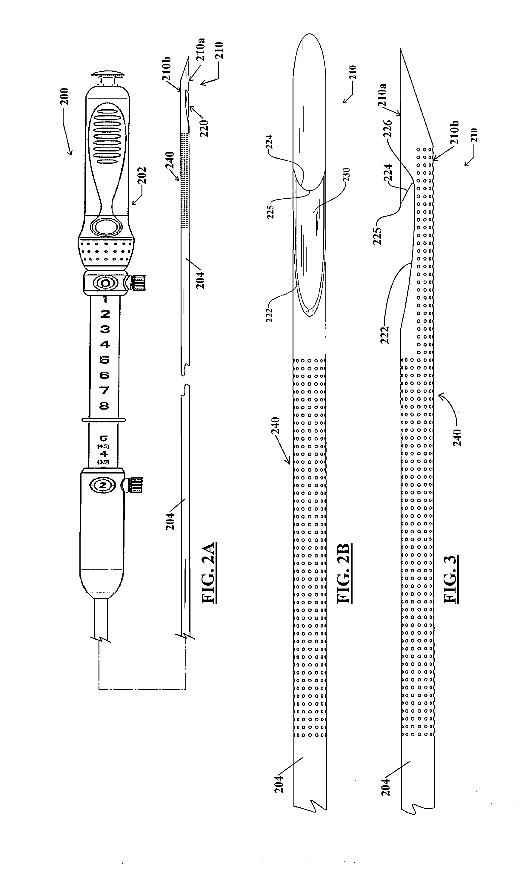
[0014]One embodiment of a tissue-sampling needle device is described with reference to FIGS. 1A-1D, which show a tissue-sampling needle device 100. As shown in the side plan view of FIG. 1A, the device includes a proximal handle or hub 102 from which an elongate tubular cannula 104 extends distally. The cannula 104 includes a cannula wall 106 that defines a cannula lumen 108. A distal end 110 of the cannula 104 is beveled, including a long side 110a substantially parallel with the central longitudinal axis of the cannula 104 and extending to its distal-most tip end. A short side 110b of th...


PUM


Abstract

A notched tissue-collection needle configured similarly to a fine-needle-aspiration needle is provided with a cutting edge disposed in the notch and configured to excise tissue into the notch for collection. A stylet may be provided through a lumen of the needle during introduction into a patient body. The needle may be provided with echogenicity-enhancing features.

Description

CROSS-REFERENCE TO RELATED APPLICATIONS[0001]This application claims priority as a Continuation under 35 USC §120 to U.S. patent application Ser. No. 13 / 079,226, originally filed Apr. 6, 2010, (as U.S. Provisional Application Ser. No. 61 / 321,243, converted under 37 CFR §1.53(c)(3)) each of which is incorporated herein by reference in its entirety.TECHNICAL FIELD[0002]The invention relates generally to endoscopic surgical devices. More particularly, the invention pertains to a biopsy needle configured for use during minimally-invasive procedures such as endoscopic procedures.BACKGROUND[0003]Fine needle aspiration (FNA) is a diagnostic biopsy procedure used to obtain a sample from a target site in a patient body. A fine needle (e.g., 19-gauge to 25-gauge) is directed to a target site, and suction is applied to the proximal end of a lumen of the needle to aspirate cells through its distal end. The procedure typically is far less invasive than other biopsy techniques, whether performed ...

Claims


Application Information

Patent Timeline
Patent Type & Authority Applications(United States)
IPC IPC(8): A61B10/02A61B1/24A61B10/04A61B8/08A61B1/018
CPCA61B10/0275A61B1/018A61B1/24A61B8/0841A61B8/481A61B10/04A61B2010/0093A61B2010/045A61B2017/3413A61B2090/3925
Inventor SCHEMBRE, DREW B.CLANCY, MICHAEL S.CHMURA, KEVIN
Owner COOK MEDICAL TECH LLC
Why Eureka
Try Eureka