Method of cancer diagnosis, progression and response to therapy using a primary xenograft mouse model for cancer serum biomarker discovery

Active Publication Date: 2015-09-10
H LEE MOFFITT CANCER CENT & RES INST INC
View PDF0 Cites 3 Cited by
  • Summary
  • Abstract
  • Description
  • Claims
  • Application Information

AI Technical Summary

Benefits of technology

[0004]The inventors have shown that patient tumor tissue that grows in immunocompromised mice allows identification of secreted tumor specific bi

Problems solved by technology

However, frequently cancer patients are diagnosed with advanced stage disease which adversely affects clinical outcome

Method used


Image

  • Method of cancer diagnosis, progression and response to therapy using a primary xenograft mouse model for cancer serum biomarker discovery
  • Method of cancer diagnosis, progression and response to therapy using a primary xenograft mouse model for cancer serum biomarker discovery
  • Method of cancer diagnosis, progression and response to therapy using a primary xenograft mouse model for cancer serum biomarker discovery

Examples


example

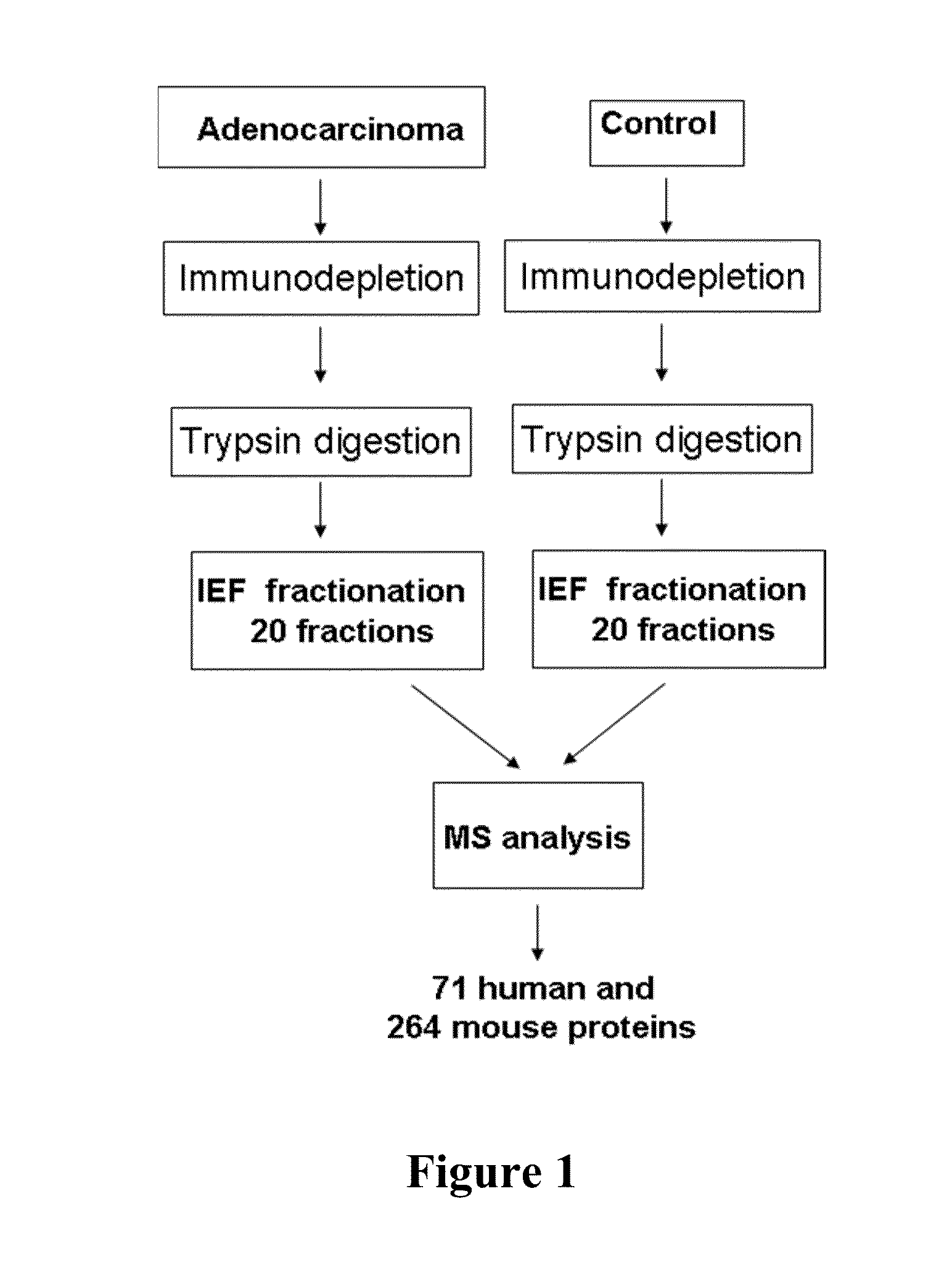
[0057]An overview of the experimental workflow is shown in FIG. 1. Mouse serum was immunodepleted of the 7 most abundant proteins using a Seppro® Mouse Spin Columns and concentrated to 300 μL using 5 kDa MWCO spin concentrators. Denaturing buffer was added and buffer exchange step was repeated twice. After digestion with porcine trypsin the samples were de-salted followed by elution process. Peptide solutions were IEF fractionated to 20 fractions. The peptides are extracted and dried. After re-suspension the samples were injected on column for MS analysis. A nanoflow liquid chromatograph (U3000, Dionex, Sunnyvale, Calif.) coupled to a quadrupole time-of-flight mass spectrometer (QSTAR® Elite, Applied Biosystems, Foster City, Calif.) was used for tandem mass spectrometry peptide sequencing experiments.

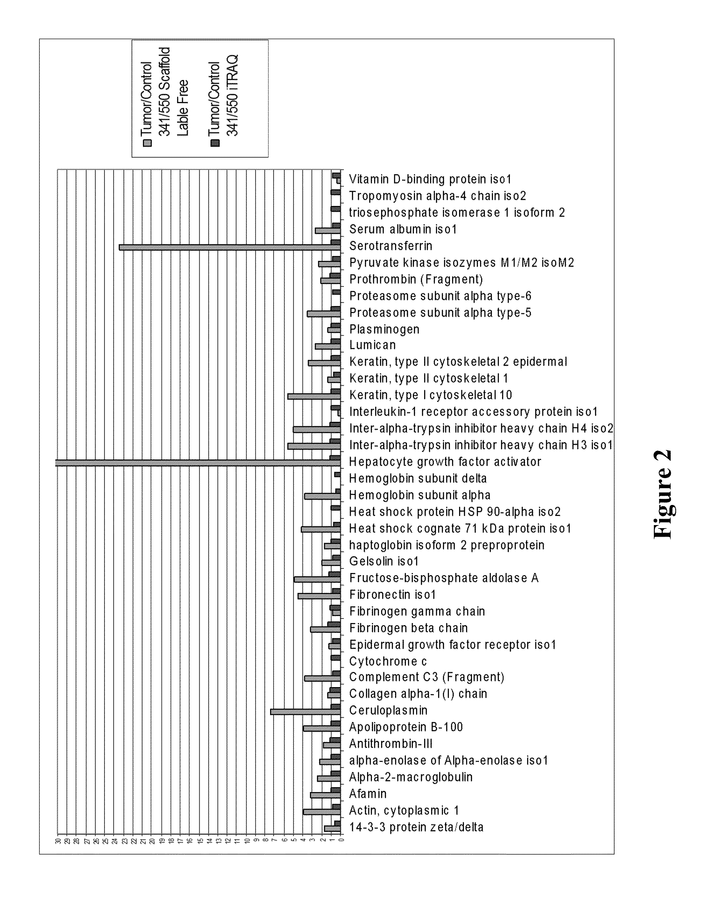
[0058]FIG. 2 is a graph illustrating that plasma / serum obtained from xenograft models of human cancer prepared with fresh human tumor can be used to identify low-abundance cancer specif...


PUM

PropertyMeasurementUnit
Gene expression profileaaaaaaaaaa
Timeaaaaaaaaaa
Levelaaaaaaaaaa

Abstract

A method of determining disease diagnosis, progression and response to therapy through the use of xenograft animal models for determining cancer biomarkers is presented. In use, a tumor from a patient is transplanted into an animal model such as a mouse. Expression levels of biomarkers for a disease are assessed in the serum of the animal model. A personalized gene expression profile or biomarker gene signature may be obtained from the expression levels of the biomarkers which can be used to determine disease recurrence, progression and response to therapy.

Description

CROSS REFERENCE TO RELATED APPLICATIONS[0001]This application is a continuation of prior filed International Application, No. PCT / US2013 / 070778 filed on Nov. 19, 2013, which claims priority to U.S. Provisional Application No. 61 / 727,870, entitled “Primary Xenograft Mouse Model of Human Cancer as a Platform for Personalized Cancer Serum Biomarker Discovery”, filed on Nov. 19, 2012, the entire contents of which are hereby incorporated by reference into this disclosure.FIELD OF INVENTION[0002]This invention relates to tumorigenic assays. Specifically, the invention provides a method of detecting tumor growth as well as monitoring tumor progression / regression and assessing the therapeutic response of the tumor to an agent using a biomarker signature derived from patient-derived mouse xenograft models.BACKGROUND OF THE INVENTION[0003]Early detection of cancer is generally recognized as the most effective strategy to reduce mortality. However, frequently cancer patients are diagnosed with...

Claims


Application Information

Patent Timeline
IPC IPC(8): G01N33/68
CPCG01N33/6893G01N2570/00G01N2800/60G01N2800/52C12Q1/6886C12Q2600/158G01N33/5088G01N33/57484
Inventor ALTIOK, SONER
Owner H LEE MOFFITT CANCER CENT & RES INST INC
Why Eureka
Try Eureka