Methods for optimizing gain of ultrasound images and automatic gain optimization apparatuses for ultrasound imaging

an optimization apparatus and ultrasound imaging technology, applied in image enhancement, instruments, applications, etc., can solve the problems of inability to obtain optimal gain curve, time-consuming manual adjustment, and inability to achieve fixed gain predetermined in the system, etc., to achieve the effect of improving scan efficiency

Inactive Publication Date: 2015-09-24
SHENZHEN MINDRAY BIO MEDICAL ELECTRONICS CO LTD
View PDF4 Cites 12 Cited by
  • Summary
  • Abstract
  • Description
  • Claims
  • Application Information

AI Technical Summary

Benefits of technology

[0047]The method for optimizing the gain of the ultrasound image can adaptively determine thresholds for different regions according to the image information and automatically divide the image into four regions (a boundary region, a tissue region, a low echo region and a noise region). The region division (also called as region identification) can be more accurate since the differences between the thresholds of different images are taken into consideration, and the region identification can be suitable for multiple kinds of ultrasound images obtained during the B-mode imaging and the contrast imaging. Besides, the gain optimization method determined based on the percentage of the tissue region in the de-noised tissue image can ensure complete display of the image information and meet the equalization demand on the brightness of the optimized image.
[0048]Embodiments of this disclosure can correct an adjustment parameter of the contrast image using a gain adjustment parameter of the tissue image. In this case, the brightness of the contrast images of different subjects can be made substantially uniform after the gain optimization, thereby greatly improving scan efficiency.

Problems solved by technology

Since ultrasonic waves may be attenuated to a different extent within each patient, however, the fixed gain predetermined in the systems may not be suitable in all cases.
Such manual adjustment is time-consuming, and an optimal gain curve may not be obtained in this case, where automatic gain optimization can overcome these drawbacks and automatic gain optimization function may thus be equipped for many ultrasound imaging systems.
Besides, it is difficult to apply the method for optimizing the gain of the conventional B-mode ultrasound image to the contrast image.
In this case, the conventional methods for automatically optimizing the gain by counting image brightness cannot be applied to the contrast image.
However, this may lead to different brightness compensation for a same patient since different optimization methods are started at each stage.
Due to different characteristics of the contrast image and the tissue image, however, the gain curve set according to the tissue image may not be suitable for the contrast image.

Method used


Image

  • Methods for optimizing gain of ultrasound images and automatic gain optimization apparatuses for ultrasound imaging
  • Methods for optimizing gain of ultrasound images and automatic gain optimization apparatuses for ultrasound imaging
  • Methods for optimizing gain of ultrasound images and automatic gain optimization apparatuses for ultrasound imaging

Examples


first embodiment

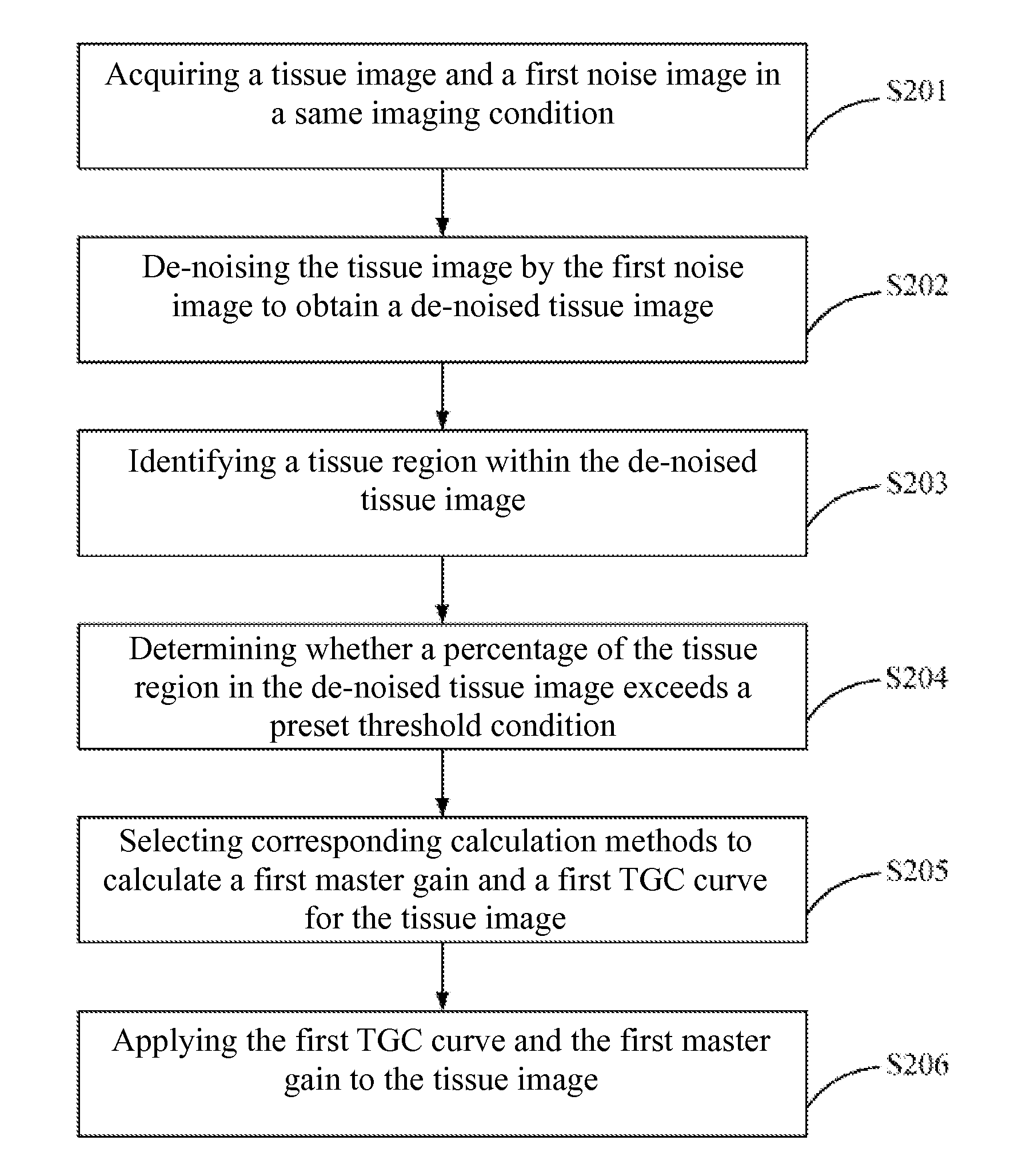
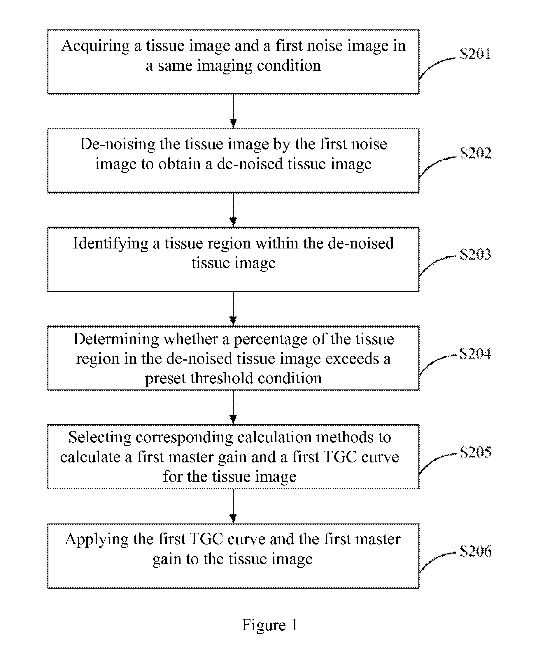
[0060]As shown in FIG. 1, a method for optimizing gain of an ultrasound image can be provided according to this disclosure. The method may include steps S201 to S206.

[0061]In step S201, a tissue image and a first noise image can be acquired under a same imaging condition.

[0062]In step S202, the tissue image can be de-noised using the first noise image to obtain a de-noised tissue image.

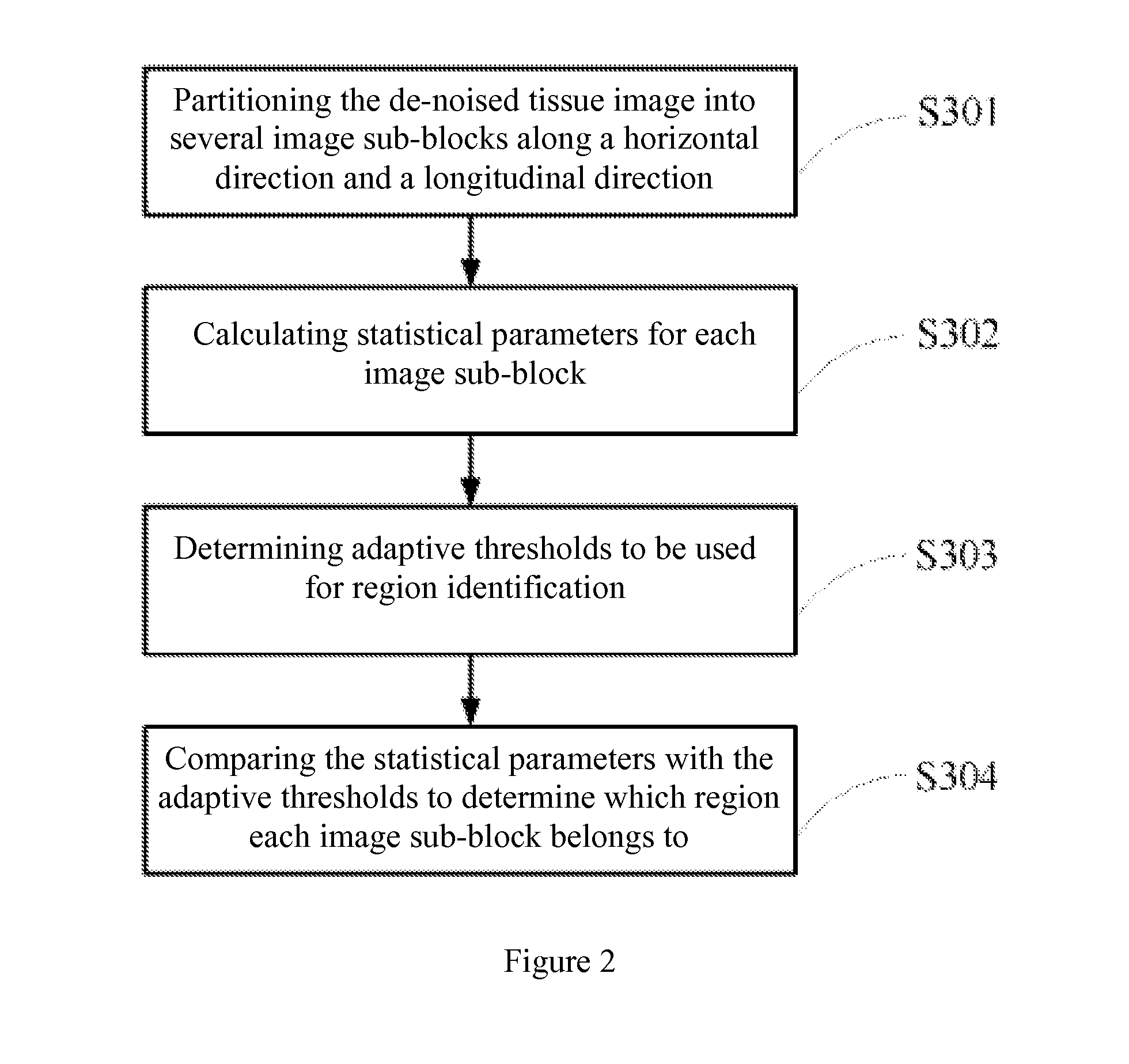
[0063]In step S203, a tissue region can be identified within the de-noised tissue image.

[0064]In step S204, it can be determined whether a percentage of the tissue region in the de-noised tissue image exceeds a preset threshold condition.

[0065]In step S205, a corresponding calculation method can be selected according to a determination result in step S204 to calculate a master gain and a TGC curve for the tissue image.

[0066]In step S206, the TGC curve and the master gain obtained through the calculation in step S205 can be applied to the tissue image acquired in step S201.

[0067]The tissue image acquir...

third embodiment

[0135]As shown in FIG. 7, an automatic gain optimization apparatus for ultrasound imaging can be provided according to this disclosure. The apparatus can include a first image processing module, a second image processing module, a first image output module and a second image output module. The first image processing module and the second image processing module can connect with an input port, the first image output module and the second image output module can connect with an output port, the first image processing module can connect with the first image output module, the second image processing module can connect with the second image output module, and the first image processing module can also connect with the second image processing module.

[0136]The first image processing module can receive image information, calculate a first master gain and a first TGC curve for the tissue image according to the above-described method in the first embodiment of this disclosure, transmit the f...

second embodiment

[0138]The second image processing module can receive the image information, calculate a second master gain and a second TGC curve of the contrast image according to the above-described method in this disclosure, and transmit the second TGC curve and the second master gain of the contrast image obtained through calculation to the second image output module.

[0139]The second image output module can apply the second master gain and the second TGC curve of the contrast image to the acquired contrast image, and output the contrast image with optimized gain through the output port.

[0140]The image information can be the fundamental image or the harmonic image and the corresponding noise image obtained during the B-mode imaging. The image information can also be the tissue image and the corresponding noise image obtained during the ultrasound contrast imaging.

[0141]In embodiments of this disclosure, when the automatic gain optimization apparatus receives the fundamental image or the harmonic...


PUM


Abstract

Methods for optimizing gain of an ultrasound image may include: acquiring a tissue image and a first noise image under a same imaging condition; de-noising the tissue image by the first noise image to obtain a de-noised tissue image; identifying a tissue region in the de-noised tissue image; determining whether a percentage of the tissue region in the de-noised tissue image exceeds a preset threshold condition; selecting, according to the determination result, a corresponding calculation method to calculate a first master gain and a first time gain compensation (TGC) curve for the tissue image; and applying the first TGC curve and the first master gain obtained through calculation to the tissue image acquired before.

Description

TECHNICAL FIELD[0001]This disclosure relates to ultrasound image optimization, and particularly to methods for optimizing gain of ultrasound images and automatic gain optimization apparatuses for ultrasound imaging.BACKGROUND[0002]Gain adjustment is important for ultrasound imaging. Correspondingly, ultrasound imaging systems may be generally provided with a fixed gain that is suitable for a majority of subjects. Since ultrasonic waves may be attenuated to a different extent within each patient, however, the fixed gain predetermined in the systems may not be suitable in all cases. At this point, users may need to manually adjust time gain compensation (TGC), lateral gain compensation (LGC) and a master gain. Such manual adjustment is time-consuming, and an optimal gain curve may not be obtained in this case, where automatic gain optimization can overcome these drawbacks and automatic gain optimization function may thus be equipped for many ultrasound imaging systems. However, automa...

Claims


Application Information

Patent Timeline
Patent Type & Authority Applications(United States)
IPC IPC(8): A61B8/08A61B8/00A61B8/14
CPCA61B8/5269A61B8/5207A61B8/461A61B8/14A61B8/481A61B8/54G01S7/52033G06T5/002G06T2207/10132G06T2207/30004
Inventor CHU, XIASANG, MAODONG
Owner SHENZHEN MINDRAY BIO MEDICAL ELECTRONICS CO LTD
Why Eureka
Try Eureka