Anti-mif antibody cell migration assay

a technology of antibody cell migration and anti-mif, which is applied in the field of anti-mif antibody cell migration assay, can solve the problems of inability to use the same for diagnosis or therapy, and no robust mif bioassay based on the inhibition of autocrine-induced cell migration of e.g. monocytic cells, and achieves the effect of reducing the number of false positives and reducing false negatives

Inactive Publication Date: 2015-10-29
BAXALTA GMBH +1
View PDF5 Cites 0 Cited by
  • Summary
  • Abstract
  • Description
  • Claims
  • Application Information

AI Technical Summary

Benefits of technology

[0022]“Potency” in this context is a measure of drug activity expressed in terms of the amount required to

Problems solved by technology

Without a clear indication of the respective potency of an anti-MIF antibody, the same cannot be used for either diagnosis or therapy.
Although it was shown in earlier assay formats that the MIF protein exhibits chemokine-like functions and that the

Method used


Image

  • Anti-mif antibody cell migration assay
  • Anti-mif antibody cell migration assay

Examples


example 1

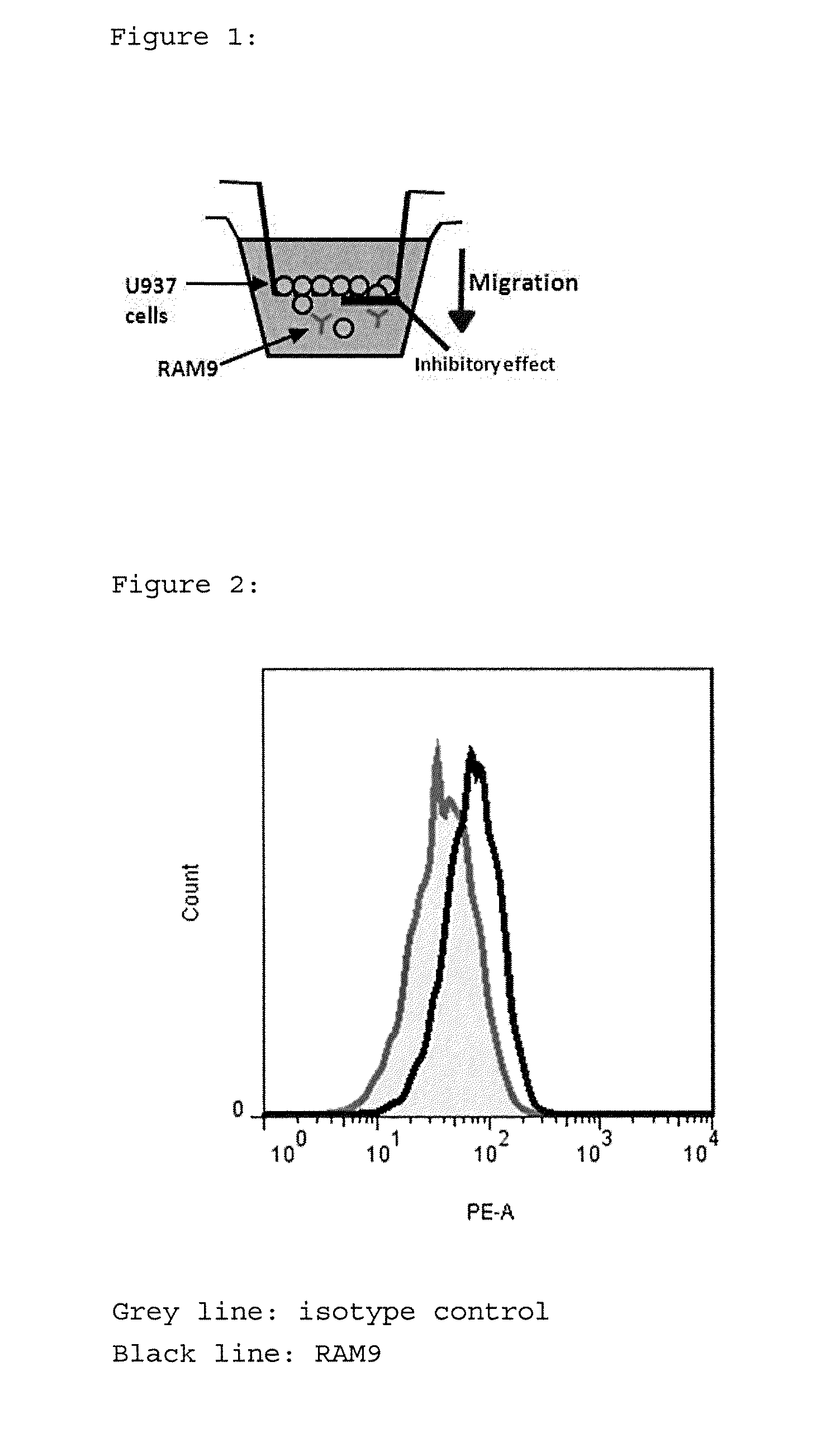
Anti-MIF Antibody RAM9 Chemokinesis Assay; Cell Based Assay

[0135]Intended Purpose:

[0136]The test was set up to test the functionality of Glycine-buffered anti-MIF RAM9 preparations (=test item) to inhibit random migration (=chemokinesis) of monocytic cells. It has been shown by the present inventors that this assay and respective method can be used as quality control test at the process step for the Final Drug Product (FDP).

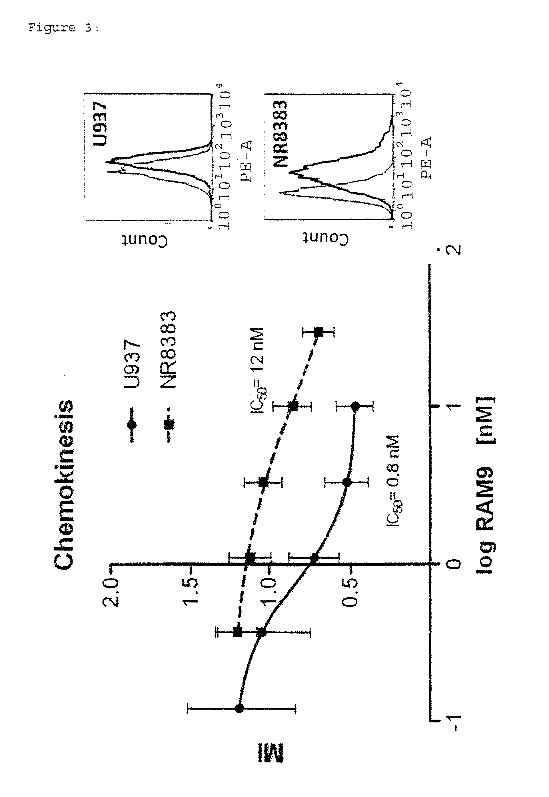
[0137]1) Rationale:[0138]MIF is constitutively expressed in 0397 (and other cancerous) monocytes and oxMIF is present on the cell surface of these cells (FACS data, see FIG. 2) where it supports migratory functions. It is an important feature of the present invention to use cells which express (ox)MIF endogenously or exogenously, like cells from disease samples. This principle is based on the earlier finding of the present inventors that oxMIF is not present in healthy cells or tissues. The U397 cell line is a preferred example to carry out the present invention....

example 2

[0249]In principle, the same assay method was used to determine whether the antibodies could be provided together with the cells in the upper chamber.

[0250]Short Summary:

[0251]Cell Migration Assay: HTS Transwell plates (Corning): 5 μm[0252]cells: U937 (10th passage), overnight starving[0253]RAM9 antibody: 30 nM-0.04 nM, pipetted into inserts[0254]Synagis® in Glycine: 30 nM; 10 nM; 3.3 nM pipetted into inserts[0255]Neg. control: Glycine; Pos. control: FBS (fetal bovine serum)[0256]Incubation: 2 plates, 16 h[0257]Cellavista® used for calculation of results

[0258]Results:

[0259]Plate 1

Conc. Antibody (nM)measuredfit01997.60.042452.72498.30.122490.82388.40.372019.22106.01.11642.01621.23.31179.61115.510688.0810.830753.7685.7Max=A2555Slope=B1.0IC 50=C1.2Min=D620sumxmy244358Fit: I % = (A − D) / (1 + (X / C)ExpB + D (=sigmoid curve)

[0260]Plate 2

CKonc. Antibody (nM)measuredfit02183.10.042562.82646.50.122685.22575.90.372354.22357.31.11872.51939.53.31621.21527.4101263.31323.0301266.71256.2Max=A2674Sl...


PUM

PropertyMeasurementUnit
Temperatureaaaaaaaaaa
Lengthaaaaaaaaaa
Timeaaaaaaaaaa

Abstract

The present invention pertains to a robust, precise and easy-to-use assay for potency of anti-MIF antibodies. In particular, the invention discloses an anti-MIF antibody-cell migration assay and respective method and kit.

Description

[0001]The present invention pertains to an assay which is suitable to test the potency of anti-MIF antibodies. In particular, the present invention is directed to an advantageous and easy-to-use cell migration assay.BACKGROUND[0002]Macrophage migration inhibitory factor (MIF) is a cytokine initially isolated based upon its ability to inhibit the in vitro random migration of peritoneal exudate cells from tuberculin hypersensitive guinea pigs (containing macrophages) (Bloom et al. Science 1966, 153, 80-2; David et al. PNAS 1966, 56, 72-7). Today, MIF is known as a critical upstream regulator of the innate and acquired immune response that exerts a pleiotropic spectrum of activities.[0003]The human MIF cDNA was cloned in 1989 (Weiser et al., PNAS 1989, 86, 7522-6), and its genomic localization was mapped to chromosome 22. The product of the human MIF gene is a protein with 114 amino acids (after cleavage of the N-terminal methionine) and an apparent molecular mass of about 12.5 kDa. MI...

Claims


Application Information

Patent Timeline
IPC IPC(8): G01N33/50C07K16/40
CPCG01N33/5047C07K16/40C07K2317/76G01N2333/4704G01N2333/99G01N33/5029G01N33/6863
Inventor THIELE, MICHAELKERSCHBAUMER, RANDOLF
Owner BAXALTA GMBH
Why Eureka
Try Eureka